

Focused Ultrasound-Enhanced Delivery of Intranasally Administered Anti-Programmed Cell Death-Ligand 1 Antibody to an Intracranial Murine Glioma Model
- PMID: 33535531
- PMCID: PMC7912734
- DOI: 10.3390/pharmaceutics13020190
Focused Ultrasound-Enhanced Delivery of Intranasally Administered Anti-Programmed Cell Death-Ligand 1 Antibody to an Intracranial Murine Glioma Model
Abstract
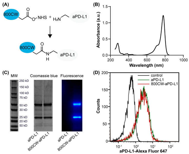
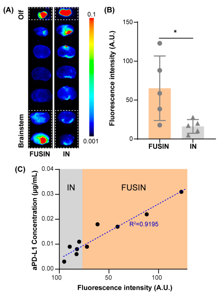
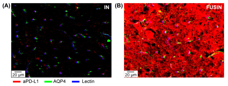
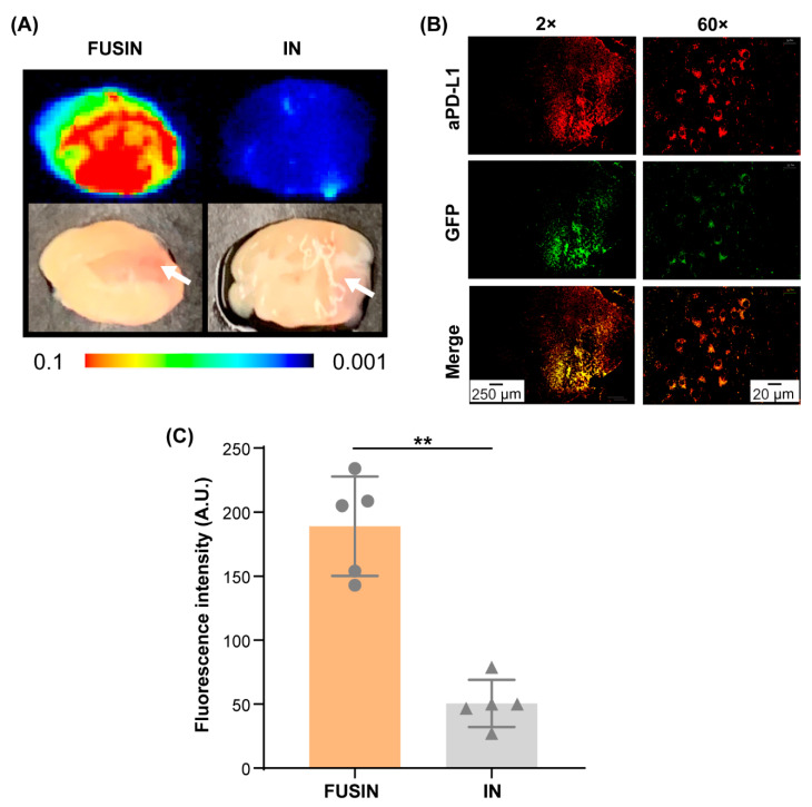
Immune checkpoint inhibitors have great potential for the treatment of gliomas; however, their therapeutic efficacy has been partially limited by their inability to efficiently cross the blood-brain barrier (BBB). The objective of this study was to evaluate the capability of focused-ultrasound-mediated intranasal brain drug delivery (FUSIN) in achieving the locally enhanced delivery of anti-programmed cell death-ligand 1 antibody (aPD-L1) to the brain. Both non-tumor mice and mice transcranially implanted with GL261 glioma cells at the brainstem were used in this study. aPD-L1 was labeled with a near-infrared fluorescence dye (IRDye 800CW) and administered to mice through the nasal route to the brain, followed by focused ultrasound sonication in the presence of systemically injected microbubbles. FUSIN enhanced the accumulation of aPD-L1 at the FUS-targeted brainstem by an average of 4.03- and 3.74-fold compared with intranasal (IN) administration alone in the non-tumor mice and glioma mice, respectively. Immunohistochemistry staining found that aPD-L1 was mainly located within the perivascular spaces after IN delivery, while FUSIN further enhanced the penetration depth and delivery efficiency of aPD-L1 to the brain parenchyma. The delivered aPD-L1 was found to be colocalized with the tumor cells after FUSIN delivery to the brainstem glioma. These findings suggest that FUSIN is a promising technique to enhance the delivery of immune checkpoint inhibitors to gliomas.
Keywords: blood-brain barrier; brain drug delivery; brainstem glioma; focused ultrasound; immune checkpoint inhibitor; intranasal delivery.
Conflict of interest statement
The authors declare no conflict of interest.
Figures
Similar articles
-
Focused ultrasound combined with microbubble-mediated intranasal delivery of gold nanoclusters to the brain.J Control Release. 2018 Sep 28;286:145-153. doi: 10.1016/j.jconrel.2018.07.020. Epub 2018 Jul 26. J Control Release. 2018. PMID: 30009893 Free PMC article.
-
Characterization of focused ultrasound-mediated brainstem delivery of intranasally administered agents.J Control Release. 2020 Dec 10;328:276-285. doi: 10.1016/j.jconrel.2020.08.053. Epub 2020 Aug 29. J Control Release. 2020. PMID: 32871204 Free PMC article.
-
Focused ultrasound-mediated intranasal brain drug delivery technique (FUSIN).MethodsX. 2021 Feb 9;8:101266. doi: 10.1016/j.mex.2021.101266. eCollection 2021. MethodsX. 2021. PMID: 34434788 Free PMC article.
-
Anticancer drug delivery by focused ultrasound-mediated blood-brain/tumor barrier disruption for glioma therapy: From benchside to bedside.Pharmacol Ther. 2023 Oct;250:108518. doi: 10.1016/j.pharmthera.2023.108518. Epub 2023 Aug 22. Pharmacol Ther. 2023. PMID: 37619931 Review.
-
Focused Ultrasound and Microbubbles-Mediated Drug Delivery to Brain Tumor.Pharmaceutics. 2020 Dec 24;13(1):15. doi: 10.3390/pharmaceutics13010015. Pharmaceutics. 2020. PMID: 33374205 Free PMC article. Review.
Cited by
-
Applications of Focused Ultrasound for the Treatment of Glioblastoma: A New Frontier.Cancers (Basel). 2022 Oct 8;14(19):4920. doi: 10.3390/cancers14194920. Cancers (Basel). 2022. PMID: 36230843 Free PMC article. Review.
-
Preclinical Research on Focused Ultrasound-Mediated Blood-Brain Barrier Opening for Neurological Disorders: A Review.Neurol Int. 2023 Feb 14;15(1):285-300. doi: 10.3390/neurolint15010018. Neurol Int. 2023. PMID: 36810473 Free PMC article. Review.
-
Mechanically manipulating glymphatic transport by ultrasound combined with microbubbles.Proc Natl Acad Sci U S A. 2023 May 23;120(21):e2212933120. doi: 10.1073/pnas.2212933120. Epub 2023 May 15. Proc Natl Acad Sci U S A. 2023. PMID: 37186852 Free PMC article.
-
Ultrasound-Targeted Microbubble Destruction: Modulation in the Tumor Microenvironment and Application in Tumor Immunotherapy.Front Immunol. 2022 Jul 1;13:937344. doi: 10.3389/fimmu.2022.937344. eCollection 2022. Front Immunol. 2022. PMID: 35844515 Free PMC article. Review.
-
Therapeutic ultrasound-enhanced immune checkpoint inhibitor therapy.Front Phys. 2021 Mar;9:636985. doi: 10.3389/fphy.2021.636985. Epub 2021 Mar 24. Front Phys. 2021. PMID: 37994329 Free PMC article.
References
-
- Bors L.A., Erdö F. Overcoming the blood-brain barrier. Challenges and tricks for CNS drug delivery. Sci. Pharm. 2019;87:6. doi: 10.3390/scipharm87010006. - DOI
Grants and funding
LinkOut - more resources
Full Text Sources
Other Literature Sources
