. 2021 Feb 1;8(2):9.
doi: 10.3390/medicines8020009.

The Infantile Leukoencephalopathy-Associated Mutation of C11ORF73/HIKESHI Proteins Generates de novo Interactive Activity with Filamin A, Inhibiting Oligodendroglial Cell Morphological Differentiation

Affiliations

The Infantile Leukoencephalopathy-Associated Mutation of C11ORF73/HIKESHI Proteins Generates de novo Interactive Activity with Filamin A, Inhibiting Oligodendroglial Cell Morphological Differentiation

Kohei Hattori et al. Medicines (Basel). .

Abstract

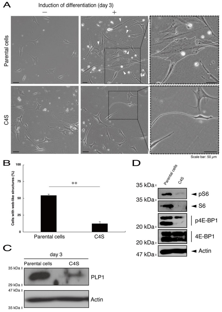
Genetic hypomyelinating diseases are a heterogeneous group of disorders involving the white matter. One infantile hypomyelinating leukoencephalopathy is associated with the homozygous variant (Cys4-to-Ser (C4S)) of the c11orf73 gene. Methods: We observed that in mouse oligodendroglial FBD-102b cells, the C4S mutant proteins but not the wild type ones of C11orf73 are microscopically localized in the lysosome. And, they downregulate lysosome-related signaling in an immunoblotting technique. Results: The C4S mutant proteins specifically interact with Filamin A, which is known to anchor transmembrane proteins to the actin cytoskeleton; the C4S mutant proteins and Filamin A are also observed in the lysosome fraction. While parental FBD-102b cells and cells harboring the wild type constructs exhibit morphological differentiation, cells harboring C4S mutant constructs do not. It may be that morphological differentiation is inhibited because expression of these C4S mutant proteins leads to defects in the actin cytoskeletal network involving Filamin A. Conclusions: The findings that leukoencephalopathy-associated C11ORF73 mutant proteins specifically interact with Filamin A, are localized in the lysosome, and inhibit morphological differentiation shed light on the molecular and cellular pathological mechanisms that underlie infantile hypomyelinating leukoencephalopathy.

Keywords: C11orf73; Filamin A; HLD13; leukoencephalopathy; oligodendrocyte differentiation.


Conflict of interest statement

The authors have declared that no competing interests exist.

Figures

Figure 1
Figure 1
The Cys4-to-Ser (C4S) mutant proteins of C11ORF73 accumulate into punctate structures in cells. (A) FBD-102b cells (oligodendroglial cell lines), which are surrounded by dotted lines, were transfected with a plasmid encoding the wild type (WT) or the C4S mutant construct of GFP-tagged C11ORF73 (green) and stained with DAPI (blue). A scan plot was performed along the white line in the direction of the arrow in the image. Graphs showing the fluorescence intensities (F.I., in an arbitrary unit) along the white lines can be seen in the right panels. Some punctate structures (indicated by arrowheads) and nuclear regions (surrounded by dotted lines) are also shown in the images and graphs. (B) Percentages of cells with punctate structures are statistically shown (**, p < 0.01 of Student’s t-test; n = 25 cells).
Figure 2
Figure 2
The C4S mutant proteins of C11ORF73 are not colocalized with the endoplasmic reticulum (ER) marker. (A) FBD-102b cells, which are surrounded by dotted lines, were transfected with a plasmid encoding the wild type (WT) GFP-tagged C11ORF73 (green) and stained with the ER marker KDEL antigen (red). A scan plot was performed along the white line in the direction of the arrow in the image. (B) Graphs showing the fluorescence intensities (F.I., in an arbitrary unit) along the white lines can be seen in the panels. (C) Cells were transfected with a plasmid encoding the C4S mutant construct of GFP-tagged C11ORF73 (green) and stained with the ER marker (red). A scan plot was performed along the white line in the direction of the arrow in the image. (D) Graphs showing the fluorescence intensities (F.I., in an arbitrary unit) along the white lines can be seen in the panels. (E) Merged percentages of mutant proteins with organelles (percentages of yellow-colored pixels/green-colored pixels) are shown in the graph (n = 3 independent experiments).
Figure 3
Figure 3
The C4S mutant proteins of C11ORF73 are not colocalized with the Golgi body marker. (A) FBD-102b cells, which are surrounded by dotted lines, were transfected with a plasmid encoding the wild type (WT) GFP-tagged C11ORF73 (green) and stained with the Golgi body marker GM130 (red). A scan plot was performed along the white line in the direction of the arrow in the image. (B) Graphs showing the fluorescence intensities (F.I., in an arbitrary unit) along the white lines can be seen in the panels. (C) Cells were transfected with a plasmid encoding the C4S mutant construct of GFP-tagged C11ORF73 (green) and stained with the Golgi body marker (red). Regions (ac) within white squares are magnified in the below panels. A scan plot was performed along the white line in the direction of the arrow in the image. (D) Graphs showing the fluorescence intensities (F.I., in an arbitrary unit) along the white lines can be seen in the panels. (E) Merged percentages of mutant proteins with organelles (percentages of yellow-colored pixels/green-colored pixels) are shown in the graph (n = 3 independent experiments).
Figure 4
Figure 4
The C4S mutant proteins of C11ORF73 are colocalized with the lysosome marker. (A) FBD-102b cells, which are surrounded by dotted lines, were transfected with a plasmid encoding the wild type (WT) GFP-tagged C11ORF73 (green) and stained with the lysosome marker LAMP1 (red). A scan plot was performed along the white line in the direction of the arrow in the image. (B) Graphs showing the fluorescence intensities (F.I., in an arbitrary unit) along the white lines can be seen in the panels. (C) Cells were transfected with a plasmid encoding the C4S mutant construct of GFP-tagged C11ORF73 (green) and stained with the lysosome marker (red). A scan plot was performed along the white line in the direction of the arrow in the image. (D) Graphs showing the fluorescence intensities (F.I., in an arbitrary unit) along the white lines can be seen in the panels. (E) Merged percentages of mutant proteins with organelles (percentages of yellow-colored pixels/green-colored pixels) are shown in the graph (n = 3 independent experiments).
Figure 5
Figure 5
The C4S as well as the V54L mutant proteins of C11ORF73 exhibit oligomeric structures in non-denaturing polyacrylamide gel electrophoresis. (A) The lysates of COS-7 cells were transfected with empty vectors or with a plasmid encoding wild type, Cys4-to-Ser (C4S), or Val54-to-Leu (V54L) C11ORF73, and then were subjected to non-denaturing polyacrylamide gel electrophoresis and immunoblotted with an anti-GFP antibody. Positions corresponding to the molecular weights of the C11ORF73 monomer, dimer, and trimer are also shown, and the oligomeric proteins of molecules with more than three units can be seen in the V54L mutant proteins. The lysates were also subjected to denaturing polyacrylamide gel electrophoresis and immunoblotted with an anti-actin or anti-GFP antibody. Immunoreactive bands below monomer could be degradative proteins of C11ORF73. (B) Immune-reactive band intensities in a range containing oligomeric proteins were quantitatively compared to those of wild-type proteins. (*, p < 0.05 of one-way analysis of variance (ANOVA) with post-hoc Fisher’s test; n = 3 blots).
Figure 6
Figure 6
Cells harboring the C4S mutant constructs fail to exhibit differentiated phenotypes. (A) FBD-102b cells stably harboring the C4S mutant constructs or their parental cells were allowed to differentiate for three days. Arrowheads indicate cells with web-like structures as an example of a differentiated phenotype. Some cells are surrounded by white dots. Square fields with dotted lines in the center panels are magnified in the right panels. (B) Cells with web-like structures are statistically shown (**, p < 0.01 of one-way ANOVA with post-hoc Fisher’s test; n = 3 fields). (C) The lysates of the respective cells were immunoblotted with an antibody against proteolipid protein 1 (PLP1) or control actin. (D) The lysates were also immunoblotted with an antibody against phosphorylated S6 (pS6), S6, phosphorylated 4E-BP1 (p4E-BP1), 4E-binding protein 1 (4E-BP1), or actin.
Figure 7
Figure 7
Cells harboring the wild type constructs exhibit the same differentiated phenotypes as their parental cells. (A) FBD-102b cells stably harboring the wild type constructs or their parental cells were allowed to differentiate for three days. Arrowheads indicate cells with web-like structures as an example of a differentiated phenotype. Some cells are surrounded by white dots. Square fields with dotted lines in the center panels are magnified in the right panels. (B) Cells with web-like structures are statistically shown (n = 3 fields).
Figure 8
Figure 8
Filamin A specifically interacts with the C4S mutant proteins which inhibit the formation of filamentous Filamin A structures at cell peripheral regions. (A) Proteins interacting with the wild type, C4S, or V54L mutant proteins (bait) were identified by protein-tag affinity-precipitation and MS analysis. (B) COS-7 cells were transfected with a plasmid encoding one of the two C11ORF73 mutant constructs or the wild type (WT). The lysates were immunoprecipitated with an anti-Filamin A antibody in preparation for immunoblotting with an anti-GFP (for C11ORF73 proteins) antibody. Total C11ORF73 and Filamin A proteins are shown. (C,D) FBD-102b cells were transfected with plasmids encoding one of the C11ORF73 constructs (green) and stained with an anti-Filamin A antibody (red). Regions within white squares in merged images are magnified in the right panels. A scan plot was performed along the white line in the direction of the arrow in the image. (E) Merged percentages of mutant proteins with organelles (percentages of yellow-colored pixels/green-colored pixels) are shown in the graph (n = 3 independent experiments). (F) Cells with peripheral filamentous structures of Filamin A proteins are statistically shown (*, p < 0.05 of one-way ANOVA with post-hoc Fisher’s test; n = 3 fields).
Figure 9
Figure 9
Formation of filamentous actin structures at cell peripheral regions is inhibited in cells expressing C4S mutant proteins. (A,B) FBD-102b cells were transfected with plasmids encoding one of the C11ORF73 mutant constructs or the wild type (WT) (green) and stained with Phalloidin (red). Cells with peripheral filamentous structures of actin proteins are statistically shown (**, p < 0.01 of one-way ANOVA with post-hoc Fisher’s test; n = 3 fields). (C,D) Cells were transfected with plasmids encoding one of the C11ORF73 constructs (green) and stained with an anti-F-actin antibody (red). Cells with peripheral filamentous structures of actin proteins are statistically shown (*, p < 0.05 of one-way ANOVA with post-hoc Fisher’s test; n = 3 fields).
Figure 10
Figure 10
The C4S mutant proteins and Filamin A are present in the lysosome fraction. COS-7 cells were transfected with a plasmid encoding the wild type (WT), C4S, or V54L mutant construct. The lysates obtained from cells destroyed with isotonic solution were immunoprecipitated with an anti-SLC38A9 antibody in preparation for immunoblotting with an antibody against Filamin A or green fluorescent protein (GFP) (for C11ORF73 proteins). Immunoprecipitates were also immunoblotted with antibodies against KDEL antigen, Golgi matrix protein of 130 kDa (GM130), and lysosomal-associated membrane protein 1 (LAMP1), whose immunoreactive bands indicated that control proteins were present in the lysates of total cells. Total Filamin A, C11ORF73, and actin proteins can be seen in the lower three blots.

Similar articles

Cited by

References

    1. Simons M., Lyons D.A. Axonal selection and myelin sheath generation in the central nervous system. Curr. Opin. Cell Biol. 2013;25:512–519. doi: 10.1016/j.ceb.2013.04.007. - DOI - PubMed
    1. Morton P.D., Ishibashi N., Jonas R.A., Gallo V. Congenital cardiac anomalies and white matter injury. Trends Neurosci. 2015;38:353–363. doi: 10.1016/j.tins.2015.04.001. - DOI - PMC - PubMed
    1. Saab A.S., Nave K.-A. Myelin dynamics: Protecting and shaping neuronal functions. Curr. Opin. Neurobiol. 2017;47:104–112. doi: 10.1016/j.conb.2017.09.013. - DOI - PubMed
    1. Abu-Rub M., Miller R.H. Emerging cellular and molecular strategies for enhancing central nervous system (CNS) remyelination. Brain Sci. 2018;8:111. doi: 10.3390/brainsci8060111. - DOI - PMC - PubMed
    1. Garbern J., Cambi F., Shy M.E., Kamholz J. The molecular pathogenesis of Pelizaeus-Merzbacher disease. Arch. Neurol. 1999;56:1210. doi: 10.1001/archneur.56.10.1210. - DOI - PubMed

LinkOut - more resources