. 2021 Feb 1;22(3):1443.
doi: 10.3390/ijms22031443.

Plant Seed Mucilage as a Glue: Adhesive Properties of Hydrated and Dried-in-Contact Seed Mucilage of Five Plant Species

Affiliations

Plant Seed Mucilage as a Glue: Adhesive Properties of Hydrated and Dried-in-Contact Seed Mucilage of Five Plant Species

Agnieszka Kreitschitz et al. Int J Mol Sci. .

Abstract

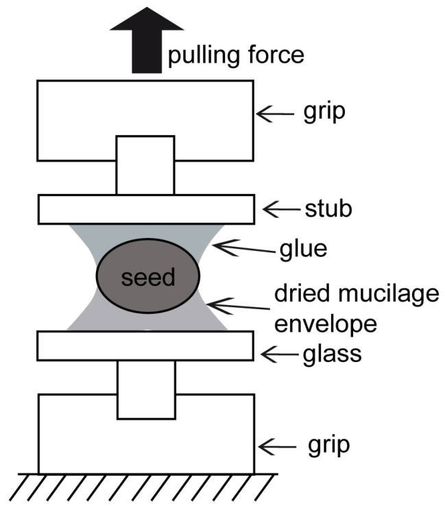
Seed and fruit mucilage is composed of three types of polysaccharides-pectins, cellulose, and hemicelluloses-and demonstrates adhesive properties after hydration. One of the important functions of the mucilage is to enable seeds to attach to diverse natural surfaces. Due to its adhesive properties, which increase during dehydration, the diaspore can be anchored to the substrate (soil) or attached to an animal's body and dispersed over varied distances. After complete desiccation, the mucilage envelope forms a thin transparent layer around the diaspore creating a strong bond to the substrate. In the present study, we examined the mucilaginous seeds of six different plant taxa (from genera Linum, Lepidium, Ocimum, Salvia and Plantago) and addressed two main questions: (1) How strong is the adhesive bond of the dried mucilage envelope? and (2) What are the differences in adhesion between different mucilage types? Generally, the dried mucilage envelope revealed strong adhesive properties. Some differences between mucilage types were observed, particularly in relation to adhesive force (Fad) whose maximal values varied from 0.58 to 6.22 N. The highest adhesion force was revealed in the cellulose mucilage of Ocimum basilicum. However, mucilage lacking cellulose fibrils, such as that of Plantago ovata, also demonstrated high values of adhesion force with a maximum close to 5.74 N. The adhesion strength, calculated as force per unit contact area (Fad/A0), was comparable between studied taxa. Obtained results demonstrated (1) that the strength of mucilage adhesive bonds strongly surpasses the requirements necessary for epizoochory and (2) that seed mucilage has a high potential as a nontoxic, natural substance that can be used in water-based glues.

Keywords: Linum; Ocimum; Plantago; cell wall; diaspores; seed mucilage.


Conflict of interest statement

The authors declare no conflict of interest.

Figures

Figure 1
Figure 1
Probe fixation in pull-off experiments. The sample with the seed was placed between two grips: the lower one was motionless and the upper one moved upwards. The lower seed surface was attached to the aluminum SEM stub by the dried mucilage envelope. The upper (mucilage free) seed surface was glued to the SEM stub by the commercial glue. The upper grip moved up and stopped when the seed was detached from the glass slide.
Figure 2
Figure 2
Mucilage morphology presented using optical microscopy. (AI) Staining with ruthenium red. Characteristic purple color indicates the presence of pectins in the mucilage; (A) L. usitatissimum; (B) L. sativum; (CE) O. basilicum; (D) Characteristic “mucilaginous tubules” with spirally coiled cellulose fibrils (cf) with enclosed inside starch grains (sg) in the mucilage of O. basilicum; (E) staining of starch grains with IKI. The grains have dark coloration; (F) S. sclarea mucilage with delicate, radially spread cellulose fibrils; (G) P. lanceolata; (H,I) P. ovata seed mucilage (H) and husk mucilage (I); (JM) detection of crystalline cellulose (cc) (polarized light microscope) in the mucilage envelope; (J) L. sativum; (K) O. basilicum; (L) S. sclarea; (M) P. lanceolata.
Figure 3
Figure 3
(AC) Adhesion force (A), contact area (B) and adhesive strength (C) of the seed mucilage dried out in contact. (A’C’) Multiple comparison graphs of the adhesion force (A’), contact area (B’) and strength force (C’) of the seed mucilage. Statistically significant differences are marked with dots. Abbreviations: Lu—L. usitatissimum, Ls—L. sativum, Ob—O. basilicum, Sh—S. hispanica, Pl—P. lanceolata, Po—P. ovata, Uh—UHU glue. In the box-and-whisker diagrams, the bottom and top of the box are the 25th and 75th percentiles, the line inside the box is the median; the ends of the whiskers are the 10th and 90th percentiles.
Figure 4
Figure 4
Adhesion force of P. ovata husk mucilage and UHU glue control sample. Abbreviations: Po-h—Plantago ovata husk mucilage, Uh—UHU glue. In the box-and-whisker diagram, the bottom and top of the box are the 25th and 75th percentiles, the line inside the box is the median; the ends of the whiskers are the 10th and 90th percentiles.
Figure 5
Figure 5
Seed mucilage structure in the fractures of the adhesive layer after pull-off measurements. (A) L. usitatissimum seed with small remains of the mucilage envelope (me, arrows) on the seed surface; (B) L. sativum; (C) O. basilicum; (D) S. hispanica; (E) P. lanceolata and visible small fragments of the mucilage envelope (me, arrows); (F) P. ovata. The mucilage envelope after drying formed a thin, transparent layer. After measurements, when the seed was detached from the glass, that thin layer stayed partially attached to the seed, which is visible as small (A,E) or larger fragments on its surface (BD,F).
Figure 6
Figure 6
Mucilage envelope fractures after pull-off tests. (A) L. usitatissimum; (B) L. sativum. In both taxa, homogenous edges are visible; (C) O. basilicum, visible cellulose fibrils (cf) protruding from the mucilage envelope; (D) S. hispanica, fragment of the mucilage with visible lamellar structure; (E) nearly homogeneous mucilage layer of P. lanceolata; (F) P. ovata seed mucilage; (G) P. ovata husk mucilage; (H) wrinkled UHU glue layer.

References

    1. Zykwinska A.W., Thibault J.-F.J., Ralet M.-C.J. Organization of pectic arabinan and galactan side chains in association with cellulose microfibrils in primary cell walls and related models envisaged. J. Exp. Bot. 2007;58:1795–1802. doi: 10.1093/jxb/erm037. - DOI - PubMed
    1. Donaldson L. Cellulose microfibril aggregates and their size variation with cell wall type. Wood. Sci. Technol. 2007;41:443–460. doi: 10.1007/s00226-006-0121-6. - DOI
    1. Jarvis M.C. Plant cell walls: Supramolecular assembly, signalling and stress. Struct. Chem. 2009;20:245–253. doi: 10.1007/s11224-009-9427-y. - DOI
    1. Kreitschitz A., Gorb S.N. How does the cell wall ‘stick’ in the mucilage? A detailed microstructural analysis of the seed coat mucilaginous cell wall. Flora. 2017;229:9–22. doi: 10.1016/j.flora.2017.02.010. - DOI
    1. Grubert M. Studies on the distribution of myxospermy among seeds and fruits of Angiospermae and its ecological importance. Acta Biol. Venez. 1974;8:315–551.

LinkOut - more resources