Review
. 2021 Feb 1;22(3):1448.
doi: 10.3390/ijms22031448.

Immune Actions on the Peripheral Nervous System in Pain

Affiliations
Review

Immune Actions on the Peripheral Nervous System in Pain

Jessica Aijia Liu et al. Int J Mol Sci. .

Abstract

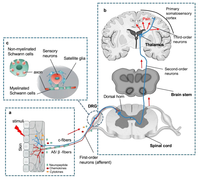
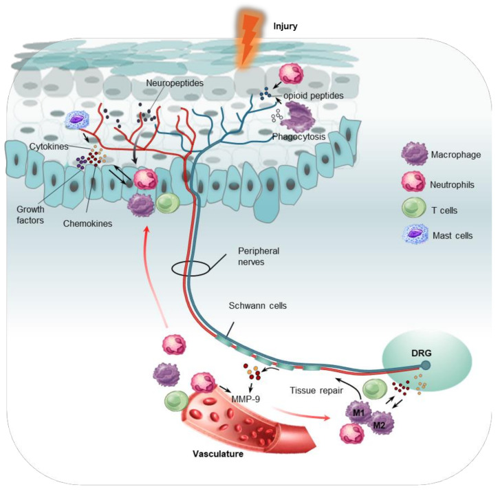
Pain can be induced by tissue injuries, diseases and infections. The interactions between the peripheral nervous system (PNS) and immune system are primary actions in pain sensitizations. In response to stimuli, nociceptors release various mediators from their terminals that potently activate and recruit immune cells, whereas infiltrated immune cells further promote sensitization of nociceptors and the transition from acute to chronic pain by producing cytokines, chemokines, lipid mediators and growth factors. Immune cells not only play roles in pain production but also contribute to PNS repair and pain resolution by secreting anti-inflammatory or analgesic effectors. Here, we discuss the distinct roles of four major types of immune cells (monocyte/macrophage, neutrophil, mast cell, and T cell) acting on the PNS during pain process. Integration of this current knowledge will enhance our understanding of cellular changes and molecular mechanisms underlying pain pathogenies, providing insights for developing new therapeutic strategies.

Keywords: immune response; inflammation; pain; peripheral nervous system.


Conflict of interest statement

The authors declare no conflict of interest.

Figures

Figure 1
Figure 1
An overview of peripheral nervous system (PNS) in the sensory pathways leading from the skin to the brain. (a) The peripheral nerve endings comprise both unmyelinated c-fibers and myelinated Aδ/β -fibers in the skin that sense a stimulus, where chemicals such as inflammatory mediators and neuropeptides released from the injury site or the nerve endings activate the receptors and channels on the adjacent peripheral nerve terminals, subsequently resulting in initiating an action potential at the initial segment of the axon. (b) The axon of the peripheral sensory neuron (first order neuron) enters the spinal cord and contacts second-order neuron in the gray matter, where an action potential is generated at the initial segment of this neuron and travels up the sensory pathway to a region of the brainstem and thalamic nuclei. The sensory signal reaches the third-order neurons from the thalamus, and these project pain signaling to several cortical and subcortical regions (red arrows). (c) Schematic of organization of dorsal root ganglion (DRG). Sensory neuronal bodies are separated and wrapped satellite glial sheath. Myelinated Schwann cells envelop large diameter axons of sensory neurons, whereas nonmyelinated Schwann cells ensheath small diameter axons forming a remark bundle (upper left in (b)).
Figure 2
Figure 2
Interactions between distinct parts of the PNS with immune cells (neutrophils, macrophages, mast cells and T cells). Upon stimuli, injury initiates the release of inflammatory mediators (cytokines and chemokines) that cause degranulation of mast cells close to the nerve terminal, resulting in proinflammatory production. Nociceptive nerve terminals secrete neuropeptides through antidromic activation of neighboring nerve terminal branches to attract immune cells. Activated Schwann cells and neutrophils mediate breakdown of the blood–nerve barrier via the secretion of matrix metalloproteinase 9(MMP-9), promoting infiltration of immune cells including macrophages, T cells to the DRG and peripheral nerve ending. These cells, once activated, release a battery of inflammatory mediators (growth factors, cytokines, and chemokines) that act on receptors expressed on adjacent nociceptor nerve terminals, leading to nociceptor sensitization. Macrophages exhibit functions in mediating phagocytosis and tissue repair. Both macrophages and neutrophils inhibit nociceptive effects by releasing opioid peptides at injury sites.

Similar articles

Cited by

References

    1. Basbaum A.I., Bautista D.M., Scherrer G., Julius D. Cellular and molecular mechanisms of pain. Cell. 2009;139:267–284. doi: 10.1016/j.cell.2009.09.028. - DOI - PMC - PubMed
    1. Baral P., Udit S., Chiu I.M. Pain and immunity: Implications for host defence. Nat. Rev. Immunol. 2019;19:433–447. doi: 10.1038/s41577-019-0147-2. - DOI - PMC - PubMed
    1. Chung G., Jung S.J., Oh S.B. Cellular and molecular mechanisms of dental nociception. J. Dent. Res. 2013;92:948–955. doi: 10.1177/0022034513501877. - DOI - PubMed
    1. Slavin K.V. Peripheral nerve stimulation for neuropathic pain. Neurotherapeutics. 2008;5:100–106. doi: 10.1016/j.nurt.2007.11.005. - DOI - PMC - PubMed
    1. Gold M.S., Gebhart G.F. Nociceptor sensitization in pain pathogenesis. Nat. Med. 2010;16:1248–1257. doi: 10.1038/nm.2235. - DOI - PMC - PubMed

MeSH terms

LinkOut - more resources