Review
. 2021 Feb 1;11(2):179.
doi: 10.3390/brainsci11020179.

Shaken Baby Syndrome: Magnetic Resonance Imaging Features in Abusive Head Trauma

Affiliations
Review

Shaken Baby Syndrome: Magnetic Resonance Imaging Features in Abusive Head Trauma

Gaia Cartocci et al. Brain Sci. .

Abstract

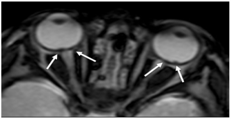
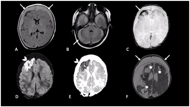
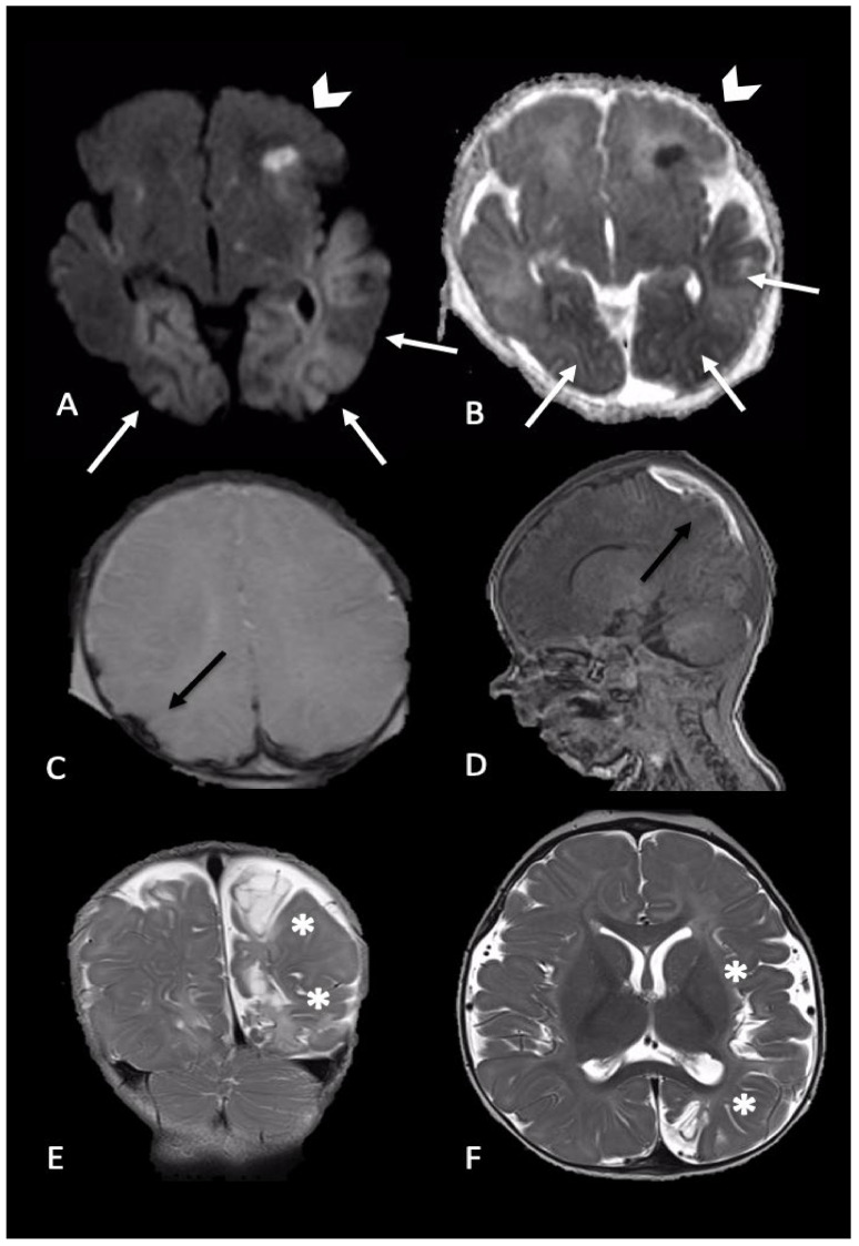
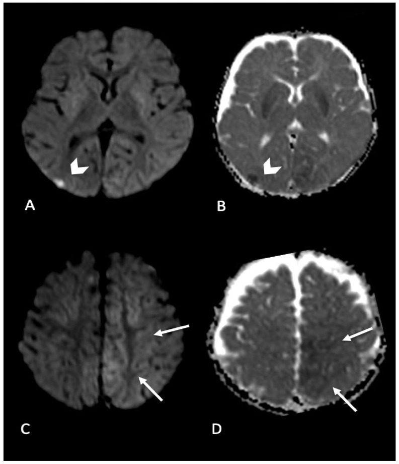
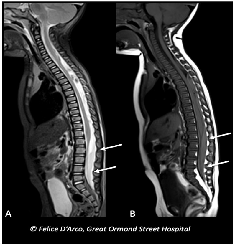
In the context of child abuse spectrum, abusive head trauma (AHT) represents the leading cause of fatal head injuries in children less than 2 years of age. Immature brain is characterized by high water content, partially myelinated neurons, and prominent subarachnoid space, thus being susceptible of devastating damage as consequence of acceleration-deceleration and rotational forces developed by violent shaking mechanism. Diagnosis of AHT is not straightforward and represents a medical, forensic, and social challenge, based on a multidisciplinary approach. Beside a detailed anamnesis, neuroimaging is essential to identify signs suggestive of AHT, often in absence of external detectable lesions. Magnetic resonance imaging (MRI) represents the radiation-free modality of choice to investigate the most typical findings in AHT, such as subdural hematoma, retinal hemorrhage, and hypoxic-ischemic damage and it also allows to detect more subtle signs as parenchymal lacerations, cranio-cervical junction, and spinal injuries. This paper is intended to review the main MRI findings of AHT in the central nervous system of infants, with a specific focus on both hemorrhagic and non-hemorrhagic injuries caused by the pathological mechanisms of shaking. Furthermore, this review provides a brief overview about the most appropriate and feasible MRI protocol to help neuroradiologists identifying AHT in clinical practice.

Keywords: MRI; abusive head trauma; child; infant; neuroimaging; shaking mechanism.


Conflict of interest statement

The authors declare no conflict of interest.

Figures

Figure 1
Figure 1
Axial GRE imaging shows irregular thickening and hypointensity of bilateral posterior ocular globes, indicative of retinal hemorrhage (arrows).
Figure 2
Figure 2
Newborn first accessed in the emergency department with fever and vomit, then was hospitalized four days later due to generalized seizures. CT showed mixed-density bilateral fluid collections (not shown). MRI demonstrated bilateral subdural CSF-blood collection, supra- and infra-tentorial (A,B axial FLAIR, arrows); subarachnoid blood products are revealed by susceptibility weighted imaging in right frontal lobe (C, axial SWI, arrow); intraparenchymal cortical-subcortical hemorrhage in the right frontal lobe (D,E, axial DWI and ADC map, arrows-head). One-month follow-up MRI (F, axial T2) showed malacic evolution of intraparenchymal damage (asterisks) and bilateral subdural hygromas (arrows).
Figure 3
Figure 3
Newborn reached the emergency department for single episode of apnea followed by generalized seizures. MRI performed 4 days later showed intraparenchymal injuries and bilateral extra-axial mixed-intensity fluid collections. In particular, (A,B) (axial DWI and ADC maps) showed bilateral temporal and occipital cortical areas of restricted diffusion (white arrows) and a focal area of restricted diffusion localized in the left frontal lobe and diffuse (arrows-head), indicative of acute intraparenchymal injury; (C,D) (axial SWI and sagittal T1-W) showed extra-axial blood collection along the parietal convexity bilaterally (black arrows). Six-month follow-up MRI ((E,F), coronal and axial T2-W) revealed malacic evolution of intraparenchymal damage and consequent expansion of extra-axial spaces due to tissue loss (asterisks).
Figure 4
Figure 4
Infant found with right side focal seizures with secondary generalization. Bruises on the right ear, retinal hemorrhages and subdural hemorrhages. MRI performed in the emergency department showed bilateral parenchymal injury. In particular, in (A,B) (axial DWI and ADC map) diffusion weighted imaging revealed focal area of restricted diffusion in right occipital cortex (arrow’s head); (C,D) (axial DWI and ADC map) showed areas of restricted diffusion in left mesial parietal cortex (arrows).
Figure 5
Figure 5
Infant with multiple long bones and ribs fractures (X-ray not shown), and bilateral subdural blood collections at the convexity (brain MRI not shown), subsequently underwent MRI spine examination showing extra-axial mixed-intensity fluid collection indicative of subdural hemorrhages at the level of lumbar spine (arrows) (A and B, sagittal T1 and T2).

References

    1. Keenan H.T., Runyan D.K., Marshall S.W., Nocera M.A., Merten D.F., Sinal S.H. A population-based study of inflicted traumatic brain injury in young children. JAMA. 2003;290:621–626. doi: 10.1001/jama.290.5.621. - DOI - PubMed
    1. Parks S.E., Kegler S.R., Annest J.L., Mercy J.A. Characteristics of fatal abusive head trauma among children in the USA: 2003–2007: An application of the CDC operational case definition to national vital statistics data. Inj. Prev. 2012;18:193–199. doi: 10.1136/injuryprev-2011-040128. - DOI - PubMed
    1. Mian M., Shah J., Dalpiaz A., Schwamb R., Miao Y., Warren K., Khan S. Shaken Baby Syndrome: A review. Fetal Pediatr. Pathol. 2015;34:169–175. doi: 10.3109/15513815.2014.999394. - DOI - PubMed
    1. Caffey J. Multiple fractures in the long bones of infants suffering from chronic subdural hematoma. Am. J. Roentgenol. Radium Ther. 1946;56:163–173. doi: 10.1007/s11999-010-1666-0. - DOI - PubMed
    1. Caffey J. On the theory and practice of shaking infants. Its potential residual effects of permanent brain damage and mental retardation. Am. J. Dis. Child. 1972;124:161–169. doi: 10.1001/archpedi.1972.02110140011001. - DOI - PubMed

LinkOut - more resources