. 2021 Feb 3;14(1):40.
doi: 10.1186/s12920-021-00889-z.

Integrated transcriptome and proteome analyses identify novel regulatory network of nucleus pulposus cells in intervertebral disc degeneration

Affiliations

Integrated transcriptome and proteome analyses identify novel regulatory network of nucleus pulposus cells in intervertebral disc degeneration

Chen Xu et al. BMC Med Genomics. .

Abstract

Background: Degeneration of intervertebral disc is a major cause of lower back pain and neck pain. Studies have tried to unveil the regulatory network using either transcriptomic or proteomic analysis. However, neither have fully elucidated the exact mechanism of degeneration process. Since post-transcriptional regulation may affect gene expression by modulating the translational process of mRNA to protein product, a combined transcriptomic and proteomic study may provide more insight into the key regulatory network of Intervertebral disc degeneration.

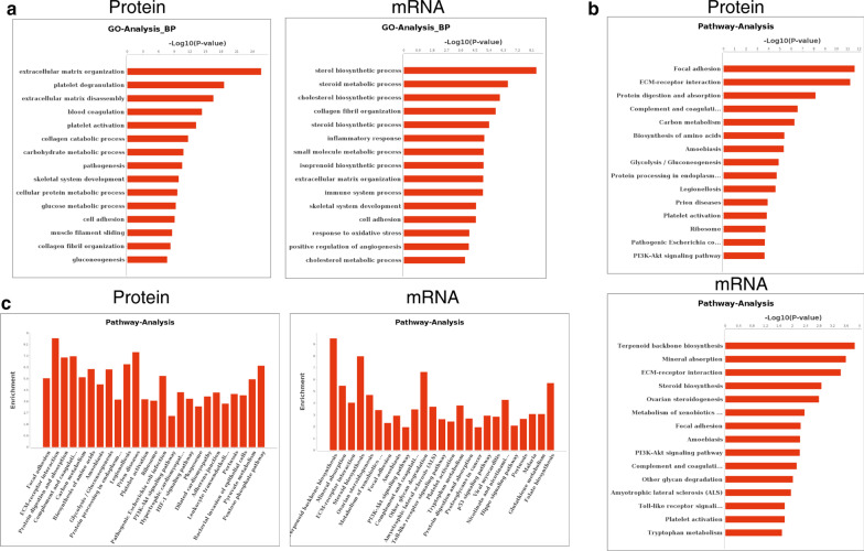
Methods: In order to obtain the proteomic and transcriptomic data, we performed label-free proteome analysis on freshly isolated nucleus pulposus cells and obtained transcriptome profiling data from the Gene Expression Omnibus repository. To identify the key regulatory network of intervertebral disc degeneration in nucleus pulposus cells, we performed bioinformatic analyses and established a protein-RNA interacting network. To validate the candidate genes, we performed in vitro experimentation and immunochemistry labeling to identify their potential function during nucleus pulposus degeneration.

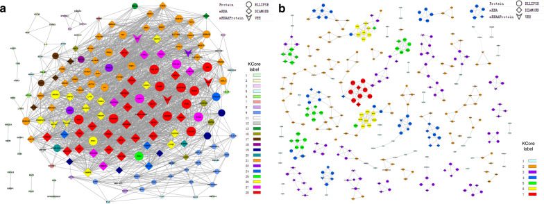
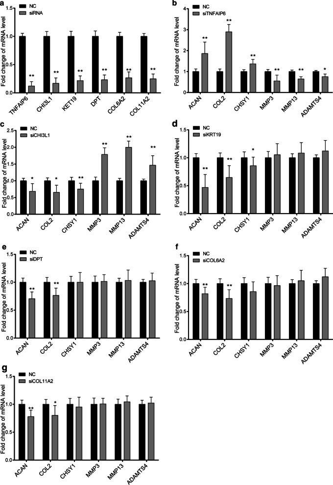
Results: The label-free proteome analysis identified altogether 656 proteins, and 503 of which were differentially expressed between nucleus pulposus cells from degenerated or normal disc cells. Using the existing nucleus pulposus transcriptomic profiling data, we integrated the proteomic and transcriptomic data of nucleus pulposus cells, and established a protein-RNA interacting network to show the combined regulatory network of intervertebral disc degeneration. In the network, we found 9 genes showed significant changes, and 6 of which (CHI3L1, KRT19, COL6A2, DPT, TNFAIP6 and COL11A2) showed concordant changes in both protein and mRNA level. Further functional analysis showed these candidates can significantly affect the degeneration of the nucleus pulposus cell when altering their expression.

Conclusions: This study is the first to use combined analysis of proteomic and transcriptomic profiling data to identify novel regulatory network of nucleus pulposus cells in intervertebral disc degeneration. Our established protein-RNA interacting network demonstrated novel regulatory mechanisms and key genes that may play vital roles in the pathogenesis of intervertebral disc degeneration.

Keywords: Bioinformatic analysis; Intervertebral disc degeneration; Nucleus pulposus; Proteomics; Transcriptome.


Conflict of interest statement

The authors have no conflict of interest to declare.

Figures

Fig.1
Fig.1
Assessing the differentially expressed mRNAs and proteins of NP cells in intervertebral disc degeneration. The unsupervised Hierarchical cluster plot of the label-free proteomic sequencing data (a) or RNA sequencing data (c) showing the differentially expressed proteins (a) or mRNAs (c) of NP cells from IVD and IDD groups. The green color indicates relatively lower expression of each gene, while the red color indicates higher expression. The volcano plot showing the degree of fold changes of each gene’s protein (b) or mRNA (d) expression in NP cells between IVD and IDD groups. e A Venn plot showing the number of genes that have significant changes in either protein level, mRNA level or both. f The list of the overlapped genes that are significantly changed during disc degeneration
Fig. 2
Fig. 2
Functional analysis of differentially expressed proteins and mRNAs in the NP cells during intervertebral disc degeneration. a Gene Ontology (GO) analysis showing the significance (P-value) of each GO function categories according to the differentially expressed genes. Pathway analysis showing the b significance (P-value) or c enrichment of different pathways according to the differentially expressed genes
Fig.3
Fig.3
Protein and mRNA Co-expression network of normal and degenerated NP cells. The gene co-expression network (GCN) of IDD (a) or IVD (b) group are constructed using the differentially expressed mRNAs and proteins according to their expression level. Different shapes of the nodes represent a gene that is either differentially expressed in protein, mRNA or both protein and mRNA level in the IDD group. The index of K-Core represents different proposed interaction groups, the importance of each color group raises with K-Core values
Fig. 4
Fig. 4
Verification of the expression of candidate genes in normal (IVD) and degenerated (IDD) NP primary cells. a The expression of TNFAIP6, CHI3L1, KRT19, DPT, COL6A2, and COL11A2 were examined in normal (IVD, n = 6) and degenerated (IDD, n = 6) NP tissues using immunohistochemistry. b The quantifications of the immunohistochemistry analysis were performed to show the relative expression level of the candidate genes. c The RNA expression level of these candidate genes in normal (IVD, n = 3) and degenerated (IDD, n = 3) NP primary cells were assessed using real-time PCR method. d The RNA expression level of these candidate genes in normal (NC, n = 3) and IL-1β (50 ng/ml, resembles the degeneration process in vitro, n = 3) treated NP primary cells were assessed using real-time PCR method. RNA level of GAPDH was used as internal reference. All Data are represented as mean ± SD, **p < 0.01
Fig. 5
Fig. 5
Functional study of candidate genes in degenerated NP primary cells. a The efficiency of TNFAIP6, CHI3L1, KRT19, DPT, COL6A2, and COL11A2 knockdown were examined in normal NP cells. NC group represents NP cell transfection using a scramble siRNA that showed no potential target to these genes. The RNA expression level of NP ECM related anabolic genes (ACAN, COL2 and CHSY1) and catabolic genes (MMP3, MMP13, ADAMTS4) were examined in TNFAIP6 (b), CHI3L1 (c), KRT19 (d), DPT (e), COL6A2 (f), and COL11A2 (g) knockdown NP cells using real-time PCR method. RNA level of GAPDH was used as internal reference. All Data are represented as mean ± SD, *p < 0.05, *p < 0.01

Similar articles

Cited by

References

    1. Lai A, Moon A, Purmessur D, Skovrlj B, Winkelstein BA, Cho SK, et al. Assessment of functional and behavioral changes sensitive to painful disc degeneration. J Orthop Res Off Publ Orthop Res Soc. 2015;33(5):755–764. doi: 10.1002/jor.22833. - DOI - PMC - PubMed
    1. Hu B, Shi C, Tian Y, Zhang Y, Xu C, Chen H, et al. TGF-beta induces up-regulation of chondroitin sulfate synthase 1 (CHSY1) in nucleus pulposus cells through MAPK signaling. Cell Physiol Biochem Int J Exp Cell Physiol Biochem Pharmacol. 2015;37(2):793–804. doi: 10.1159/000430396. - DOI - PubMed
    1. Hu B, Xu C, Tian Y, Shi C, Zhang Y, Deng L, et al. Inflammatory microRNA-194 and -515 attenuate the biosynthesis of chondroitin sulfate during human intervertebral disc degeneration. Oncotarget. 2017;8(30):49303–49317. doi: 10.18632/oncotarget.17571. - DOI - PMC - PubMed
    1. Nakayama E, Matsumoto T, Kazama T, Kano K, Tokuhashi Y. Transplantation of dedifferentiation fat cells promotes intervertebral disc regeneration in a rat intervertebral disc degeneration model. Biochem Biophys Res Commun. 2017;493(2):1004–1009. doi: 10.1016/j.bbrc.2017.09.101. - DOI - PubMed
    1. Nikkhoo M, Wang JL, Parnianpour M, El-Rich M, Khalaf K. Biomechanical response of intact, degenerated and repaired intervertebral discs under impact loading—ex-vivo and in-silico investigation. J Biomech. 2018;70:26–32. doi: 10.1016/j.jbiomech.2018.01.026. - DOI - PubMed

LinkOut - more resources