. 2021 Mar 25;95(8):e01907-20.
doi: 10.1128/JVI.01907-20. Epub 2021 Feb 3.

Prominent Neutralizing Antibody Response Targeting the Ebolavirus Glycoprotein Subunit Interface Elicited by Immunization

Affiliations

Prominent Neutralizing Antibody Response Targeting the Ebolavirus Glycoprotein Subunit Interface Elicited by Immunization

Yimeng Wang et al. J Virol. .

Abstract

The severe death toll caused by the recent outbreak of Ebola virus disease reinforces the importance of developing ebolavirus prevention and treatment strategies. Here, we have explored the immunogenicity of a novel immunization regimen priming with vesicular stomatitis virus particles bearing Sudan Ebola virus (SUDV) glycoprotein (GP) that consists of GP1 & GP2 subunits and boosting with soluble SUDV GP in macaques, which developed robust neutralizing antibody (nAb) responses following immunizations. Moreover, EB46, a protective nAb isolated from one of the immune macaques, is found to target the GP1/GP2 interface, with GP-binding mode and neutralization mechanism similar to a number of ebolavirus nAbs from human and mouse, indicating that the ebolavirus GP1/GP2 interface is a common immunological target in different species. Importantly, selected immune macaque polyclonal sera showed nAb specificity similar to EB46 at substantial titers, suggesting that the GP1/GP2 interface region is a viable target for ebolavirus vaccine.Importance: The elicitation of sustained neutralizing antibody (nAb) responses against diverse ebolavirus strains remains as a high priority for the vaccine field. The most clinically advanced rVSV-ZEBOV vaccine could elicit moderate nAb responses against only one ebolavirus strain, EBOV, among the five ebolavirus strains, which last less than 6 months. Boost immunization strategies are desirable to effectively recall the rVSV vector-primed nAb responses to prevent infections in prospective epidemics, while an in-depth understanding of the specificity of immunization-elicited nAb responses is essential for improving vaccine performance. Here, using non-human primate animal model, we demonstrated that booster immunization with a stabilized trimeric soluble form of recombinant glycoprotein derived from the ebolavirus Sudan strain following the priming rVSV vector immunization led to robust nAb responses that substantially map to the subunit interface of ebolavirus glycoprotein, a common B cell repertoire target of multiple species including primates and rodents.


Figures

FIG 1
FIG 1
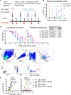
Immunization and isolation of SUDV GP-specific monoclonal antibodies from immunized NHPs. (A) Schematic presentation of the immunization and serum/PBMC sampling schedule. Rhesus macaques were primed with rVSV-SUDV GP, followed by three booster immunizations of protein SUDV GPΔmuc with adjuvant; serum and PBMCs from animals were collected 1 week after each inoculation, indicated with a red arrow. (B) Serum neutralization titers (ID50) of the macaques against rVSV-SUDV GP-Luc or rVSV–EBOV GP-Luc pseudotype virus particles during immunization course. Mean of ID50 titers of four NHPs is shown, with the error bar showing standard deviation. Neutralization assays were performed in triplicate. (C) Neutralization capacity of the week 7 serum of 4 NHPs. Neutralization assays were performed in triplicate. (D) Single B cell sorting of SUDV GP-specific mAbs by flow cytometry. Week 7 PBMCs of immune macaques were incubated with antibody-fluorochrome conjugates for cell markers and antigen sorting probes consisting of SUDV GPΔmuc. SUDV GP-specific memory B cells with the phenotype of CD20+IgG+Aqua blueCD14CD3CD8CD27+IgM and SUDV GPΔmuchi were sorted for Ig gene amplification. Shown is the flow cytometry sorting of GP-specific memory B cells of NHP 133503, with the gated cell population frequency in corresponding parental cell population depicted in red font. (E) Validation of SUDV GP-specific mAb sorting and cloning. SUDV or EBOV GPΔmuc binding with representative mAbs assessed by ELISA is shown.
FIG 2
FIG 2
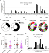
Genetic features of the SUDV GPΔmuc-sorted NHP Ig repertoire. (A) VH gene segment frequencies of the SUDV GPΔmuc-sorted Ig sequences. Above the dashed line (>10% frequency) are overexpressed segments in the Ig repertoire. Ig sequences sorted from two animals were combined for analysis. (B) Light chain usage of SUDV GPΔmuc-sorted Ig repertoire in two NHPs. (C) Clonotype analysis of SUDV GPΔmuc-sorted Ig repertoire VH sequences defined by the criteria that clones using the same V and J gene segments and having identical CDR3 length with CDR3 AA homology >83% are considered to belong to the same clonotype (lineage). Predominant lineages representing more than 5% of the sequences were marked as exploded portions with an asterisk. N stands for number of sorted Ig and C for number of clonal types. (D) SHM level of both heavy and light chain variable regions in SUDV GPΔmuc-sorted Ig repertoire in two animals, with means of SHM indicated by red lines. (E) Analysis of heavy chain CDR3 (CDRH3) in SUDV GPΔmuc-sorted Ig repertoire. CDRH3 region is delineated with the IMGT CDR3 definition. The individual CDRH3 length (number of amino acid [AA] residues) frequency out of the total number of CDRH3 region was calculated and the mean of CDRH3 length in this Ig repertoires is 13.4 AA residues.
FIG 3
FIG 3
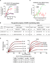
Characterization of SUDV neutralizing mAbs (A) Neutralization activity of SUDV GPΔmuc-specific mAbs EA97 and EB46 determined using rVSV-SUDV-Bon GP-Luc pseudotype virus. 16F6 serves as positive control. Two recovered mAbs, EB32 and EA85, are also shown for comparison. Shown is mean value plus or minus standard deviation of the experiment, performed in triplicate. (B) Genetic properties of two SUDV pseudovirus neutralizing mAbs, EA97 and EB46, including V(D)J gene segment usage, CDRH3 and CDRL3 (complementarity-determining region of the heavy and light [lambda] chain, respectively) amino acid residue sequence and length, and somatic hypermutation rate (% SHM) of V gene segment compared with that of corresponding rhesus macaque Ig germ line sequence (https://www.ncbi.nlm.nih.gov/igblast) shown in percentage at nucleotide (nt) and amino acid residue (AA) level. (C) Neutralization activity of EA97 or EB46 determined using authentic SUDV virus (Gulu strain). Shown is mean value plus or minus standard deviation of the experiment, performed in duplicate. (D) Kinetics of mAb-GP binding analyzed by BLI. SUDV GPΔmuc protein containing 6-His tag was loaded onto Ni-NTA sensors and tested for binding to mAb at 5 different concentrations in 2-fold serial dilution starting from 50 nM. On-rate (kon), off-rate (koff), and KD values are shown below the sensograms.
FIG 4
FIG 4
SUDV GP-specific mAbs epitope mapping by competition ELISA. The competition for GP binding between the biotinylated test mAb, EB46 or EA97, and the competitor mAbs of known binding epitopes. (A) Competition ELISA binding curves showing the biotin-labeled EB46 (30 ng/ml) or EA97 (15 ng/ml) binding to GP in the presence of competitor mAbs at various concentrations. The competitor mAbs were titrated at various concentrations to evaluate the effect on EB46 or EA97 binding. (B) The remaining binding of biotin-labeled mAb (EB46 or EA97) in the presence of the competitor mAb compared to the binding of biotin-labeled mAb alone. mAbs were considered to compete if maximum binding of the biotin-labeled mAb in the presence of competitor was reduced to <35% of its uncompeted binding (yellow), while mAbs were considered noncompeting if maximum binding of the biotin-labeled mAb was >65% of its uncompeted binding (white). mAbs with intermediate phenotype (between 35% and 65% of uncompeted binding) are labeled with blue background.
FIG 5
FIG 5
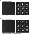
Single-particle electron microscopy (EM) data of EB46 or EA97 Fab bound to SUDV GPΔmuc. (A) EA97-GP complex. (B) EB46-GP complex. Left, raw micrograph of Fab in complex with SUDV GPΔmuc. Right, 2D classes of complex.
FIG 6
FIG 6
Epitope mapping of SUDV GP-specific mAbs by 3D EM. (A) 3D EM reconstruction of EA97 Fab (purple): SUDV GPΔmuc complex is shown in transparent surface representation with GPΔmuc trimer (gray) (PDB: 3CSY) fitted into the density. (B) 3D EM reconstruction of EB46 Fab (blue): SUDV GPΔmuc (gray) complex.
FIG 7
FIG 7
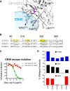
Functional analysis of EB46 epitope. (A) Potential EB46 contact residues revealed by the EB46 Fab (blue): SUDV GPΔmuc (gray) complex 3D EM reconstruction, with GPΔmuc trimer (PDB: 3CSY) fitted into the density. Side chains of GP residues overlapping with EB46 density are shaded in light blue. GP residues as potential contacts confirmed with loss-of-function mutations (E44, N552, and L561) in C and D are highlighted in magenta. (B) Homology between ebolavirus GP sequences within the regions encompassing the EB46 epitope. The residues on SUDV GP overlapping with EB46 Fab density in the EM 3D reconstruction are highlighted in yellow. SUDV and EBOV GP residues involved in 16F6 (3VE0) and KZ52 (3CSY) contact, respectively, are underlined. (C) Capacity of EB46 to neutralize rVSV-GP-eGFP pseudotype viruses bearing GP WT or neutralization escape variant (E44K). Means plus or minus standard deviation for six replicates are shown. (D) Binding of RBS binder FVM04, GP base binder 16F6, and EB46 to SUDV GP WT and mutants expressed on the surface of 293T cells.
FIG 8
FIG 8
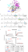
Similarity between NHP mAb EB46 and mouse mAb 16F6. (A) mAbs EB46 and 16F6 share the similar GP binding mode illustrated by homology modeling. EB46-Fab binding to GP was modeled using mouse mAb 16F6-GP complex as the template (PDB: 3VE0). The structural elements of mAb 16F6 (orange), mAb EB46 heavy chain (blue) and light chain (cyan), GP1 (magenta), and GP2 (gray) are depicted, respectively. The shared contact residues Y32, R98, and Y101 on 16F6 are labeled in brown, and residues on GP1 and GP2 affecting EB46 binding are labeled in magenta and gray, respectively. As the major different contact region compared to 16F6, EB46 CDRL2 is indicated. (B) Amino acid alignment of V(D)J sequences of the heavy (H) and light (L) chains of 16F6 and EB46. The CDRs, delineated with Kabat system, are highlighted in gray and contact residues of 16F6 highlighted in cyan, and the residues in the EB46 CDRs identical to the counterpart contact residues in 16F6 are labeled in yellow. (C) Kinetics of H/L chain swapped EB46-16F6 mAbs binding to SUDV GPΔmuc assessed by BLI. The V(D)J sequences encoding humanized 16F6 heavy and light chains were inserted into human IgG1 cassettes to generate full-length IgG consisting of 16F6 heavy and light chains (16F6-H+L). The swapped mAb, 16F6H-EB46L or EB46H-16F6L, was generated by cotransfection of humanized 16F6 heavy chain and EB46 light chain or EB46 heavy chain with humanized 16F6 light chain, respectively. Purified antibody IgGs were loaded to anti-human Fc sensors and tested for binding with SUDV GPΔmuc at 3 different concentrations (250, 125, and 62.5 nM). On-rate (kon), off-rate (koff), and KD values are shown below the sensograms.
FIG 9
FIG 9
Mechanistic basis of mAb-dependent blockage of viral entry. (A) Reactivity of 3 mAbs to SUDV GPΔTM determined by ELISA at neutral (pH 7.4) and acidic (pH 5.5) conditions. (B) Comparison of neutralization capacity of mAbs, including EB46, EA97, 16F6, and CA45 against rVSV-SUDV GP-eGFP virus particles bearing uncleaved (SUDV) or thermolysin-cleaved (SUDVcl) GP. Means plus or minus standard deviation for three replicates are shown. (C) Capacity of mAbs blocking SUDV GP→GPcl cleavage. SUDV GPΔmuc protein was incubated with antibodies, followed by thermolysin treatment. GP1 was visualized by Western blotting by HRP-21D10 antibody conjugate. Left two lanes were set in the absence of IgG antibody with or without thermolysin treatment. Control IgG is anti-HIV-1 mAb, VRC01. ADI-15946 serves as positive control for blocking this cleavage.
FIG 10
FIG 10
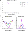
In vivo protective efficacy of mAbs in mice. C57BL/6J IFNAR−/− mice (n = 10/group) were challenged with SUDV Boniface strain followed by treatment with 200 μg of EA97 or EB46 at 1 dpi (d + 1) or 1 and 3 dpi (d + 1,3) by i.p. injection. Pan-ebola cocktail (CA45 and FVM04, with each 100 μg) and PBS were used as positive and negative controls, respectively. (A) Survival rate comparison between animal groups treated with PBS and mAbs, Mantel-Cox log-rank test with significance *, P < 0.05; ns, not significant. (B) Weight change of animals. (C) Clinical score of animals. Statistical analysis in B and C are performed for comparison between animal groups treated with PBS and mAbs by t tests of Wilcoxon (matched-pairs signed rank test), *, P < 0.05, **, P < 0.01; ns, not significant.
FIG 11
FIG 11
In vivo protective efficacy of EB46 in guinea pigs. Female Hartley guinea pigs (n = 6/group) were challenged with 1,000 PFU of GPA-SUDV and treated with 5 mg of EB46 or PBS at 3 dpi (d + 3) or 1 and 5 dpi (d + 1,5). (A) Survival rate comparison between animal groups with EB46 and PBS treatment, Mantel-Cox log-rank test with *, P < 0.05. (B) Weight change of animals. (C) Temperature. (D) Clinical score of animals. Statistical analysis in B, C, and D are performed for comparison between animal groups treated with EB46 and PBS by two-way analysis of variance (ANOVA) test, *, P < 0.05, **, P < 0.01, ***, P < 0.001, ****, P < 0.0001.
FIG 12
FIG 12
mAb and week 7 NHP immune serum neutralization specificity characterization. (A) mAb-mediated neutralization phenotype of rVSV-SUDV GP-Luc (WT and interface mutant) pseudotype viruses. Neutralization assays were performed in triplicate. Mean of mAb neutralization activity at each concentration is shown, with the error bar showing standard deviation. (B) mAb neutralization IC50 titer against mutant E44K or N552A compared to WT neutralization, calculated as (IC50 mutant/IC50 WT) × 100. IC50 value represents the reciprocal of mAb concentration that results in 50% inhibition of pseudotype virus entry. (C) Neutralizing epitope specificity of week 7 NHP immune serum delineated by rVSV-SUDV GP-Luc pseudotype virus bearing WT or mutant GPs. Neutralization assays were performed in triplicate, repeated twice. Mean of NHP serum neutralization activity at each dilution point of representative data set is shown, with the error bar showing standard deviation. (D) Serum neutralization capacity against rVSV-SUDV GP-Luc pseudotype virus bearing WT compared to mutant GPs. ID50 value represent the reciprocal serum dilution factor that resulted in 50% inhibition of rVSV-SUDV GP entry. Serum 1/ID50 for neutralizing mutant E44K or N552A relative to WT pseudotype virus, calculated as (ID50 WT/ID50 mutant) × 100, is shown.

Similar articles

Cited by

References

    1. Feldmann H, Geisbert TW. 2011. Ebola haemorrhagic fever. Lancet 377:849–862. doi:10.1016/S0140-6736(10)60667-8. - DOI - PMC - PubMed
    1. Team WIS. 1978. Ebola haemorrhagic fever in Sudan, 1976. Report of a WHO/International Study Team. Bull World Health Organ 56:247–270. - PMC - PubMed
    1. Albarino CG, Shoemaker T, Khristova ML, Wamala JF, Muyembe JJ, Balinandi S, Tumusiime A, Campbell S, Cannon D, Gibbons A, Bergeron E, Bird B, Dodd K, Spiropoulou C, Erickson BR, Guerrero L, Knust B, Nichol ST, Rollin PE, Stroher U. 2013. Genomic analysis of filoviruses associated with four viral hemorrhagic fever outbreaks in Uganda and the Democratic Republic of the Congo in 2012. Virology 442:97–100. doi:10.1016/j.virol.2013.04.014. - DOI - PMC - PubMed
    1. Bowen ET, Lloyd G, Harris WJ, Platt GS, Baskerville A, Vella EE. 1977. Viral haemorrhagic fever in southern Sudan and northern Zaire. Preliminary studies on the aetiological agent. Lancet 1:571–573. doi:10.1016/S0140-6736(77)92001-3. - DOI - PubMed
    1. Sanchez A, Rollin PE. 2005. Complete genome sequence of an Ebola virus (Sudan species) responsible for a 2000 outbreak of human disease in Uganda. Virus Res 113:16–25. doi:10.1016/j.virusres.2005.03.028. - DOI - PubMed

LinkOut - more resources