. 2021 Mar 12;371(6534):1139-1142.
doi: 10.1126/science.abf6950. Epub 2021 Feb 3.

Recurrent deletions in the SARS-CoV-2 spike glycoprotein drive antibody escape

Affiliations

Recurrent deletions in the SARS-CoV-2 spike glycoprotein drive antibody escape

Kevin R McCarthy et al. Science. .

Abstract

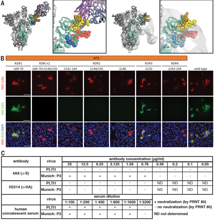
Zoonotic pandemics, such as that caused by severe acute respiratory syndrome coronavirus 2 (SARS-CoV-2), can follow the spillover of animal viruses into highly susceptible human populations. The descendants of these viruses have adapted to the human host and evolved to evade immune pressure. Coronaviruses acquire substitutions more slowly than other RNA viruses. In the spike glycoprotein, we found that recurrent deletions overcome this slow substitution rate. Deletion variants arise in diverse genetic and geographic backgrounds, transmit efficiently, and are present in novel lineages, including those of current global concern. They frequently occupy recurrent deletion regions (RDRs), which map to defined antibody epitopes. Deletions in RDRs confer resistance to neutralizing antibodies. By altering stretches of amino acids, deletions appear to accelerate SARS-CoV-2 antigenic evolution and may, more generally, drive adaptive evolution.


Figures

Fig. 1
Fig. 1. Deletions in SARS-CoV-2 spike glycoprotein arise during persistent infections of immunosuppressed patients.
(A) Top: Sequences of viruses isolated from PLTI1 (PT) and viruses from patients with deletions in the same NTD region. Chromatograms are shown for sequences from PLTI1, which include sequencing of bulk reverse transcription products (CON) and individual cDNA clones. Bottom: Sequences from other long-term infections from individuals AM (18), MA-JL (MA) (19), and a MSK cohort (M) with individuals 3, 4, 6, 8, and 11 (13). Letters (A and B) designate different variants from the same patient. (B) Sequences of viruses from two patients (M2 and M13) with deletions in a different region of the NTD. All sequences are aligned to reference sequence (REF) MN985325 (WA-1). See fig. S1 for genetic analysis of patient isolates. Amino acid abbreviations: A, Ala; D, Asp; F, Phe; G, Gly; H, His; K, Lys; L, Leu; N, Asn; P, Pro; R, Arg; S, Ser; V, Val; Y, Tyr.
Fig. 2
Fig. 2. Identification and characterization of recurrent deletion regions in SARS-CoV-2 spike protein.
(A) Positional quantification of deleted nucleotides in S among GISAID sequences. We designate the four clusters as recurrent deletion regions (RDRs) 1 to 4. (B) Length distribution of deletions. (C) The percentage of deletion events at the indicated site that either maintain the open reading frame (ORF) or introduce a frameshift or premature stop codon (F.S./Stop). (D) Phylogenetic analysis of deletion variants (red branches) and genetically diverse nondeletion variants (black branches). Specific deletion clades/lineages are identified. Maximum likelihood phylogenetic trees, rooted on NC_045512, were calculated with 1000 bootstrap replicates. Trees with branch labels are in fig. S2. (E) Abundance of nucleotide (nt) deletions in each RDR. Positions are defined by reference sequence MN985325, by codon (top) and nucleotide (below). Amino acid abbreviations: A, Ala; D, Asp; F, Phe; G, Gly; H, His; I, Ile; L, Leu; N, Asn; P, Pro; R, Arg; S, Ser; T, Thr; V, Val; W, Trp; Y, Tyr.
Fig. 3
Fig. 3. Geographic, genetic, and temporal abundance of RDR variants.
(A and B) Geographic (A) and genetic (B) distributions of RDR variants compared to the GISAID database (sequences from 1 December 2019 to 24 October 2020). GISAID clade classifications are used in (B). (C) Frequency of RDR variants among all complete genomes deposited in GISAID. (D) Frequency of specific RDR deletion variants (numbered according to spike amino acids) among all GISAID variants. The plot of RDR3/Δ210 has been adjusted by 0.02 units on the y axis for visualization in (C) because of its overlap with RDR2, and this adjustment has been retained in (D) to enable direct comparisons between panels.
Fig. 4
Fig. 4. Deletions in the spike NTD alter its antigenicity; RDRs map to defined antigenic sites.
(A) Left: A structure of antibody 4A8 (17) (PDB ID 7C21) (purple) bound to one protomer (green) of a SARS-CoV-2 spike trimer (gray). RDRs 1 to 4 are colored red, orange, blue, and yellow, respectively, and are shown as spheres. The boxed image is a close-up of the interaction site. Right: The electron microscopy density of COV57 serum Fabs (18) (EMDB emd_22125) fit to SARS-CoV-2 S glycoprotein trimer (PDB ID 7C21). The boxed image is a close-up of the interaction site. (B) S glycoprotein distribution in Vero E6 cells at 24 hours after transfection with S protein deletion mutants, visualized by indirect immunofluorescence in permeabilized cells. A monoclonal antibody to SARS-CoV-2 S protein receptor-binding domain (RBD mAb; red) detects all mutant forms of the protein (Δ69–70, Δ69–70+Δ141–144, Δ141–144, Δ144/145, Δ146, Δ210, and Δ243–244) and the unmodified protein (wild type), whereas 4A8 mAb (green) does not detect mutants containing deletions in RDR2 or RDR4 (Δ69–70+Δ141–144, Δ141–144, Δ144/145, Δ146, and Δ243–244). Overlay images (RBD/4A8/DAPI) depict colocalization of the antibodies; nuclei were counterstained with 4′,6-diamidino-2-phenylindole (DAPI; blue). Scale bars, 100 μm. (C) Virus isolated from PLTI1 resists neutralization by 4A8. A nondeletion variant (Munich) is neutralized by 4A8, both are neutralized by convalescent serum, and neither is neutralized by H2214, an influenza hemagglutinin binding antibody (29).

Comment in

References

    1. Zhu N., Zhang D., Wang W., Li X., Yang B., Song J., Zhao X., Huang B., Shi W., Lu R., Niu P., Zhan F., Ma X., Wang D., Xu W., Wu G., Gao G. F., Tan W., A Novel Coronavirus from Patients with Pneumonia in China, 2019. N. Engl. J. Med. 382, 727–733 (2020). 10.1056/NEJMoa2001017 - DOI - PMC - PubMed
    1. Wu F., Zhao S., Yu B., Chen Y.-M., Wang W., Song Z.-G., Hu Y., Tao Z.-W., Tian J.-H., Pei Y.-Y., Yuan M.-L., Zhang Y.-L., Dai F.-H., Liu Y., Wang Q.-M., Zheng J.-J., Xu L., Holmes E. C., Zhang Y.-Z., A new coronavirus associated with human respiratory disease in China. Nature 579, 265–269 (2020). 10.1038/s41586-020-2008-3 - DOI - PMC - PubMed
    1. Zhou H., Chen X., Hu T., Li J., Song H., Liu Y., Wang P., Liu D., Yang J., Holmes E. C., Hughes A. C., Bi Y., Shi W., A Novel Bat Coronavirus Closely Related to SARS-CoV-2 Contains Natural Insertions at the S1/S2 Cleavage Site of the Spike Protein. Curr. Biol. 30, 3896 (2020). 10.1016/j.cub.2020.09.030 - DOI - PMC - PubMed
    1. Lam T. T., Jia N., Zhang Y.-W., Shum M. H.-H., Jiang J.-F., Zhu H.-C., Tong Y.-G., Shi Y.-X., Ni X.-B., Liao Y.-S., Li W.-J., Jiang B.-G., Wei W., Yuan T.-T., Zheng K., Cui X.-M., Li J., Pei G.-Q., Qiang X., Cheung W. Y.-M., Li L.-F., Sun F.-F., Qin S., Huang J.-C., Leung G. M., Holmes E. C., Hu Y.-L., Guan Y., Cao W.-C., Identifying SARS-CoV-2-related coronaviruses in Malayan pangolins. Nature 583, 282–285 (2020). 10.1038/s41586-020-2169-0 - DOI - PubMed
    1. Boni M. F., Lemey P., Jiang X., Lam T. T.-Y., Perry B. W., Castoe T. A., Rambaut A., Robertson D. L., Evolutionary origins of the SARS-CoV-2 sarbecovirus lineage responsible for the COVID-19 pandemic. Nat. Microbiol. 5, 1408–1417 (2020). 10.1038/s41564-020-0771-4 - DOI - PubMed

Publication types