. 2021 Feb 2;30(159):200193.
doi: 10.1183/16000617.0193-2020. Print 2021 Mar 31.

Hermansky-Pudlak syndrome pulmonary fibrosis: a rare inherited interstitial lung disease

Affiliations

Hermansky-Pudlak syndrome pulmonary fibrosis: a rare inherited interstitial lung disease

Tadafumi Yokoyama et al. Eur Respir Rev. .

Abstract

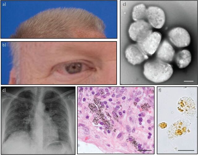
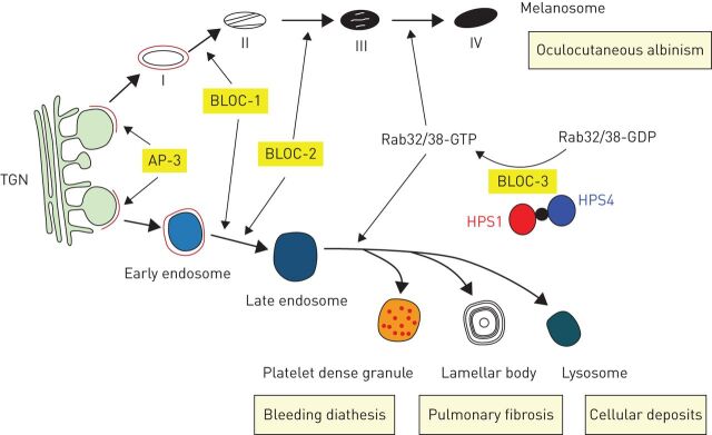
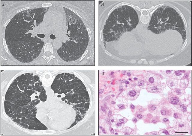
Pulmonary fibrosis is a progressive interstitial lung disease of unknown aetiology with a poor prognosis. Studying genetic diseases associated with pulmonary fibrosis provides insights into the pathogenesis of the disease. Hermansky-Pudlak syndrome (HPS), a rare autosomal recessive disorder characterised by abnormal biogenesis of lysosome-related organelles, manifests with oculocutaneous albinism and excessive bleeding of variable severity. Pulmonary fibrosis is highly prevalent in three out of 10 genetic types of HPS (HPS-1, HPS-2 and HPS-4). Thus, genotyping of individuals with HPS is clinically relevant. HPS-1 tends to affect Puerto Rican individuals due to a genetic founder effect. HPS pulmonary fibrosis shares some clinical features with idiopathic pulmonary fibrosis (IPF), including dyspnoea, cough, restrictive lung physiology and computed tomography (CT) findings of fibrosis. In contrast to IPF, HPS pulmonary fibrosis generally affects children (HPS-2) or middle-aged adults (HPS-1 or HPS-4) and may be associated with ground-glass opacification in CT scans. Histopathology of HPS pulmonary fibrosis, and not IPF, shows vacuolated hyperplastic type II cells with enlarged lamellar bodies and alveolar macrophages with lipofuscin-like deposits. Antifibrotic drugs approved as treatment for IPF are not approved for HPS pulmonary fibrosis. However, lung transplantation has been performed in patients with severe HPS pulmonary fibrosis. HPS pulmonary fibrosis serves as a model for studying fibrotic lung disease and fibrosis in general.


Conflict of interest statement

Conflict of interest: T. Yokoyama has nothing to disclose. Conflict of interest: B.R. Gochuico has nothing to disclose.

Figures

FIGURE 1
FIGURE 1
Clinical features of Hermansky–Pudlak syndrome (HPS). a, b) Hypopigmentation of a) hair and b) skin and iris of a Puerto Rican male. c) δ-granules are absent in platelets from a patient with HPS-1 imaged using whole-mount transmission electron microscopy. Scale bar: 2 μm. d) Bilateral interstitial infiltrates are found in a chest radiograph of a 50-year-old female with HPS pulmonary fibrosis. e) Fibrotic lung tissue stained with haematoxylin and eosin and f) unstained renal epithelial cells isolated from urine derived from a subject with HPS-1 show abnormal intracellular deposits. Scale bar: 20 μm.
FIGURE 2
FIGURE 2
Relationships of protein complexes involved in Hermansky–Pudlak syndrome (HPS), biogenesis of intracellular organelles and clinical manifestations of disease. Four stages (I–IV) of melanosome maturation and platelet δ-granule (dense granule), lamellar body and lysosome formation via the endosomal pathway from the TGN are shown. Defects in these vesicles are associated with oculocutaneous albinism, bleeding diathesis, pulmonary fibrosis and excessive cellular deposits in HPS. Protein complexes associated with HPS, including AP-3, BLOC-1, BLOC-2 and BLOC-3, and a Rab32/38 guanine nucleotide exchange factor comprised of HPS1 and HPS4 protein subunits, are involved in the formation of melanosomes, platelet δ-granules, lamellar bodies and lysosomes. TGN: trans-Golgi network; AP: adaptor protein; BLOC: biogenesis of lysosome-related organelles complex.
FIGURE 3
FIGURE 3
Computed tomography (CT) scan and histopathological images of Hermansky–Pudlak syndrome (HPS) pulmonary fibrosis. a) Supine and b) prone CT scan images of a 47-year-old subject with moderate HPS pulmonary fibrosis show bilateral ground-glass opacities, reticulations and honeycombing. c) CT scan image of a 38-year-old subject with HPS pulmonary fibrosis demonstrates bilateral ground-glass opacities, traction bronchiectasis, reticulations and honeycombing. d) Aggregates of foamy alveolar macrophages, vacuolated hyperplastic type II alveolar epithelial cells containing enlarged lamellar bodies, and interstitial accumulation of cells and extracellular matrix are found in lung tissue from a subject with HPS pulmonary fibrosis. Haematoxylin and eosin staining. Scale bar: 20 μm.

References

    1. Lederer DJ, Martinez FJ. Idiopathic pulmonary fibrosis. N Engl J Med 2018; 378: 1811–1823. doi:10.1056/NEJMra1705751 - DOI - PubMed
    1. Martinez FJ, Collard HR, Pardo A, et al. . Idiopathic pulmonary fibrosis. Nat Rev Dis Primers 2017; 3: 17074–17093. doi:10.1038/nrdp.2017.74 - DOI - PubMed
    1. Richeldi L, Collard HR, Jones MG. Idiopathic pulmonary fibrosis. Lancet 2017; 389: 1941–1952. doi:10.1016/S0140-6736(17)30866-8 - DOI - PubMed
    1. Wynn TA. Integrating mechanisms of pulmonary fibrosis. J Exp Med 2011; 208: 1339–1350. doi:10.1084/jem.20110551 - DOI - PMC - PubMed
    1. Sisson TH, Mendez M, Choi K, et al. . Targeted injury of type II alveolar epithelial cells induces pulmonary fibrosis. Am J Respir Crit Care Med 2010; 181: 254–263. doi:10.1164/rccm.200810-1615OC - DOI - PMC - PubMed

MeSH terms

LinkOut - more resources