. 2021 Feb 3;13(579):eabb6576.
doi: 10.1126/scitranslmed.abb6576.

Eosinophils attenuate hepatic ischemia-reperfusion injury in mice through ST2-dependent IL-13 production

Affiliations

Eosinophils attenuate hepatic ischemia-reperfusion injury in mice through ST2-dependent IL-13 production

Yaochun Wang et al. Sci Transl Med. .

Abstract

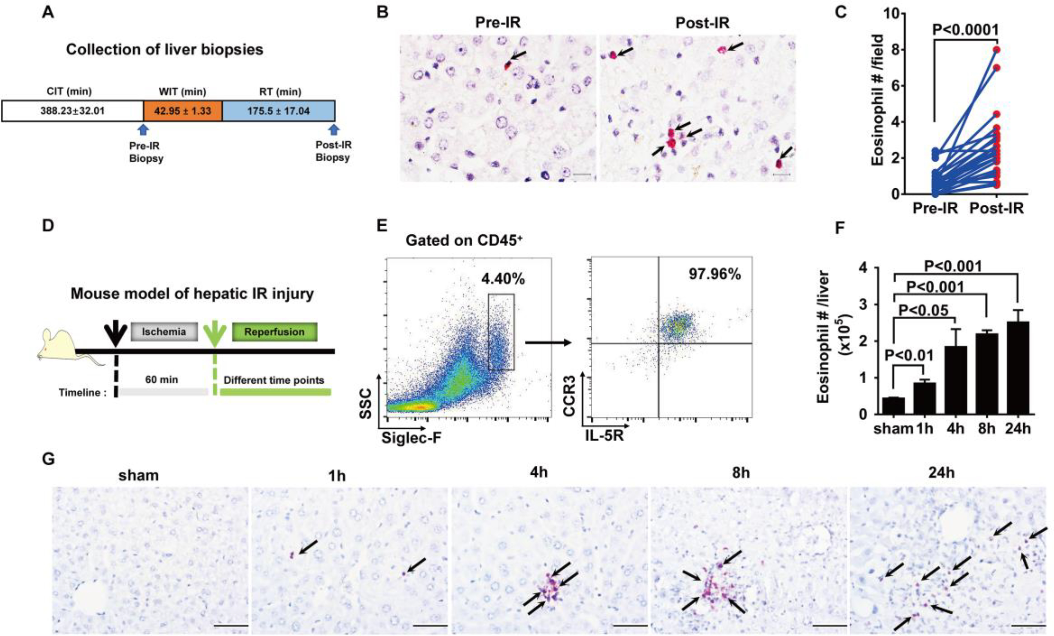
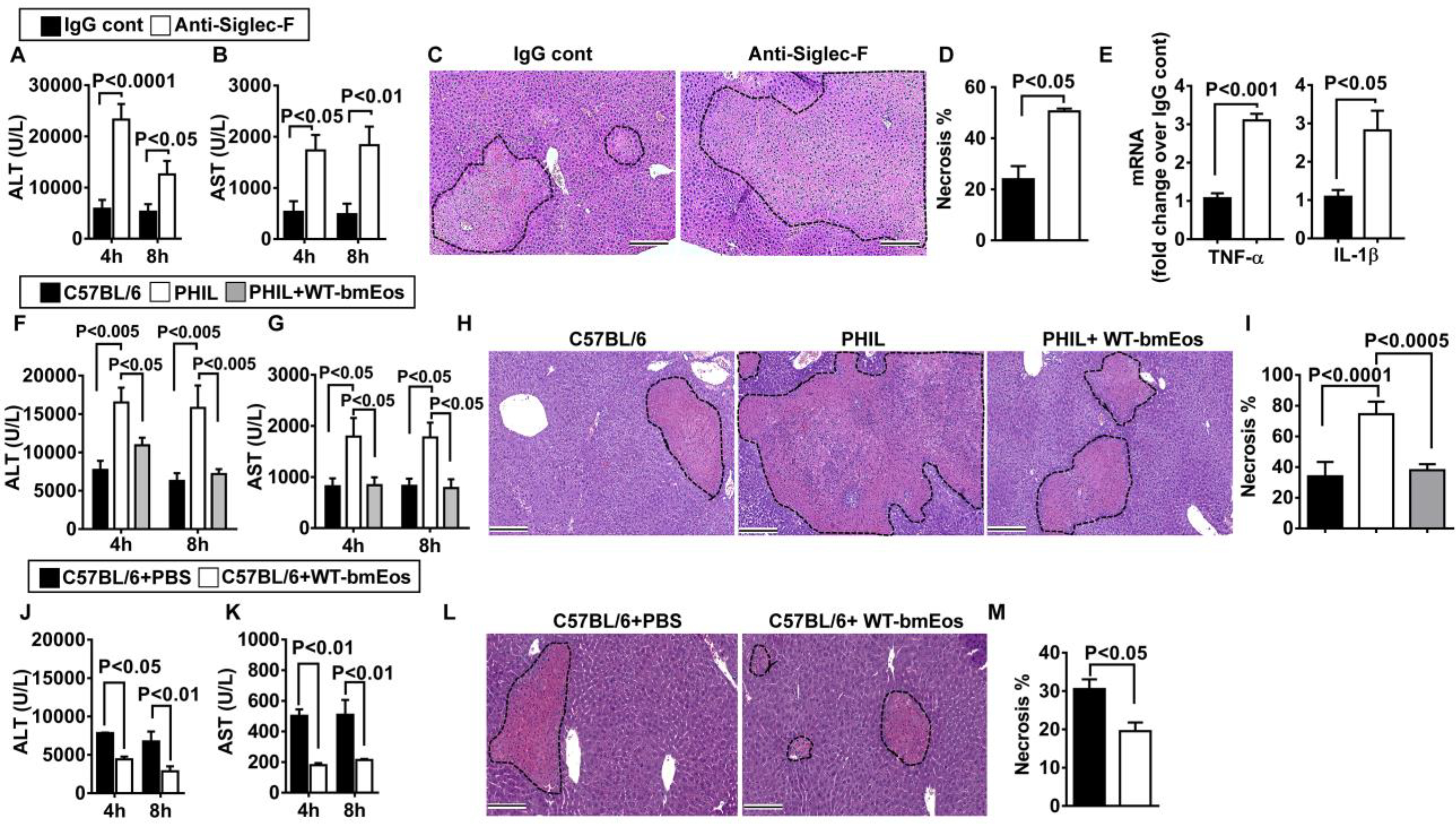
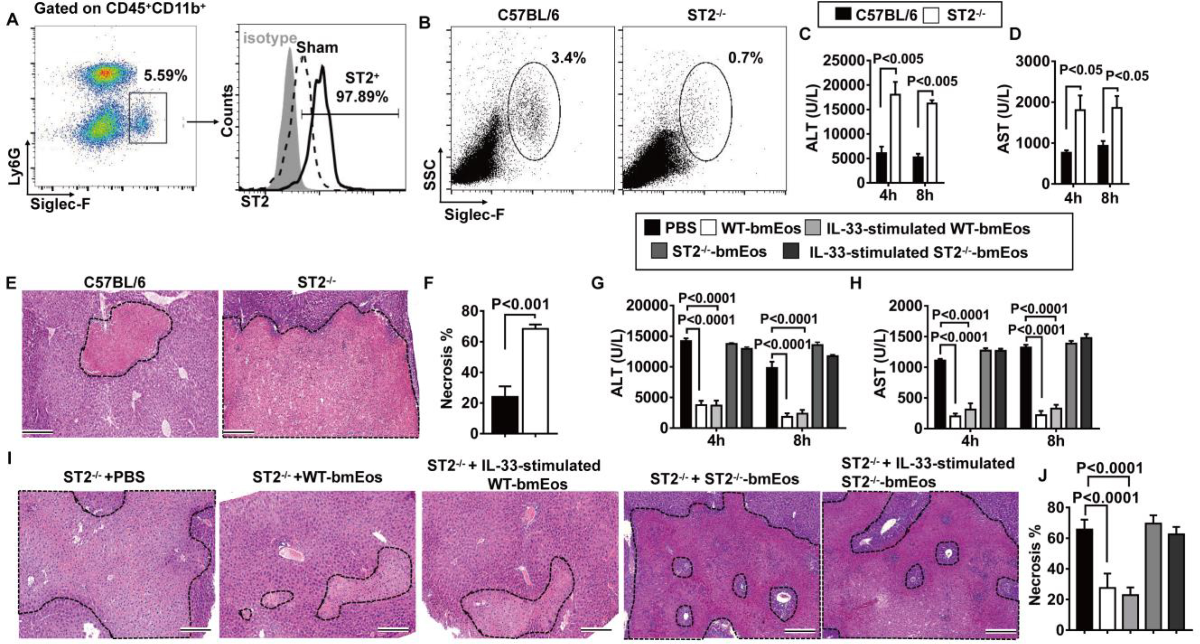
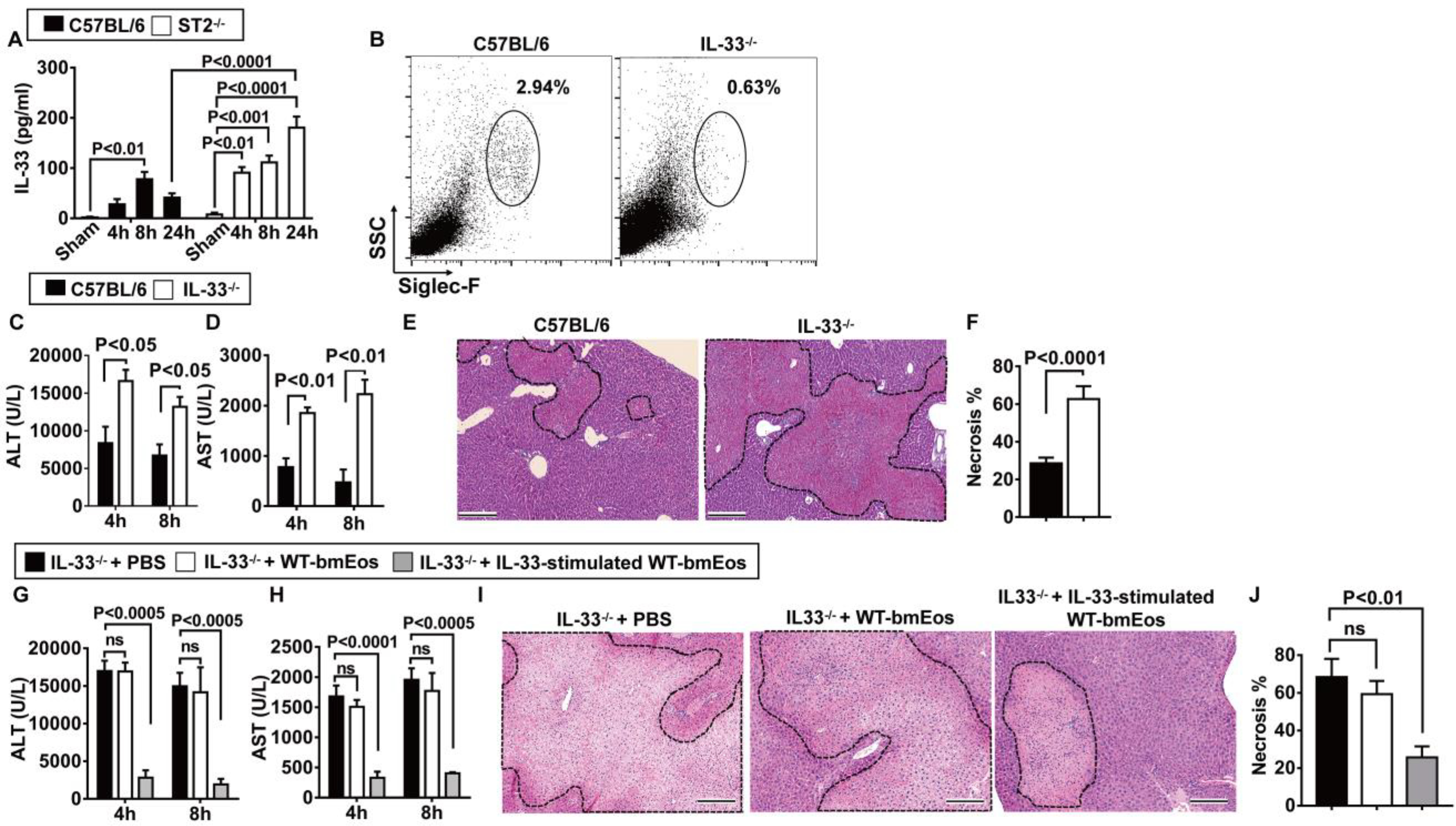
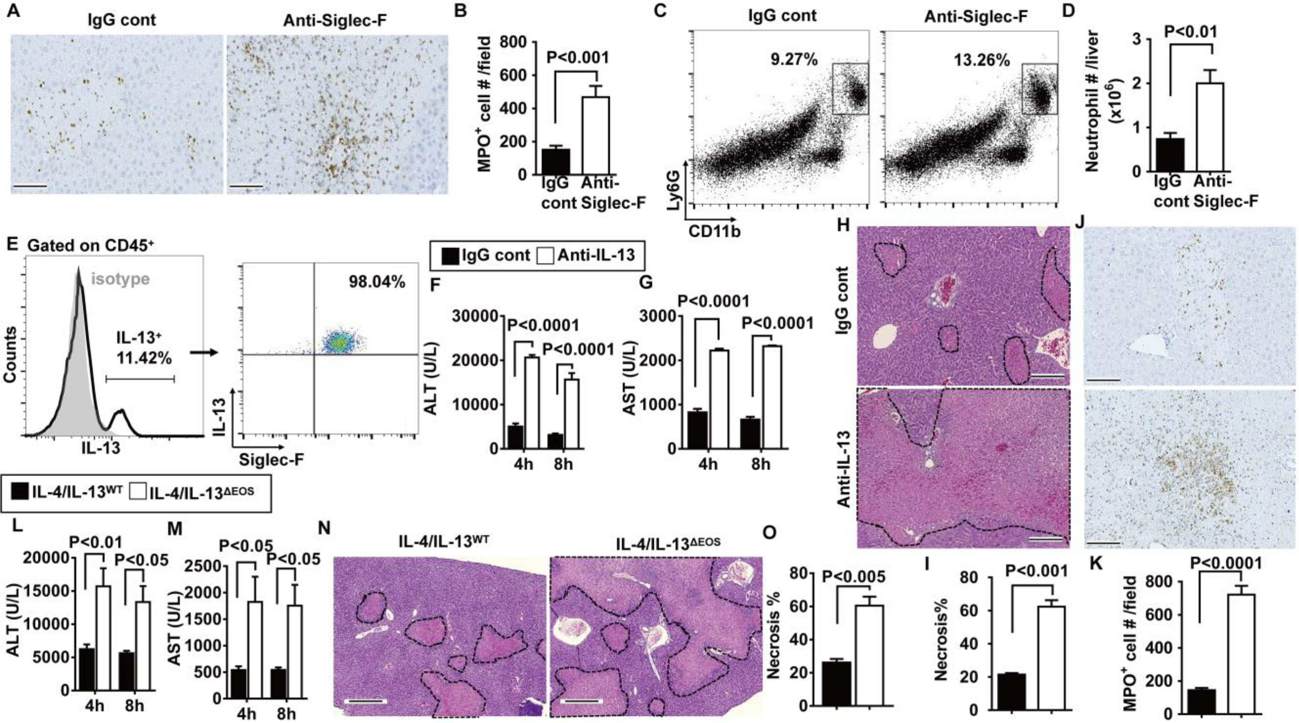
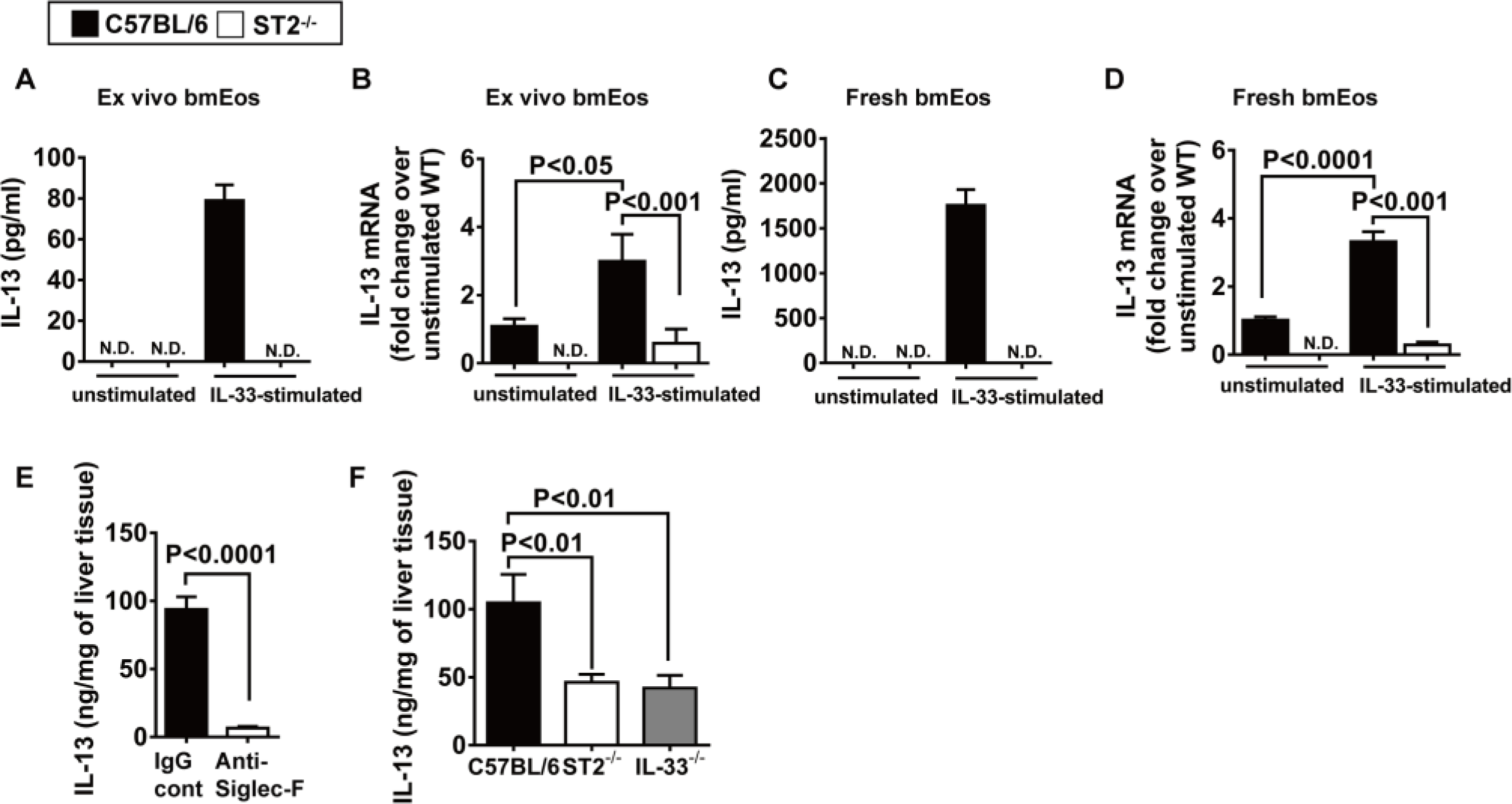
Eosinophils are a myeloid cell subpopulation that mediates type 2 T helper cell immune responses. Unexpectedly, we identified a rapid accumulation of eosinophils in 22 human liver grafts after hepatic transplantation. In contrast, no eosinophils were detectable in healthy liver tissues before transplantation. Studies with two genetic mouse models of eosinophil deficiency and a mouse model of antibody-mediated eosinophil depletion revealed exacerbated liver injury after hepatic ischemia and reperfusion. Adoptive transfer of bone marrow-derived eosinophils normalized liver injury of eosinophil-deficient mice and reduced hepatic ischemia and reperfusion injury in wild-type mice. Mechanistic studies combining genetic and adoptive transfer approaches identified a critical role of suppression of tumorigenicity (ST2)-dependent production of interleukin-13 by eosinophils in the hepatoprotection against ischemia-reperfusion-induced injury. Together, these data provide insight into a mechanism of eosinophil-mediated liver protection that could serve as a therapeutic target to improve outcomes of patients undergoing liver transplantation.


Conflict of interest statement

Competing interests: The authors declare that they have no competing interests.

Figures

Fig. 1.
Fig. 1.. Eosinophils accumulate in the liver during hepatic IR injury.
(A) Collection of liver biopsies. (B) Eosinophils in human donor liver biopsies (pre-IR) and biopsies from the same tissues after liver transplantation (post-IR) were detected by IHC staining using anti-human EPX antibody. Scale bars, 25 μm. Red indicates EPX staining, and arrows point to EPX-positive eosinophils. (C) The numbers of eosinophils were quantified (n = 22 patients per group). CIT, cold ischemia time; WIT, warm ischemia time; RT, reperfusion time. (D) Male C57BL/6 mice were subjected to hepatic ischemia for 60 min, and the numbers of eosinophils in the liver were measured at 1, 4, 8, or 24 hours after reperfusion. (E) Representative flow cytometry plots and (F) quantification demonstrate the numbers of eosinophils (SSChiSiglec-F+CCR3+IL-5R+cells) in the liver after IR injury (n = 4 per group). SSC, side scatter. (G) IHC staining of eosinophils using anti-mouse MBP antibody. Arrows point to MBP-positive eosinophils stained red. Scale bars, 200 μm. Images shown are representative of n = 4 mice per time point. A two-tailed paired Student’s t test with Welch’s correction was performed in (C). A one-way ANOVA was performed in (F).
Fig. 2.
Fig. 2.. Eosinophils protect against hepatic IR injury.
(A to E) Eosinophils were depleted by anti–Siglec-F antibody in male C57BL/6 mice, and control mice were treated with IgG (n = 5 in IgG control and n = 3 in anti–Siglec-F treatment group), and serum concentrations of ALT (A) and AST (B) were measured at 4 and 8 hours after reperfusion. (C) Liver necrosis (scale bars, 200 μm) was evaluated and quantified (D) at 24 hours after reperfusion. (E) mRNA expression of cytokines in liver nonparenchymal cells was measured at 4 hours after reperfusion (n = 6 mice per group). (F to I) WT littermates, eosinophil-deficient PHIL mice, and PHIL mice receiving WT-bmEos (2 × 10⁷) were subjected to hepatic IR injury (n = 4 mice per group). Serum concentrations of ALT (F) and AST (G) were measured at 4 and 8 hours after reperfusion. (H) Liver necrosis (scale bars, 200 μm) was examined and quantified (I) at 24 hours after reperfusion. (J to M) WT C57BL/6 mice (n = 4 mice per group) received WT-bmEos or PBS as a control. Serum concentrations of ALT (J) and AST (K) were measured at 4 and 8 hours after reperfusion. (L) Liver necrosis (scale bars, 200 μm) was examined and quantified (M) 24 hours after reperfusion. A two-tailed unpaired Student’s t test with Welch’s correction was performed in (D), (E), and (M). A one-way ANOVA was performed in (I). A two-way ANOVA was performed in (A), (B), (F), (G), (J), and (K).
Fig. 3.
Fig. 3.. ST2 plays a critical role in the hepatoprotective effect of eosinophils.
(A) Liver nonparenchymal cells were isolated at 24 hours after reperfusion. ST2 expressions on eosinophils were measured by flow cytometry. Data shown represent samples from four mice. (B to F) WT and ST2−/− mice were subjected to hepatic IR surgery (n = 4 mice per group). Hepatic eosinophil accumulation, shown as proportions among CD45+ nonparenchymal cells, was measured at 4 hours after reperfusion (B). Serum concentrations of ALT (C) and AST (D) were measured at 4 and 8 hours after reperfusion. Liver necrosis (scale bars, 200 μm) was examined at 24 hours after reperfusion (E) and quantified (F). (G to J) ST2−/− mice were adoptively transferred with WT-bmEos, IL-33–stimulated WT-bmEos, ST2−/−-bmEos, IL-33–stimulated ST2−/−-bmEos, or PBS as control at 24 hours before hepatic IR surgery (n = 3 in the ST2−/− + PBS, n = 4 in all remaining groups). Serum concentrations of ALT (G) and AST (H) were measured at 4 and 8 hours after reperfusion. Liver necrosis (scale bars, 200 μm) was examined at 24 hours after reperfusion (I) and quantified (J). A two-tailed unpaired Student’s t test with Welch’s correction was performed in (F). A two-way ANOVA was performed in (C), (D), (G), (H), and (J).
Fig. 4.
Fig. 4.. IL-33 signaling through ST2 in eosinophils protects against hepatic IR injury.
(A) Serum concentrations of IL-33 were measured in C57BL/6 and ST2−/− mice at various times after reperfusion (n = 4 in sham group, n = 6 in all remaining groups). (B to F) WT and IL-33−/− mice were subjected to hepatic IR surgery. Hepatic eosinophil accumulation, shown as proportions among CD45+ nonparenchymal cells, was measured at 4 hours after reperfusion (n = 8 in WT and n = 6 in IL-33−/− group) (B). Serum concentrations of ALT (C) and AST (D) were measured at 4 and 8 hours after reperfusion. Liver necrosis (scale bars, 200 μm) was examined at 24 hours after reperfusion (E) and quantified (F) (n = 4 in WT and n = 3 in IL-33−/− group). (G to J) IL-33−/− mice were adoptively transferred with WT-bmEos, IL-33–stimulated WT-bmEos, or PBS as control at 24 hours before hepatic IR surgery (n = 3 in IL-33−/− + PBS, n = 4 in all remaining groups). Serum concentrations of ALT (G) and AST (H) were measured at 4 and 8 hours after reperfusion. Liver necrosis (scale bars, 200 μm) was examined at 24 hours after reperfusion (I) and quantified (J). A two-tailed unpaired Student’s t test with Welch’s correction was performed in (F). A one-way ANOVA was performed in (J). A two-way ANOVA was performed in (A), (C), (D), (G), and (H).
Fig. 5.
Fig. 5.. Eosinophils suppress neutrophils through IL-13 production.
(A) Eosinophils were depleted by anti–Siglec-F antibody in male C57BL/6 mice at 24 hours before hepatic IR surgery, and neutrophils were stained for MPO (brown; scale bars, 200 μm) and (B) quantified in the liver (n = 3 mice per group). (C) Representative flow cytometry plots and (D) quantification of Ly6G+CD11b+ cells at 24 hours after reperfusion are shown (n = 4 mice per group). (E) Liver nonparenchymal cells were isolated from WT mice at 4 hours after reperfusion and stained for intracellular IL-13. IL-13–positive cells were gated, and the proportions of eosinophils (Siglec-F+) that express IL-13 among total IL-13+ cells are shown (n = 4 mice per group). (F to K) WT mice were injected with anti–IL-13–neutralizing antibody or IgG control 1 hour before IR surgery (n = 3 mice per group). Serum concentrations of ALT (F) and AST (G) were measured at 4 and 8 hours after reperfusion. Liver necrosis (scale bars, 200 μm) was evaluated (H) and quantified (I). Neutrophil accumulation was examined at 24 hours after reperfusion by myeloperoxidase (MPO) staining (brown; scale bars, 200 μm) (J) and quantified (K). (L to N) IL-4/IL-13ΔEOS mice and IL-4/IL-13WT littermates were subjected to IR surgery (n = 3 mice per group). Serum concentrations of ALT (L) and AST (M) were measured at 4 and 8 hours after reperfusion. Liver necrosis (scale bars, 200 μm) was examined (N) and quantified (O) at 24 hours after reperfusion. A two-tailed unpaired Student’s t test with Welch’s correction was performed in (B), (D), (I), (K), and (O). A two-way ANOVA was performed in (F), (G), (L), and (M).
Fig. 6.
Fig. 6.. -mediated IL-33 signaling stimulates eosinophils to produce IL-13.
ST2 (A and B) bmEos were generated ex vivo from C57BL/6 and ST2−/− mice (n = 5 mice per group). IL-13 concentrations in culture supernatants (5 × 10⁶ cells per well) (A) and mRNA expression (B) were measured after stimulation with recombinant mouse IL-33 for 24 hours by ELISA and qPCR, respectively. N.D. indicates not detectable. (C and D) Eosinophils (1 × 10⁶ cells per well) were freshly isolated from the bone marrow of C57BL/6 and ST2−/− mice (n = 4 mice per group) and stimulated with IL-33 for 24 hours. IL-13 protein in culture supernatants (C) and mRNA (D) expression were measured by ELISA and qPCR, respectively. (E) WT C57BL/6 mice were treated with anti–Siglec-F antibody to deplete eosinophils or IgG as control at 24 hours before hepatic IR surgery (n = 4 mice per group). IL-13 concentration in the livers was measured at 4 hours after reperfusion by ELISA. (F) C57BL/6, ST2−/−, and IL-33−/− mice were subjected to IR surgery (n = 5 in C57BL/6, n = 7 in ST2−/−, and n = 5 IL-33−/− mice). IL-13 concentrations in the livers were measured at 4 hours after reperfusion by ELISA. A two-tailed unpaired Student’s t test with Welch’s correction was performed in (E). A one-way ANOVA was performed in (F). A two-way ANOVA was performed in (A), (B), (C), and (D).

Similar articles

Cited by

References

    1. Hilmi I, Horton CN, Planinsic RM, Sakai T, Nicolau-Raducu R, Damian D, Gligor S, Marcos A, The impact of postreperfusion syndrome on short-term patient and liver allograft outcome in patients undergoing orthotopic liver transplantation. Liver. Transpl 14, 504–508 (2008). - PubMed
    1. Watt KDS, Lyden ER, Gulizia JM, McCashland TM, Recurrent hepatitis C posttransplant: Early preservation injury may predict poor outcome. Liver Transpl. 12, 134–139 (2006). - PubMed
    1. Busuttil RW, Tanaka K, The utility of marginal donors in liver transplantation. Liver Transpl. 9, 651–663 (2003). - PubMed
    1. Kageyama S, Nakamura K, Fujii T, Ke B, Sosa RA, Reed EF, Datta N, Zarrinpar A, Busuttil RW, Kupiec-Weglinski JW, Recombinant relaxin protects liver transplants from ischemia damage by hepatocyte glucocorticoid receptor: From bench-to-bedside. Hepatology 68, 258–273 (2018). - PMC - PubMed
    1. Liu Y, Ji H, Zhang Y, Shen X, Gao F, He X, Li GA, Busuttil RW, Kuchroo VK, Kupiec-Weglinski JW, Recipient T cell TIM-3 and hepatocyte galectin-9 signalling protects mouse liver transplants against ischemia-reperfusion injury. J. Hepatol 62, 563–572 (2015). - PMC - PubMed

Publication types