

Mapping proteolytic neo-N termini at the surface of living cells
- PMID: 33536314
- PMCID: PMC7923363
- DOI: 10.1073/pnas.2018809118
Mapping proteolytic neo-N termini at the surface of living cells
Abstract
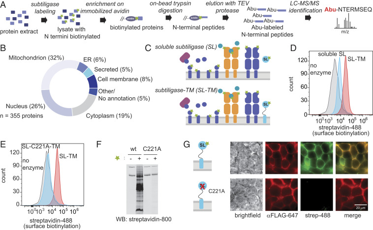
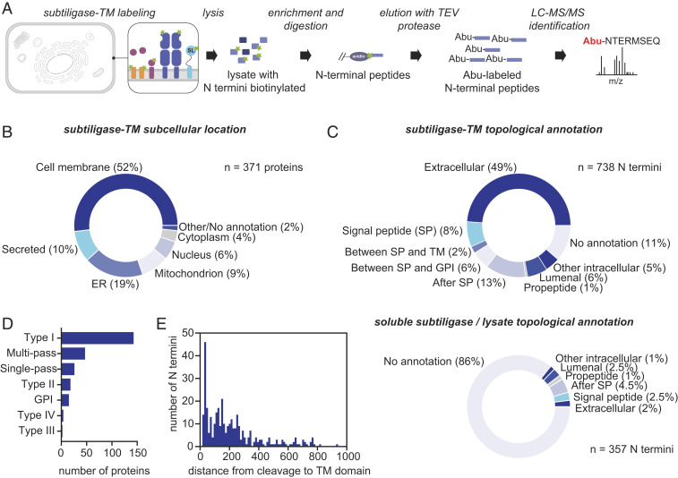
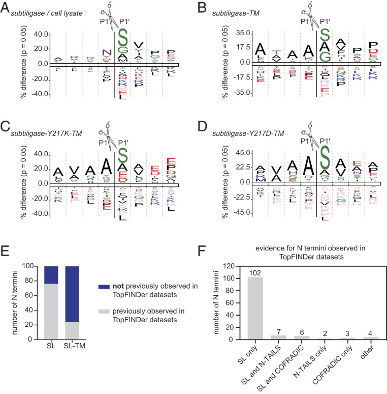
N terminomics is a powerful strategy for profiling proteolytic neo-N termini, but its application to cell surface proteolysis has been limited by the low relative abundance of plasma membrane proteins. Here we apply plasma membrane-targeted subtiligase variants (subtiligase-TM) to efficiently and specifically capture cell surface N termini in live cells. Using this approach, we sequenced 807 cell surface N termini and quantified changes in their abundance in response to stimuli that induce proteolytic remodeling of the cell surface proteome. To facilitate exploration of our datasets, we developed a web-accessible Atlas of Subtiligase-Captured Extracellular N Termini (ASCENT; http://wellslab.org/ascent). This technology will facilitate greater understanding of extracellular protease biology and reveal neo-N termini biomarkers and targets in disease.
Keywords: N terminomics; proteolysis; proteomics.
Conflict of interest statement
Competing interest statement: A.M.W. and J.A.W. and the Regents of the University of California have filed a patent application (US Provisional Patent Application 62/398,898) related to engineered subtiligase variants.
Figures

Similar articles
-
Spatially Resolved Tagging of Proteolytic Neo-N termini with Subtiligase-TM.J Membr Biol. 2021 Apr;254(2):119-125. doi: 10.1007/s00232-021-00171-4. Epub 2021 Feb 18. J Membr Biol. 2021. PMID: 33599828
-
Mapping Cell Surface Proteolysis with Plasma Membrane-Targeted Subtiligase.Methods Mol Biol. 2022;2456:71-83. doi: 10.1007/978-1-0716-2124-0_6. Methods Mol Biol. 2022. PMID: 35612736 Free PMC article.
-
N-Terminal Modification of Proteins with Subtiligase Specificity Variants.Curr Protoc Chem Biol. 2020 Mar;12(1):e79. doi: 10.1002/cpch.79. Curr Protoc Chem Biol. 2020. PMID: 32074409
-
Subtiligase-Catalyzed Peptide Ligation.Chem Rev. 2020 Mar 25;120(6):3127-3160. doi: 10.1021/acs.chemrev.9b00372. Epub 2019 Oct 30. Chem Rev. 2020. PMID: 31663725 Review.
-
N-Terminomics Strategies for Protease Substrates Profiling.Molecules. 2021 Aug 3;26(15):4699. doi: 10.3390/molecules26154699. Molecules. 2021. PMID: 34361849 Free PMC article. Review.
Cited by
-
Spatially Resolved Tagging of Proteolytic Neo-N termini with Subtiligase-TM.J Membr Biol. 2021 Apr;254(2):119-125. doi: 10.1007/s00232-021-00171-4. Epub 2021 Feb 18. J Membr Biol. 2021. PMID: 33599828
-
Cell-surface tethered promiscuous biotinylators enable comparative small-scale surface proteomic analysis of human extracellular vesicles and cells.Elife. 2022 Mar 8;11:e73982. doi: 10.7554/eLife.73982. Elife. 2022. PMID: 35257663 Free PMC article.
-
SheddomeDB 2023: A Revision of an Ectodomain Shedding Database Based on a Comprehensive Literature Review and Online Resources.J Proteome Res. 2023 Aug 4;22(8):2570-2576. doi: 10.1021/acs.jproteome.3c00001. Epub 2023 Jul 17. J Proteome Res. 2023. PMID: 37458416 Free PMC article. Review.
-
Direct Identification of Proteolytic Cleavages on Living Cells Using a Glycan-Tethered Peptide Ligase.ACS Cent Sci. 2022 Oct 26;8(10):1447-1456. doi: 10.1021/acscentsci.2c00899. Epub 2022 Oct 11. ACS Cent Sci. 2022. PMID: 36313159 Free PMC article.
-
Sensitive and High-Throughput Exploration of Protein N-Termini by TMT-TAILS N-Terminomics.Methods Mol Biol. 2023;2718:111-135. doi: 10.1007/978-1-0716-3457-8_7. Methods Mol Biol. 2023. PMID: 37665457
References
-
- Puente X. S., Sánchez L. M., Overall C. M., López-Otín C., Human and mouse proteases: A comparative genomic approach. Nat. Rev. Genet. 4, 544–558 (2003). - PubMed
-
- Kleifeld O., et al. ., Isotopic labeling of terminal amines in complex samples identifies protein N-termini and protease cleavage products. Nat. Biotechnol. 28, 281–288 (2010). - PubMed
-
- Staes A., et al. ., Selecting protein N-terminal peptides by combined fractional diagonal chromatography. Nat. Protoc. 6, 1130–1141 (2011). - PubMed
Publication types
MeSH terms
Substances
Grants and funding
LinkOut - more resources
Full Text Sources
Other Literature Sources
Research Materials
