. 2021 Feb 3;6(1):e01220-20.
doi: 10.1128/mSphere.01220-20.

Investigation of Heterochromatin Protein 1 Function in the Malaria Parasite Plasmodium falciparum Using a Conditional Domain Deletion and Swapping Approach

Affiliations

Investigation of Heterochromatin Protein 1 Function in the Malaria Parasite Plasmodium falciparum Using a Conditional Domain Deletion and Swapping Approach

Hai T N Bui et al. mSphere. .

Abstract

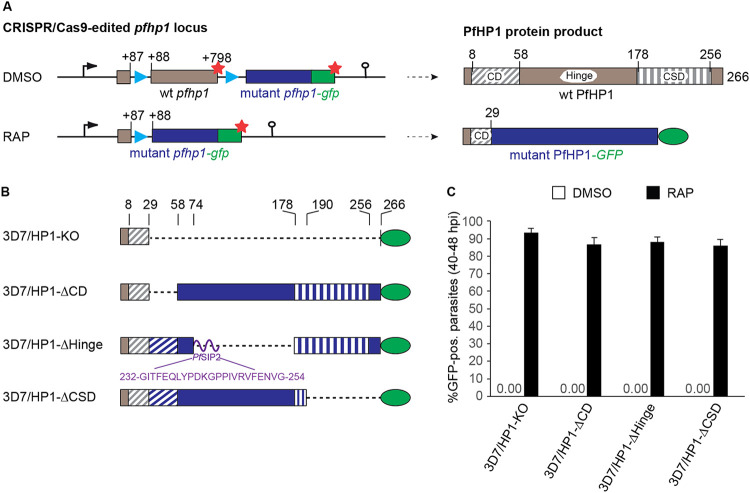
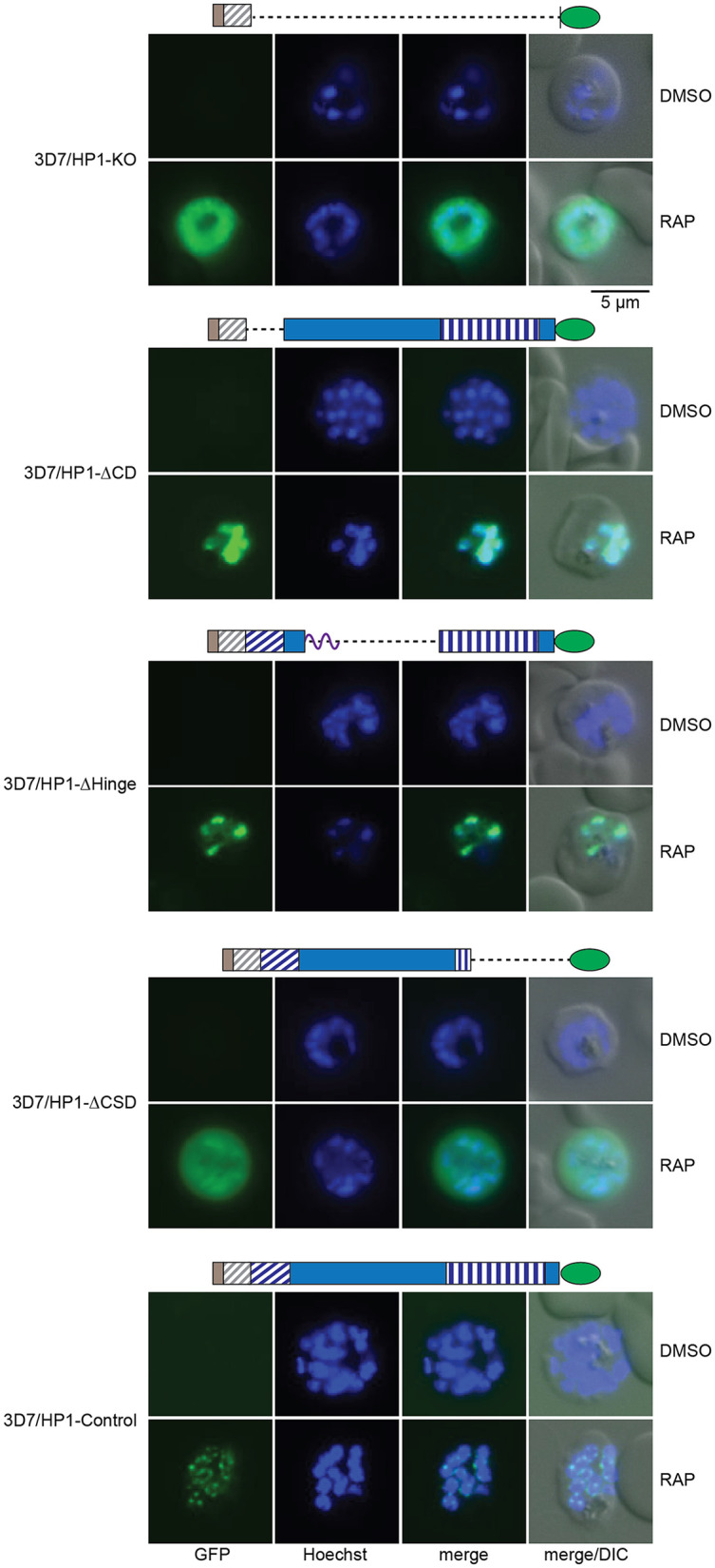
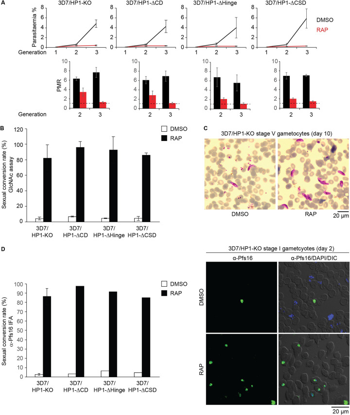
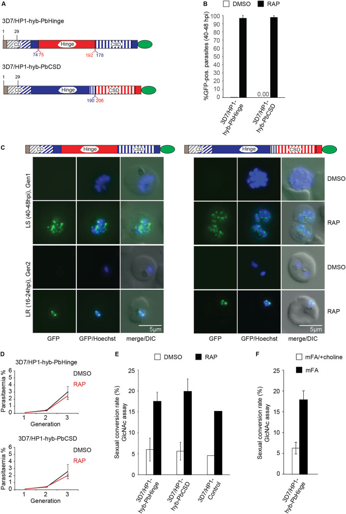
The human malaria parasite Plasmodium falciparum encodes a single ortholog of heterochromatin protein 1 (PfHP1) that plays a crucial role in the epigenetic regulation of various survival-related processes. PfHP1 is essential for parasite proliferation and the heritable silencing of genes linked to antigenic variation, host cell invasion, and sexual conversion. Here, we employed CRISPR/Cas9-mediated genome editing combined with the DiCre/loxP system to investigate how the PfHP1 chromodomain (CD), hinge domain, and chromoshadow domain (CSD) contribute to overall PfHP1 function. We show that the 76 C-terminal residues are responsible for targeting PfHP1 to the nucleus. Furthermore, we reveal that each of the three functional domains of PfHP1 are required for heterochromatin formation, gene silencing, and mitotic parasite proliferation. Finally, we discovered that the hinge domain and CSD of HP1 are functionally conserved between P. falciparum and P. berghei, a related malaria parasite infecting rodents. In summary, our study provides new insights into PfHP1 function and offers a tool for further studies on epigenetic regulation and life cycle decision in malaria parasites.IMPORTANCE Malaria is caused by unicellular Plasmodium species parasites that repeatedly invade and replicate inside red blood cells. Some blood-stage parasites exit the cell cycle and differentiate into gametocytes that are essential for malaria transmission via the mosquito vector. Epigenetic control mechanisms allow the parasites to alter the expression of surface antigens and to balance the switch between parasite multiplication and gametocyte production. These processes are crucial to establish chronic infection and optimize parasite transmission. Here, we performed a mutational analysis of heterochromatin protein 1 (HP1) in P. falciparum We demonstrate that all three domains of this protein are indispensable for the proper function of HP1 in parasite multiplication, heterochromatin formation, and gene silencing. Moreover, expression of chimeric proteins revealed the functional conservation of HP1 proteins between different Plasmodium species. These results provide new insight into the function and evolution of HP1 as an essential epigenetic regulator of parasite survival.

Keywords: CRISPR/Cas9; DiCre; HP1; Plasmodium falciparum; epigenetics; heterochromatin; malaria.


Figures

FIG 1
FIG 1
Generation of DiCre-inducible PfHP1 truncation mutants. (A) Schematics of the CRISPR/Cas9-edited pfhp1 locus (left) and corresponding PfHP1 protein products (right) before (DMSO) and after (RAP) rapamycin-induced DiCre-dependent excision of the floxed wild-type (wt) pfhp1 locus. Blue arrowheads indicate the position of sera2 intron:loxP elements. Red stars indicate stop codons. Brown and blue boxes represent the wild-type and the replacing mutant sequences pfhp1/PfHP1, respectively. Green boxes represent the gfp/GFP sequence. The CD, hinge domain, and CSD within wild-type PfHP1 are indicated. Numbers in the gene and protein schematics refer to nucleotide and amino acid positions, respectively. (B) Diagrams showing the organization of the PfHP1 truncation mutants expressed in the 3D7/HP1-KO, 3D7/HP1-ΔCD, 3D7/HP1-ΔHinge, and 3D7/HP1-ΔCSD lines after RAP treatment. Dashed lines represent corresponding deletions in the mutant PfHP1 protein sequences. Brown and blue colors represent the remaining wild-type PfHP1 N terminus and the replacing mutant protein sequences, respectively. The CD and CSD are indicated by diagonal and vertical dashed stripes, respectively. The purple curved line represents the short PfSIP2-derived linker polypeptide between the CD and CSD in the PfHP1-ΔHinge mutant. The amino acid sequence of this linker and its position within the PfSIP2 protein are indicated. Numbers on top indicate amino acid positions within wild-type PfHP1. (C) Proportion of GFP-positive parasites observed 40 h after treatment with RAP or DMSO (control). Values represent the means of results from three independent biological replicates (error bars indicate SD). For each sample, >200 iRBCs were counted. pos., positive.
FIG 2
FIG 2
Subcellular localization of PfHP1 truncation mutants. Representative live-cell fluorescence images showing the localization of the PfHP1-GFP fusions in DMSO- and RAP-treated 3D7/HP1-KO, 3D7/HP1-ΔCD, 3D7/HP1-ΔHinge, 3D7/HP1-ΔCSD, and 3D7/HP1-Control lines at late schizont stage (LS [40 to 48 hpi]; generation 1, 40 h after RAP treatment). Nuclei were stained with Hoechst dye. DIC, differential interference contrast. Scale bar, 5 μm.
FIG 3
FIG 3
Phenotypes of PfHP1 truncation mutants. (A, top) Growth curves of the DMSO- and RAP-treated PfHP1 truncation mutants over three consecutive generations. (Bottom) Parasite multiplication rates (PMRs) reflect the fold increase in parasitemia observed in generations 2 and 3. Values are the means of results from three biological replicates (error bars indicate SD). For each sample, >3,000 RBCs were counted. (B) Sexual conversion rates of PfHP1 truncation mutants in DMSO- and RAP-treated parasites, assessed by inspection of Giemsa-stained blood smears of GlcNAc-treated cultures on day 6 of gametocytogenesis. Results are the means of results from at least three replicates (error bars indicate SD). For each sample, >3,000 RBCs were counted. (C) Representative overview images from Giemsa-stained blood smears showing stage V gametocytes (day 10 of gametocytogenesis) obtained from DMSO- and RAP-treated 3D7/HP1-KO parasites. Scale bar, 20 μm. (D) Sexual conversion rates of PfHP1 truncation mutants in DMSO- and RAP-treated parasites, assessed by anti-Pfs16 IFAs performed on stage I gametocytes on day 2 of gametocytogenesis. The result from the 3D7/HP1-KO line represents the mean of results from four replicates (error bar indicates SD). All other values derive from a single experiment. For each sample, >200 iRBCs were counted. Representative overview images of anti-Pfs16 IFAs used to quantify stage I gametocytes in DMSO- and RAP-treated populations of the 3D7/HP1-KO line are shown on the right. Scale bar, 20 μm.
FIG 4
FIG 4
Generation of DiCre-inducible PfHP1-PbHP1 hybrid mutants. (A) Diagrams showing the GFP-tagged PfHP1-PbHP1 hybrid proteins expressed in the 3D7/HP1-hyb-PbHinge and 3D7/HP1-hyb-PbCSD cell lines after RAP treatment. Brown and blue colors represent the remaining wild-type PfHP1 N terminus and the replacing PfHP1 protein sequences, respectively. Red colors identify the hinge domain and CSD derived from PbHP1. The CD and CSD are indicated by diagonal and vertical dashed stripes, respectively. Numbers in blue and red refer to amino acid positions within the PfHP1 and PbHP1 sequences, respectively. (B) Proportions of GFP-positive parasites observed 40 h after treatment with RAP or DMSO (control). Values represent the means from three independent biological replicates (error bars indicate SD). For each sample, >140 iRBCs were counted. (C) Representative live-cell fluorescence images showing the localization of GFP-tagged PfHP1-PbHP1 hybrid proteins in 3D7/HP1-hyb-PbHinge and 3D7/HP1-hyb-PbCSD parasites in late schizonts (LS) (40 to 48 hpi; generation 1 [Gen1], 40 h after RAP treatment) and in late-ring-stage progeny (LR) (16 to 24 hpi, generation 2). Nuclei were stained with Hoechst dye. DIC, differential interference contrast. Scale bar, 5 μm. (D) Growth curves of the DMSO- and RAP-treated 3D7/HP1-hyb-PbHinge and 3D7/HP1-hyb-PbCSD parasites over three consecutive generations. Values are the means from at least three independent replicate experiments (error bars represent SD). For each sample, >3,000 RBCs were counted. (E) Sexual conversion rates of the DMSO- and RAP-treated 3D7/HP1-hyb-PbHinge and 3D7/HP1-hyb-PbCSD mutants and the 3D7/HP1-Control line, assessed by inspection of Giemsa-stained blood smears of GlcNAc-treated cultures on day 6 of gametocytogenesis. Values represent the means from at least three independent replicate experiments (error bars represent SD). The values for the 3D7/HP1-Control line derive from a single experiment and are consistent with previously published data (69). For each sample, >3,000 RBCs were counted. (F) Sexual conversion rates of 3D7/HP1-hyb-PbHinge parasites cultured in minimal fatty acid medium (mFA) or mFA supplemented with 2 mM choline (mFA/+choline), assessed by inspection of Giemsa-stained blood smears of GlcNAc-treated cultures on day 6 of gametocytogenesis. Values represent the means from two independent replicate experiments (error bars represent SD). For each sample, >1,800 RBCs were counted.

Similar articles

Cited by

References

    1. James TC, Elgin SC. 1986. Identification of a nonhistone chromosomal protein associated with heterochromatin in Drosophila melanogaster and its gene. Mol Cell Biol 6:3862–3872. doi:10.1128/mcb.6.11.3862. - DOI - PMC - PubMed
    1. Eissenberg JC, James TC, Foster-Hartnett DM, Hartnett T, Ngan V, Elgin SC. 1990. Mutation in a heterochromatin-specific chromosomal protein is associated with suppression of position-effect variegation in Drosophila melanogaster. Proc Natl Acad Sci U S A 87:9923–9927. doi:10.1073/pnas.87.24.9923. - DOI - PMC - PubMed
    1. Lachner M, O'Carroll D, Rea S, Mechtler K, Jenuwein T. 2001. Methylation of histone H3 lysine 9 creates a binding site for HP1 proteins. Nature 410:116–120. doi:10.1038/35065132. - DOI - PubMed
    1. Bannister AJ, Zegerman P, Partridge JF, Miska EA, Thomas JO, Allshire RC, Kouzarides T. 2001. Selective recognition of methylated lysine 9 on histone H3 by the HP1 chromo domain. Nature 410:120–124. doi:10.1038/35065138. - DOI - PubMed
    1. Schotta G, Ebert A, Krauss V, Fischer A, Hoffmann J, Rea S, Jenuwein T, Dorn R, Reuter G. 2002. Central role of Drosophila SU(VAR)3-9 in histone H3-K9 methylation and heterochromatic gene silencing. EMBO J 21:1121–1131. doi:10.1093/emboj/21.5.1121. - DOI - PMC - PubMed

Publication types

MeSH terms

Substances

LinkOut - more resources