. 2021 Feb 9;118(6):e2015654118.
doi: 10.1073/pnas.2015654118.

SLFN11 promotes CDT1 degradation by CUL4 in response to replicative DNA damage, while its absence leads to synthetic lethality with ATR/CHK1 inhibitors

Affiliations

SLFN11 promotes CDT1 degradation by CUL4 in response to replicative DNA damage, while its absence leads to synthetic lethality with ATR/CHK1 inhibitors

Ukhyun Jo et al. Proc Natl Acad Sci U S A. .

Abstract

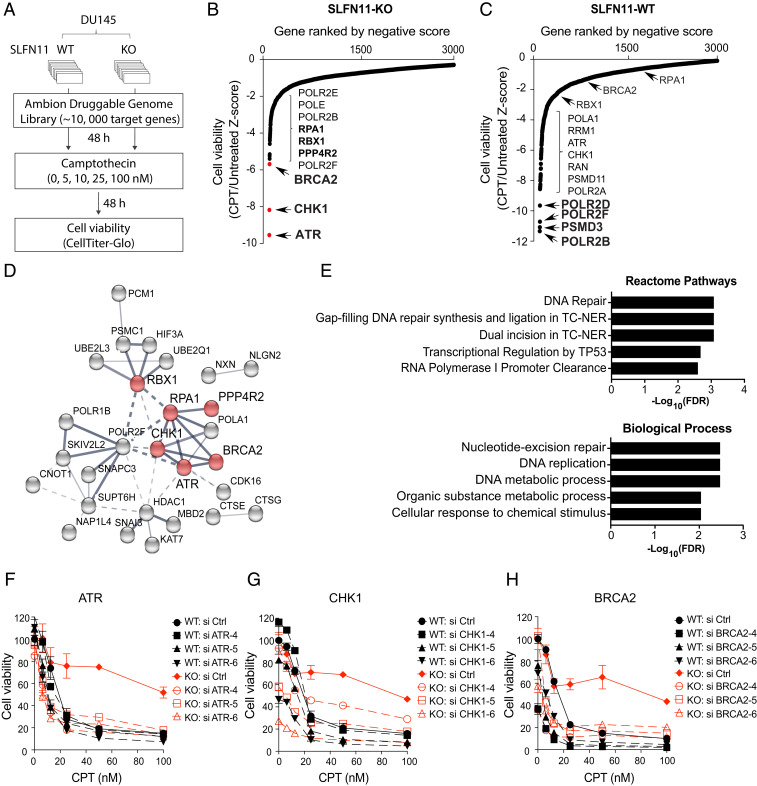
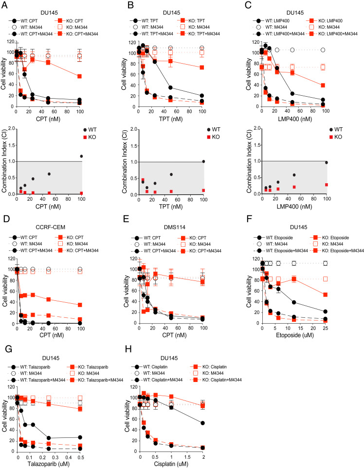
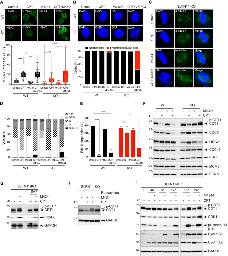
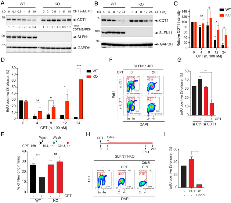
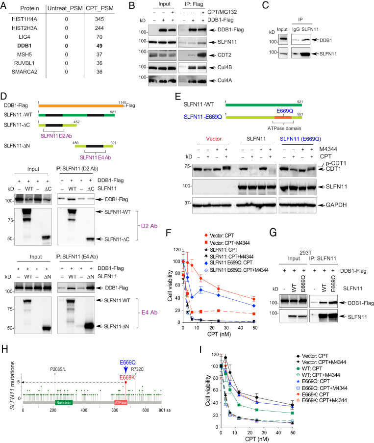
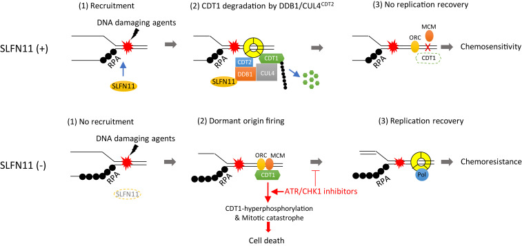
Schlafen-11 (SLFN11) inactivation in ∼50% of cancer cells confers broad chemoresistance. To identify therapeutic targets and underlying molecular mechanisms for overcoming chemoresistance, we performed an unbiased genome-wide RNAi screen in SLFN11-WT and -knockout (KO) cells. We found that inactivation of Ataxia Telangiectasia- and Rad3-related (ATR), CHK1, BRCA2, and RPA1 overcome chemoresistance to camptothecin (CPT) in SLFN11-KO cells. Accordingly, we validate that clinical inhibitors of ATR (M4344 and M6620) and CHK1 (SRA737) resensitize SLFN11-KO cells to topotecan, indotecan, etoposide, cisplatin, and talazoparib. We uncover that ATR inhibition significantly increases mitotic defects along with increased CDT1 phosphorylation, which destabilizes kinetochore-microtubule attachments in SLFN11-KO cells. We also reveal a chemoresistance mechanism by which CDT1 degradation is retarded, eventually inducing replication reactivation under DNA damage in SLFN11-KO cells. In contrast, in SLFN11-expressing cells, SLFN11 promotes the degradation of CDT1 in response to CPT by binding to DDB1 of CUL4CDT2 E3 ubiquitin ligase associated with replication forks. We show that the C terminus and ATPase domain of SLFN11 are required for DDB1 binding and CDT1 degradation. Furthermore, we identify a therapy-relevant ATPase mutant (E669K) of the SLFN11 gene in human TCGA and show that the mutant contributes to chemoresistance and retarded CDT1 degradation. Taken together, our study reveals new chemotherapeutic insights on how targeting the ATR pathway overcomes chemoresistance of SLFN11-deficient cancers. It also demonstrates that SLFN11 irreversibly arrests replication by degrading CDT1 through the DDB1-CUL4CDT2 ubiquitin ligase.

Keywords: ATR/CHK1 inhibitor; CDT1; CUL4; SLFN11.


Conflict of interest statement

The authors declare no competing interest.

Figures

Fig. 1.
Fig. 1.
Genome-wide RNAi screen identifies ATR pathway as a synergistic target to overcome chemoresistance in SLFN11-deficient cells. (A) Schematic overview of genome-wide RNAi screen workflow in DU145 WT and SLFN11 KO cells. (B and C) Ranked distribution plots of z-scores obtained from cell viability (CPT-treated/untreated) in SLFN11-KO (B) and -WT (C) cells (Dataset S1). Black dots: Individual siRNA targeted genes. Red dots: Hit genes selected for further validation. (D) Protein interaction network with RNAi screen hits in SLFN11-KO cells generated by STRING analysis. Line thickness represents the strength of data confidence. (E) GO analysis of molecular network in SLFN11-KO cells. (FH) Validation of chemosensitivity in SLFN11-WT and KO cells. Cells were transfected with three siRNAs targeting ATR, CHK1, and BRCA2 and then treated with the indicated concentrations of CPT for 72 h. Cell viability was analyzed by CellTiter-Glo (Promega). Error bars represent SD (n = 3).
Fig. 2.
Fig. 2.
Clinical inhibitor of the ATR pathway consistently resensitizes SLFN11-deficient cells to clinical DNA damaging agents. (AC, Upper) DU145 SLFN11-WT and -KO cells were treated with M4344 (25 nM) and the indicated concentrations of the TOP1 inhibitors CPT, TPT, and indotecan (LMP400) for 72 h. Cell viability was analyzed by CellTiter-Glo. Error bars represent SD (n = 3). (Lower) Combination Index (CI) versus Fa (fraction affected) calculated from cell viability data. (D) Combination treatment of M4344 (1 nM) and the indicated concentrations of CPT in the isogenic WT and SLFN11-KO leukemic lymphoblasts CCRF-CEM cells. (E) Combination treatment of M4344 (25 nM) and the indicated concentrations of CPT in small-cell lung cancer DMS114 cells. (FH) DU145 cells were treated with M4344 (25 nM) and the indicated concentrations of etoposide, talazoparib, and cisplatin for 72 h. Cell viability was analyzed by CellTiter-Glo. Error bars represent SD (n = 3).
Fig. 3.
Fig. 3.
Inhibition of ATR leads to increased DNA damage and chromosomal defects with CDT1 hyperphosphorylation in SLFN11-deficient cells treated with CPT. (A) Confocal immunofluorescence staining of γH2AX in DU145 SLFN11-WT and -KO cells treated with CPT (100 nM) and M4344 (25 nM) for 24 h. (Upper) Representative images (γH2AX green). (Magnification, 63×). (Lower) γH2AX signal quantified by ImageJ. Error bars represent SD (n ≥ 50); ****P < 0.0001 Student's t test; ns, not significant. (B) Confocal immunofluorescence staining of nuclei in cells treated with CPT and M4344 for 24 h. (Upper) Representative images (magnification, 63×) of nuclei/single cell, labeled with DAPI (blue). (Lower) Percentages of multinucleated cells after drug treatment (n = 100). (C) Confocal immunofluorescence staining of metaphase alignment in SLFN11-KO cells after treatment with CPT and M4344 for 16 h. Mitotic spindles were stained with α-tubulin (green) and chromosomes with DAPI (blue). (Magnification, 63×). (D) Cell cycle distribution analyzed by flow cytometry after treatment with CPT and M4344 for 24 h. Data are presented as mean values. Error bars represent SD (n = 3). (E) Relative EdU incorporation analyzed by flow cytometry. Cells were treated with CPT and M4344 for 24 h and pulsed with Edu (10 µM) for 30 min prior to harvesting. Error bars represent SD (n = 3). **P < 0.009, ****P < 0.0001 Student’s t test. (F) Protein expression levels of DNA replication initiation factors after treatment with CPT and M4344 for 24 h. (G) Retardation of CDT1 electrophoretic migration in response to combined treatment with CPT and M4344 in SLFN11-KO cells is due to hyperphosphorylation. SLFN11-KO cells were treated with CPT and M4344 for 24 h and then incubated with SAP (1 U/µL). (H) CDT1 hyperphosphorylation in response to combined treatment with CPT and M4344 is mediated by CDK. SLFN11-KO cells were treated with CPT, M4344 and roscovitine (20 µM) for 24 h and then analyzed by Western blotting. (I) CDT1 is hyperphosphorylated in time-dependent manner in response to combined treatment with CPT and M4344.
Fig. 4.
Fig. 4.
Defective CDT1 degradation causes replication recovery in SLFN11-deficient cells. (A) DU145 SLFN11-WT and -KO cells were treated with the indicated concentrations of CPT for 4 h. Protein levels were analyzed by Western blotting. (B) DU145 SLFN11-WT and -KO cells were treated with CPT (100 nM) for the indicated times. Levels of protein expression were analyzed by Western blotting. (C) Quantitation of CDT1 as shown in B. Bars show the mean band intensity from triplicate experiments, normalized to GAPDH. *P < 0.0274, **P < 0.0077. (D) Percentage of EdU+ S-phase cells in the time course treatment of CPT was determined by flow cytometry. Error bars represent SD (n = 3). *P < 0.0335, **P < 0.0025, ***P < 0.0005 Student’s t test. (E, Upper) Labeling protocols for the DNA combing assay. Cells were treated with CPT (100 nM) for 16 h and then pulse-labeled sequentially with IdU (100 µM) and CldU (100 µM) for 1 h. (Lower) Frequencies of new origins only labeled by the CldU pulse. Error bars represent SD (n = 3). ***P < 0.0009 Student’s t test. (F) CDT1-dependent replication recovery. SLFN11-KO cells were transfected with siControl (Ctrl) or siCDT1 for 48 h, and then treated with CPT for 24 h. Cells were incubated with EdU (10 µM) for 30 min prior to harvest. The percentage of EdU+ and DAPI+ cells was determined by flow cytometry. (G) Quantification of the EdU+ cells in S-phase. Error bars represent SD (n = 3). ***P < 0.0009 Student’s t test. (H, Upper) Treatment protocol. Cells were treated with CPT for 24 h and with the Cdc7 inhibitor (PHA-767491, 5 µM) after 4 h of CPT treatment. Cells were incubated with EdU (10 µM) for 30 min prior to harvest. Percentage of EdU+ and DAPI+ cells was determined by flow cytometry. (I) Quantification of the EdU+ cells in S-phase. Error bars represent SD (n = 3). **P < 0.008 Student’s t test.
Fig. 5.
Fig. 5.
SLFN11 promotes CDT1 degradation by binding to the CUL4–DDB1 complex in response to replicative DNA damage. (A) Representative interactors of SLFN11. DU145 cells were treated with CPT for 16 h. Nuclear cell lysates were immunoprecipitated with anti-SLFN11 antibody and analyzed by mass spectrometry. (B) HEK293 cells transfected with a DDB1-Flag construct for 2 d were treated with CPT (10 µM) and MG132 (20 µM) for 4 h were lysed and immunoprecipitated with anti-Flag antibody. Interacting proteins were profiled by Western blotting. (C) Interaction between endogenous SLFN11 and DDB1 was assessed by immunoprecipitation with anti-SLFN11 (D2) antibody after treatment with CPT (10 µM) and MG132 (20 µM) for 4 h in DU145 cells. (D, Top) Diagram of DDB1-Flag and SLFN11 deletion mutants. (Middle) 293T cells transfected with DDB1-Flag and indicated SLFN11 constructs were treated with CPT (10 µM) and MG132 (20 µM) for 4 h. Cells were immunoprecipitated with anti-SLFN11 antibody (D2) that detects the N terminus region of SLFN11. (Bottom) Same as above but using anti-SLFN11 antibody (E4) that detects the C terminus region of SLFN11. Interacting proteins were identified by Western blotting. (E) K562 cells stably transfected with vector, SLFN11 and SLFN11-E669Q constructs were treated with CPT (100 nM) and M4344 for 24 h. Proteins were identified by Western blotting. (F) K562 cells stably transfected with vector, SLFN11-WT or SLFN11-E669Q constructs were treated with CPT and M4344 for 72 h. Cell viability was analyzed by CellTiter-Glo. (G) SLFN11-E669Q interacts with DDB1. 293T cells were transiently transfected with DDB1 Flag, SLFN11-WT, and -E669Q for 48 h. Following treatment with CPT (10 µM) and MG132 (20 µM) for 4 h, protein interactions were assessed by IP with anti-SLFN11 (D2) antibody. (H) Diagram of SLFN11 mutations in TCGA. (I) 293T cells transfected with SLFN11-WT, SLFN11-E669Q, or SLFN11-E669K constructs for 48 h, were treated with CPT and M4344 for 72 h. Cell viability was analyzed by CellTiter-Glo.
Fig. 6.
Fig. 6.
Proposed model for SLFN11-mediated replication reactivation block and synthetic lethality of ATR/CHK1 inhibitors in SLFN11-negative cancer cells. In SLFN11-proficient cells, SLFN11 is recruited to chromatin by DNA damage. It subsequently binds to DDB1 and promotes the degradation of CDT1 and other substrates as a cofactor of activated DDB1–CUL4CDT2. Consequently, CDT1-mediated replication recovery is irreversibly blocked, leading to sensitivity to DNA-damaging agents. In SLFN11-deficient cells, conversely, DDB1–CUL4CDT2-mediated degradation of CDT1 is reduced. CDT1 then promotes replication recovery by activating dormant origins, resulting in chemoresistance. Treatment of ATR/CHK1 inhibitor reverses chemoresistance of SLFN11-deficient cells by inducing premature replication and mitosis through CDT1 phosphorylation, which induces mitotic catastrophe and cell death.

References

    1. Barretina J., et al. ., The Cancer Cell Line Encyclopedia enables predictive modelling of anticancer drug sensitivity. Nature 483, 603–607 (2012). - PMC - PubMed
    1. Zoppoli G., et al. ., Putative DNA/RNA helicase Schlafen-11 (SLFN11) sensitizes cancer cells to DNA-damaging agents. Proc. Natl. Acad. Sci. U.S.A. 109, 15030–15035 (2012). - PMC - PubMed
    1. Mu Y., et al. ., SLFN11 inhibits checkpoint maintenance and homologous recombination repair. EMBO Rep. 17, 94–109 (2016). - PMC - PubMed
    1. Murai J., et al. ., Resistance to PARP inhibitors by SLFN11 inactivation can be overcome by ATR inhibition. Oncotarget 7, 76534–76550 (2016). - PMC - PubMed
    1. Murai J., et al. ., SLFN11 blocks stressed replication forks independently of ATR. Mol. Cell 69, 371–384.e6 (2018). - PMC - PubMed

Publication types

MeSH terms

LinkOut - more resources