. 2021 Feb 9;118(6):e2018355118.
doi: 10.1073/pnas.2018355118.

ARMC12 regulates spatiotemporal mitochondrial dynamics during spermiogenesis and is required for male fertility

Affiliations

ARMC12 regulates spatiotemporal mitochondrial dynamics during spermiogenesis and is required for male fertility

Keisuke Shimada et al. Proc Natl Acad Sci U S A. .

Abstract

The mammalian sperm midpiece has a unique double-helical structure called the mitochondrial sheath that wraps tightly around the axoneme. Despite the remarkable organization of the mitochondrial sheath, the molecular mechanisms involved in mitochondrial sheath formation are unclear. In the process of screening testis-enriched genes for functions in mice, we identified armadillo repeat-containing 12 (ARMC12) as an essential protein for mitochondrial sheath formation. Here, we engineered Armc12-null mice, FLAG-tagged Armc12 knock-in mice, and TBC1 domain family member 21 (Tbc1d21)-null mice to define the functions of ARMC12 in mitochondrial sheath formation in vivo. We discovered that absence of ARMC12 causes abnormal mitochondrial coiling along the flagellum, resulting in reduced sperm motility and male sterility. During spermiogenesis, sperm mitochondria in Armc12-null mice cannot elongate properly at the mitochondrial interlocking step which disrupts abnormal mitochondrial coiling. ARMC12 is a mitochondrial peripheral membrane protein and functions as an adherence factor between mitochondria in cultured cells. ARMC12 in testicular germ cells interacts with mitochondrial proteins MIC60, VDAC2, and VDAC3 as well as TBC1D21 and GK2, which are required for mitochondrial sheath formation. We also observed that TBC1D21 is essential for the interaction between ARMC12 and VDAC proteins in vivo. These results indicate that ARMC12 uses integral mitochondrial membrane proteins VDAC2 and VDAC3 as scaffolds to link mitochondria and works cooperatively with TBC1D21. Thus, our studies have revealed that ARMC12 regulates spatiotemporal mitochondrial dynamics to form the mitochondrial sheath through cooperative interactions with several proteins on the sperm mitochondrial surface.

Keywords: infertility; mitochondrial sheath formation; sperm mitochondrial dynamics; spermatogenesis.


Conflict of interest statement

The authors declare no competing interest.

Figures
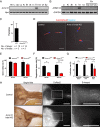
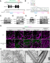
Fig. 1.
Fig. 1.
Armc12 indel KO male mice are sterile due to reduced sperm motility and abnormal sperm morphology. (A and B) RT-PCR for Armc12 from various mouse tissues (A) and RT-PCR for ARMC12 from various human tissues (B) (He, heart; Li, liver; Sp, spleen; Lu, lung; Ki, kidney; Br, brain; St, stomach; In, intestine; Te, testis; Ov, ovary; Ut, uterus; Ep, epididymis; Cap, caput; Cor, corpus; Cau, Cauda). Hprt and GAPDH were used as controls. (C) Pregnancy rate of control and Armc12 indel KO male mice. (D) Sperm morphology of control and Armc12 indel KO mice with RBGS transgenes, which express mitochondria-targeted DsRed2 (red). Nuclei were stained with Hoechst 33342 (blue). (Scale bars, 20 µm.) (E) Sperm motility from control and Armc12 indel KO mice. Error bars represent SD (n = 3). (F) Progressive sperm rate of sperm from control and Armc12 indel KO mice. **P < 0.01, Student’s t test; error bars represent SD (n = 3). (G) Fertilization rate of IVF using control and Armc12 indel KO spermatozoa. Three types of oocytes (cumulus-intact, cumulus-free, and zona-free) were used for IVF. *P < 0.05, **P < 0.01, Student’s t test; error bars represent SD (n = 3). (H) Imaging of spermatozoa inside the female reproductive tract 2 to 3 h after observing vaginal plugs. (Left) Reproductive organs under normal bright-field conditions. (Middle) Red fluorescence of RBGS spermatozoa in the female reproductive tract. (Right) Magnified images of the boxed areas. Fluorescent images were false-colored using ImageJ software. (Scale bars, 500 µm.)
Fig. 2.
Fig. 2.
ARMC12 is essential for proper mitochondrial elongation and subsequent coiling along the flagellum during spermiogenesis. (A) Ultrastructural images of step 16 spermatids analyzed by TEM. Arrows indicate aberrant location of sperm mitochondria. (Scale bars, 1.0 μm.) (B) Development of mitochondrial sheath during spermiogenesis observed by SEM. Arrows indicate exposed mitochondrial tips. (Scale bars, 1.0 μm.) (C) Sperm morphology of control and Armc12 indel KO spermatozoa collected from the cauda epididymis. Samples were observed by SEM. Arrow indicates abnormal uncoiled mitochondria that shed from the midpiece. (Scale bars, 5.0 µm.)
Fig. 3.
Fig. 3.
ARMC12 localizes to the mitochondrial outer membrane of spermatids. (A) Schematic diagram of FLAG-tagged alleles of endogenous Armc12. KI allele has a three-amino-acid spacer before the FLAG tag. (B) Control and Armc12FLAG alleles. (C) WB analysis using lysates prepared from the testis and mature spermatozoa from WT and FLAG-tagged Armc12 KI mice. ACTB was used as a loading control. (D) WB analysis using aqueous and detergent fractions of Triton X-114 extracts of the testicular cells. COX IV and ZPBP were used as membrane protein and cytosolic protein markers, respectively. (E) WB analysis using isolated mitochondrial and cytosolic fractions from FLAG-tagged Armc12 KI testicular cells. COX IV and GAPDH were used as mitochondrial protein and cytosolic protein markers, respectively. (F) Mitochondria purified from Armc12-FLAG KI testicular cells were incubated with isotonic buffer or high salt (1 M NaCl) followed by centrifugation to isolate pellet (P) and supernatant (SN) fractions. Equal volumes of protein from each fraction were subjected to WB analysis. VDAC2 and VDAC3 were used as control of integral mitochondrial membrane protein. (G) Immunostaining of FLAG-tagged ARMC12 in Armc12-FLAG KI mouse testis and epididymis. Arrowheads indicate ARMC12 that are not localized in the sperm midpiece. Fluorescent images were false colored using ImageJ software. Spermatogenic stages were identified by the nuclear morphology. (Scale bars, 20 µm.) (H) Detection of immunolabeled FLAG-tagged ARMC12 in testis by TEM using anti-FLAG antibody incubated with 1.4-nm gold-particle–conjugated secondary antibody (arrowheads). (Scale bars, 500 nm.)
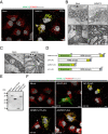
Fig. 4.
Fig. 4.
ARMC12 functions as an adherence factor between mitochondria in cultured cells. (A) COS-7 cells transiently expressing FLAG-tagged ARMC12 protein were stained with FLAG (green) and TOM20 (red) to visualize ARMC12 and mitochondria, respectively. Hoechst 33342 (white) was used to visualize the nuclei. (Scale bars, 10 µm.) The numbers in the upper right corner indicate the number of cells with mitochondrial aggregation out of the number of cells with FLAG fluorescence. (B) TEM micrographs of COS-7 cells overexpressing a control vector (Left) or ARMC12 protein (Right). (Lower) Magnified images of the boxed areas. Arrows indicate mitochondria with reduced cristae. Arrowheads indicate the border between mitochondria. (Scale bars, 2.0 μm and 500 nm, respectively.) (C) Immunolabeled detection of transiently expressing FLAG-tagged ARMC12 in COS-7 cells was observed by TEM using anti-FLAG antibody combined with 1.4 nm gold-particle–conjugated secondary antibody (arrowheads). (Scale bars, 200 nm.) (D) Schematic of various truncated ARMC12 vectors. FLAG-tag was linked posterior to the C-terminal of ARMC12. Green and yellow boxes show the N terminus and C terminus of ARMC12, respectively. Light blue boxes indicate FLAG tag. A five-amino-acid spacer is inserted between the C terminus and FLAG tag. (E) HEK293T cells were transfected with plasmids encoding FLAG-tagged full length ARMC12 and truncated forms of ARMC12. Cell lysates were analyzed by immunoblotting using an anti-FLAG antibody. (F) COS-7 cells transiently expressing truncated ARMC12 were stained with FLAG (green) and TOM20 (red) to visualize ARMC12 and mitochondria, respectively. Hoechst 33342 (white) was used to visualize the nuclei. (Scale bars, 10 µm.) The numbers in the lower left corner indicate the number of cells with mitochondrial aggregation out of the number of cells with FLAG fluorescence.
Fig. 5.
Fig. 5.
ARMC12 interacts with several proteins to form the mitochondrial sheath during spermiogenesis. (A and B) RT-PCR for Tbc1D15, Tbc1d21, and Fis1 from various mouse tissues (A) and RT-PCR for TBC1D15, TBC1D21, and FIS1 from various human tissues (B) (He, heart; Li, liver; Sp, spleen; Lu, lung; Ki, kidney; Br, brain; St, stomach; In, intestine; Te, testis; Ov, ovary; Ut, uterus; Ep, epididymis; Cap, caput; Cor, corpus; Cau, Cauda). Hprt and GAPDH were used as controls. (C and D) The co-IP followed by Western blot analysis was performed using lysates collected from WT or FLAG-tagged Armc12 KI mouse TGC. Immunoprecipitated proteins by anti-FLAG antibody (C) or anti-FIS1 antibody (D) were analyzed by WB using anti-TBC1D15 and TBC1D21 antibodies. IgG was used as a negative control for the IP. (E) List of identified proteins by MS analysis in vivo. Mitochondria were isolated from WT or FLAG-tagged Armc12 KI testis, and proteins were extracted from the isolated mitochondria. The proteins were immunoprecipitated using anti-FLAG antibody and identified by MS analysis. This list shows proteins either identified only in FLAG-tagged Armc12 KI mice (quantitative value >3.0) or highly identified in KI mice (ratio >3.0 as compared with WT) are listed. (F) Co-IP followed by WB analysis were performed using lysates collected from WT or FLAG-tagged Armc12 KI mice TGC. Immunoprecipitated proteins by anti-FLAG antibody were analyzed by WB using anti-GK2, MIC60, SPERT, TOM70, VDAC2, and VDAC3 antibodies.
Fig. 6.
Fig. 6.
Disruption of TBC1D21 causes disorganization of mitochondrial sheath formation. (A) WB analysis of TBC1D21 using proteins obtained from control and Tbc1d21 KO mouse. ACTB was used as a loading control. (B) Pregnancy rate of control and Tbc1d21 KO male mice. (C) Sperm morphology of control and Tbc1d21 KO mice with RBGS transgenes, which express mitochondria-targeted DsRed2 (red). Nuclei were stained with Hoechst 33342 (blue). (Scale bars, 20 µm.) (D) Mitochondrial sheath development during spermiogenesis observed by SEM. Arrows indicate exposed mitochondrial tips. (Scale bars, 1.0 µm.) (E) The co-IP followed by WB analysis was performed using lysates collected from Tbc1d21 heterozygous or homozygous KO mouse TGC with a FLAG-tagged Armc12 KI allele. Immunoprecipitated proteins by anti-FLAG antibody were analyzed by WB using anti-VDAC2 and VDAC3 antibodies. WB analysis using anti-FLAG and TBC1D21 was performed to confirm the existence of FLAG-tagged KI allele and disappearance of TBC1D21.
Fig. 7.
Fig. 7.
Schematic models for the function of ARMC12. (A) Schematic model of mitochondrial sheath formation observed in WT spermatids. (B) Schematic model of abnormal mitochondrial sheath formation observed in Armc12 KO spermatids.

Similar articles

Cited by

References

    1. Fawcett D. W., The mammalian spermatozoon. Dev. Biol. 44, 394–436 (1975). - PubMed
    1. Ho H.-C., Wey S., Three dimensional rendering of the mitochondrial sheath morphogenesis during mouse spermiogenesis. Microsc. Res. Tech. 70, 719–723 (2007). - PubMed
    1. Shimada K., Kato H., Miyata H., Ikawa M., Glycerol kinase 2 is essential for proper arrangement of crescent-like mitochondria to form the mitochondrial sheath during mouse spermatogenesis. J. Reprod. Dev. 65, 155–162 (2019). - PMC - PubMed
    1. Mi Y., Shi Z., Li J., Spata19 is critical for sperm mitochondrial function and male fertility. Mol. Reprod. Dev. 82, 907–913 (2015). - PubMed
    1. Imai H., et al. ., Depletion of selenoprotein GPx4 in spermatocytes causes male infertility in mice. J. Biol. Chem. 284, 32522–32532 (2009). - PMC - PubMed

Publication types

MeSH terms

Substances