. 2021 Feb 3;12(1):761.
doi: 10.1038/s41467-021-21090-x.

Conserved arginine residues in synaptotagmin 1 regulate fusion pore expansion through membrane contact

Affiliations

Conserved arginine residues in synaptotagmin 1 regulate fusion pore expansion through membrane contact

Sarah B Nyenhuis et al. Nat Commun. .

Abstract

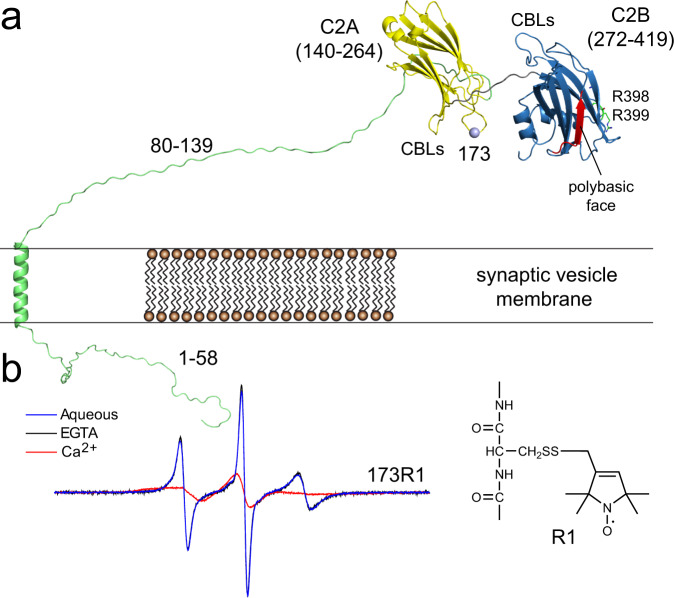
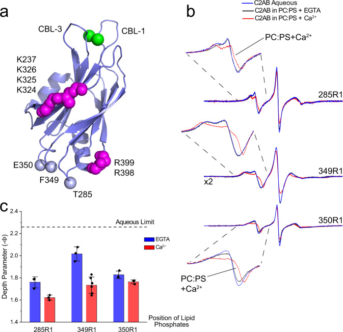
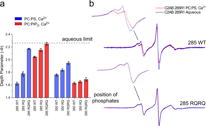
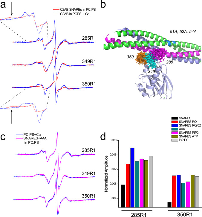
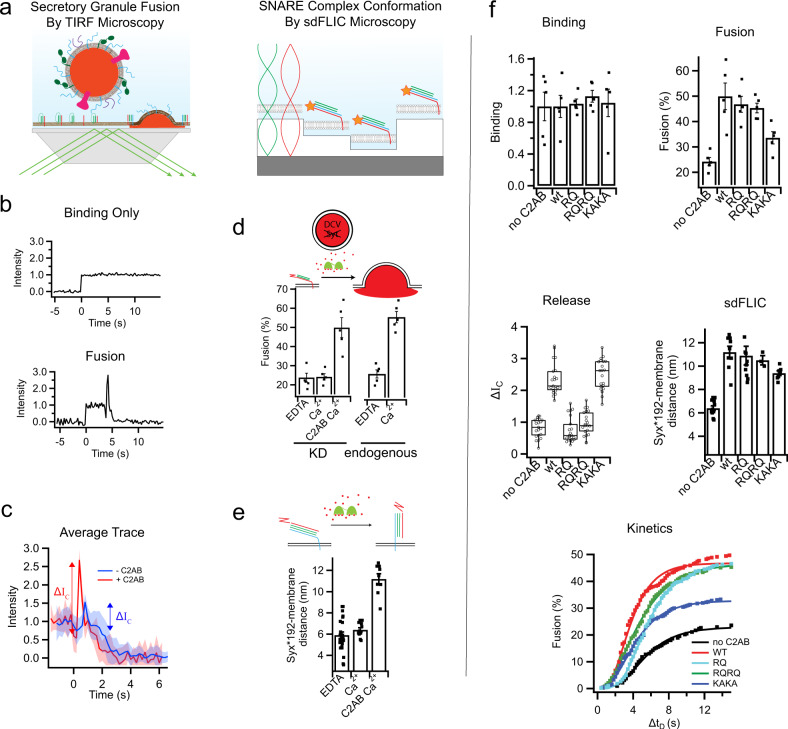
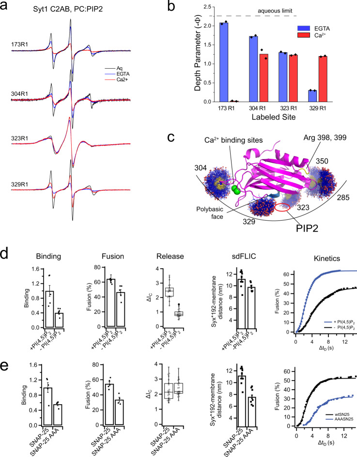
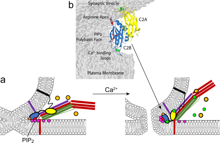
Synaptotagmin 1 is a vesicle-anchored membrane protein that functions as the Ca2+ sensor for synchronous neurotransmitter release. In this work, an arginine containing region in the second C2 domain of synaptotagmin 1 (C2B) is shown to control the expansion of the fusion pore and thereby the concentration of neurotransmitter released. This arginine apex, which is opposite the Ca2+ binding sites, interacts with membranes or membrane reconstituted SNAREs; however, only the membrane interactions occur under the conditions in which fusion takes place. Other regions of C2B influence the fusion probability and kinetics but do not control the expansion of the fusion pore. These data indicate that the C2B domain has at least two distinct molecular roles in the fusion event, and the data are consistent with a model where the arginine apex of C2B positions the domain at the curved membrane surface of the expanding fusion pore.


Conflict of interest statement

The authors declare no competing interests.

Figures

Fig. 1
Fig. 1. EPR spectroscopy is sensitive to membrane proximity and insertion of labeled Syt1.
a Model for Syt1 on the vesicle membrane surface showing position 173 in the 1st Ca2+-binding loop (CBL) of the C2A domain (yellow). The position of the polybasic face (red) and R398, 399 side chains are shown for C2B (blue). b EPR spectra from an R1 label attached to site 173. In the presence of Ca2+, the domain inserts into the bilayer, which alters the sampling of rotamers by the R1 side chain and broadens the EPR spectrum.
Fig. 2
Fig. 2. The arginine apex of C2B contacts the membrane interface.
a Model of C2B showing conserved arginine residues in the apex, lysine residues in the polybasic face and the three sites to which R1 was attached. The arginine apex is on the opposite surface of the domain from two Ca2+-binding loops (CBL) that insert into bilayers in the presence of Ca2+. b EPR spectra from the three labeled sites in the absence of membranes or the presence of POPC:POPS (80:20) lipid vesicles. c Membrane depth parameters obtained with and without Ca2+ for labels near the apex. The bars indicate the mean value with ±standard deviation. Three independent samples and power saturation measurements were made, except for 349R1 with Ca2+ where six samples were measured.
Fig. 3
Fig. 3. Mutating the arginine apex reduces or eliminates membrane contact by the arginine apex.
a power saturation indicates that the RQ and RQRQ mutations reduce or eliminate membrane contact. Vesicles were composed of POPC:POPS (80:20) or POPC:PIP2 (95:5). The bars indicate mean values with outlying points. The points represent independent samples that were power saturated. Two independent measurements were made for POPC:PIP2 samples. The 285R1 samples in POPC:POPS were repeated three times with the 285RQ sample repeated four times. The 350R1 WT sample in POPC:POPS was repeated three times. b EPR spectra obtained from site 285 for the pseudo wild-type (WT) and RQRQ mutant. EPR spectra are identical in the absence or presence of membranes for the RQRQ mutant. A depth parameter of 2 is obtained for an R1 label that lies ~4 Å from the lipid phosphate. When the label is further than 6 or 7 Å from the phosphates, the depth parameter becomes independent of label position.
Fig. 4
Fig. 4. The arginine apex contacts membrane reconstituted SNAREs, but ATP or PIP2 in the bilayer, eliminate the interaction.
a EPR spectra in the absence (blue trace) or presence (red trace) of membrane reconstituted SNARE complex composed of full-length Syx, SNAP-25, and soluble Syb. The broadened components in the spectra (arrows) are due to incompletely averaged axial components of the hyperfine tensor and indicate tertiary contact of the R1 side chain with SNAREs. b Model (PDB ID 5CCH) with available rotamers for the R1 side chains at the three sites. c Comparison of EPR spectra with and without membrane reconstituted SNAREs containing the AAA mutation in SNAP-25. d Normalized amplitudes for EPR spectra from sites 285R1 and 350R1. The normalized amplitudes provide a semi-quantitative measure of R1 motion where larger amplitudes indicate loss of R1 tertiary contact with the SNAREs.
Fig. 5
Fig. 5. The effects of synaptotagmin mutations in C2B on secretory granule fusion and SNARE orientation.
a Single granule supported membrane TIRF fusion assay (left) and sdFLIC microscopy of the ternary SNARE complex (right). b Fluorescence intensity traces of single granules interacting with supported membranes in a TIRF microscope. After a sudden increase in fluorescence, indicating binding, the intensity stays constant if the granule never fuses (top) or the trace shows a characteristic dip and peak after different delay times (bottom) if the granule membrane fuses with the reconstituted supported membrane. c Averaged traces (20 events per trace) showing a fast mode of fusion in the presence of C2AB (red) or slow mode of fusion in the absence of C2AB (blue). Both conditions were in the presence of 100 µM Ca2+. d Granules depleted of synaptotagmins (Syt1 and Syt9) do not fuse in response to calcium. 0.4 μM soluble C2AB stimulates fusion of synaptotagmin knockdown granules to a similar level as granules containing endogenous synaptotagmin in the presence of 100 µM Ca2+ (membrane composition was 32:32:15:20:1 bPC:bPE:bPS: Chol:PIP2). e Structural changes in the orientation of the ternary SNARE complex in the presence of calcium and 0.4 μM C2AB. f The effects of RQ, RQRQ, or KAKA mutations in C2AB on granule binding, fusion, release characteristics, SNARE orientation, and fusion kinetics in the presence of 100 µM Ca2+ (membrane composition was 32:32:15:20:1 bPC:bPE:bPS:Chol:PIP2). The bar plots show mean ± standard error or the mean. The boxplots show mean, 2nd and 3rd quartile, and outliers of the data.
Fig. 6
Fig. 6. The C2B domains makes simultaneous membrane contact at multiple sites that cannot be explained by docking to a planar bilayer surface.
a EPR spectra in an aqueous state compared with spectra in the presence of POPC:PIP2 (95:5) bilayers in the presence of EGTA or Ca2+. b Membrane depth parameters indicate that site 173 inserts into the PIP2 bilayer in the presence of Ca2+. Sites on C2B including 304, 323, and 329 also contact with bilayer, with or without Ca2+. Bar plots show mean values from the power saturation of two samples. c Allowable rotamers for labeled sites that contact the membrane interface including two near the arginine apex. The membrane contact seen at these sites could be accommodated by a curved membrane surface. This would place a PIP2 headgroup in a binding pocket at the polybasic face. d The effect of PIP2 on granule binding, fusion, release profile, SNARE orientation, and fusion kinetics in the presence of 100 µM Ca2+ (membrane composition was 25:25:15:30 bPC:bPE:bPS:Chol and either 5% PI or 4% PI with 1% PIP2). e The effect of the AAA mutation in SNAP-25 on granule binding, fusion, release profile, SNARE orientation, and fusion kinetics in the presence of 100 µM Ca2+ (membrane composition 32:32:15:20:1 bPC:bPE:bPS:Chol:PIP2). The bar plots show mean ± standard error of the mean. The boxplots show mean, 2nd and 3rd quartile, and outliers of the data.
Fig. 7
Fig. 7. Model for the membrane interactions of Syt1.
In a after docking to the bilayer and in the presence of ATP/Mg2+ (orange) the SNARE complex, syntaxin (red), SNAP-25 (green), synaptobrevin (purple), and Syt1 C2B (blue), promote partial lipid mixing by either zippering or contacting plasma membrane PIP2 at the arginine apex (orange) and the polybasic face (red). Upon calcium influx (green), Syt1 C2A inserts into the synaptic vesicle membrane, PS, and C2B reorients, binding the Ca2+-binding loops at a 3rd membrane contact point. The C2B domain of Syt1 might position at the site of fusion as shown in b. This orientation would sequester PIP2 into the strained membrane stalk, further destabilizing the membrane and promoting pore opening. The C2A domain is shown inserted into the vesicle surface. This orientation of C2A is preferred in full-length Syt1; however, an interaction of C2A with the plasma membrane may also take place. The Ca2+-binding loops of C2B are shown directed towards the plasma membrane surface because C2B has a high affinity towards PIP2 containing membranes in a low Ca2+ state and the loops are likely to insert into the plasma membrane.

Similar articles

Cited by

References

    1. Jahn R, Scheller RH. SNAREs–engines for membrane fusion. Nat. Rev. Mol. Cell Biol. 2006;7:631–643. doi: 10.1038/nrm2002. - DOI - PubMed
    1. Jahn R, Fasshauer D. Molecular machines governing exocytosis of synaptic vesicles. Nature. 2012;490:201–207. doi: 10.1038/nature11320. - DOI - PMC - PubMed
    1. Rizo J, Sudhof TC. The membrane fusion enigma: SNAREs, Sec1/Munc18 proteins, and their accomplices–guilty as charged? Annu. Rev. Cell Dev. Biol. 2012;28:279–308. doi: 10.1146/annurev-cellbio-101011-155818. - DOI - PubMed
    1. Chapman ER. How does synaptotagmin trigger neurotransmitter release? Annu. Rev. Biochem. 2008;77:615–641. doi: 10.1146/annurev.biochem.77.062005.101135. - DOI - PubMed
    1. Bai J, Tucker WC, Chapman ER. PIP2 increases the speed of response of synaptotagmin and steers its membrane-penetration activity toward the plasma membrane. Nat. Struct. Mol. Biol. 2004;11:36–44. doi: 10.1038/nsmb709. - DOI - PubMed

Publication types

LinkOut - more resources