. 2021 Feb 3;12(1):763.
doi: 10.1038/s41467-021-21012-x.

A Sarcina bacterium linked to lethal disease in sanctuary chimpanzees in Sierra Leone

Affiliations

A Sarcina bacterium linked to lethal disease in sanctuary chimpanzees in Sierra Leone

Leah A Owens et al. Nat Commun. .

Erratum in

Abstract

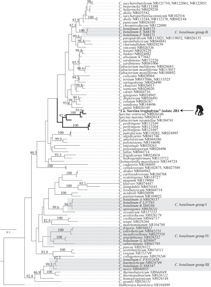
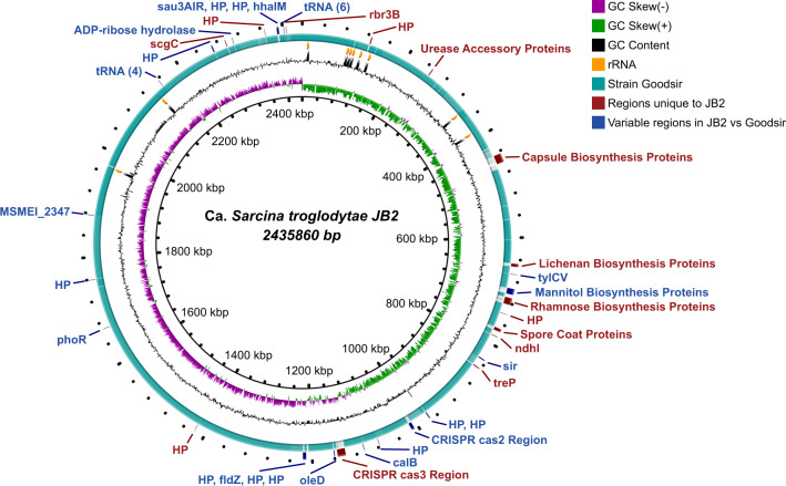
Human and animal infections with bacteria of the genus Sarcina (family Clostridiaceae) are associated with gastric dilation and emphysematous gastritis. However, the potential roles of sarcinae as commensals or pathogens remain unclear. Here, we investigate a lethal disease of unknown etiology that affects sanctuary chimpanzees (Pan troglodytes verus) in Sierra Leone. The disease, which we have named "epizootic neurologic and gastroenteric syndrome" (ENGS), is characterized by neurologic and gastrointestinal signs and results in death of the animals, even after medical treatment. Using a case-control study design, we show that ENGS is strongly associated with Sarcina infection. The microorganism is distinct from Sarcina ventriculi and other known members of its genus, based on bacterial morphology and growth characteristics. Whole-genome sequencing confirms this distinction and reveals the presence of genetic features that may account for the unusual virulence of the bacterium. Therefore, we propose that this organism be considered the representative of a new species, named "Candidatus Sarcina troglodytae". Our results suggest that a heretofore unrecognized complex of related sarcinae likely exists, some of which may be highly virulent. However, the potential role of "Ca. S. troglodytae" in the etiology of ENGS, alone or in combination with other factors, remains a topic for future research.


Conflict of interest statement

The authors declare no competing interests.

Figures

Fig. 1
Fig. 1. Graphic representation of epizootic neurologic and gastroenteric syndrome (ENGS) cases and study samples.
Individual chimpanzees are represented by rectangles and colors denote sex and case/control status (red, female ENGS case, n = 30; blue, male ENGS case, n = 26; pink, female ENGS control, n = 7; light blue, male ENGS control, n = 7). Post-mortem findings regarding gastric dilation are shown for those cases where documentation was available (n = 17; black circle, gastric dilation, n = 14; open circle, no gastric dilation, n = 1; filled gray circle, inconclusive for gastric dilation, n = 2). “Analyzed” indicates an individual from whom we obtained at least one sample used in this study (n = 32) and the asterisk indicates the single individual from whom we obtained samples pre- and post-ENGS. Pie charts are scaled to sample size with green representing individuals positive by diagnostic PCR and white representing individuals negative by diagnostic PCR.
Fig. 2
Fig. 2. Deaths attributed to epizootic neurologic and gastroenteric syndrome (ENGS) at Tacugama Chimpanzee Sanctuary from 2005 through 2018.
a Annual chimpanzee deaths from ENGS (black, overall total = 56) and other causes (gray, overall total = 32). Numbers above the bars indicate the yearly total chimpanzee population and the horizontal bracket below the x-axis denotes the period during which we obtained samples for analysis. b Summed totals of ENGS fatalities by month over 2005‒2018 (n = 56). Source data are provided as a Source Data file.
Fig. 3
Fig. 3. Gross and histopathologic images of a chimpanzee that died of epizootic neurologic and gastroenteric syndrome (ENGS).
Photographic images from an adult male chimpanzee who died of ENGS showing moderate to severe gastric dilation (a), hemorrhagic diathesis (a), and emphysematous typhlocolitis (b; arrows point to gas-filled pockets within the cecum wall, which has reddened areas [in inset, white arrowheads point to gas bubbles in the cut surfaces of the formalin-fixed cecum]; scale bars = 1 cm). On histology (c), gas-filled space (*) in the cecal submucosa, surrounded by inflammatory infiltrates (arrows) and hemorrhage (h) were visualized by hematoxylin and eosin staining (inset depicts inflammatory infiltrates, which include eosinophils [arrow] and multinucleate giant cells [arrowheads]; scale bars = 300 µm [main image] and 30 µm [inset]; micrograph is a representative image of three similar sections obtained from the same individual).
Fig. 4
Fig. 4. Bacterial 16S rDNA microbiome analysis of epizootic neurologic and gastroenteric syndrome (ENGS) case samples.
a The percentage of total reads (n = 5900 per sample) from 9 ENGS case samples at genus-level OTU with % reads mapped to the Clostridiacea OTU shown on top of each bar. b Percent abundance of reads from 23 ENGS case samples classified to genus-level OTUs Sarcina (% above each bar) and other, arranged by tissue type.
Fig. 5
Fig. 5. Comparative morphology of type strain Sarcina ventriculi “Goodsir” ATCC 29068 and “Ca. S. troglodytae” isolate JB1.
ac Cells were imaged live (a) or heat-fixed and stained with Gram stain (b) or methylene blue stain (c). Micrographs are from a single experiment and are representative of three independent experiments with similar results. Scale bars = 10 µm. d Live cell diameters were compared, with lines representing median diameters among seven distinct bacterial colonies. Source data are provided as a Source Data file. *Calculated using a Mann–Whitney U test, two-tailed.
Fig. 6
Fig. 6. Characteristic cuboidal packets of Sarcina-like organisms in tissues of ENGS-affected chimpanzees.
a Basophilic packets of cells in a tetrad formation can be seen amongst and within alveoli of hematoxylin and eosin-stained lung tissue of one individual (“Jumu”). b Unstained brain tissue homogenate from another individual (“Joko”) contains highly refractile, cuboid packets of cells. Scale bars = 10 µm. Micrographs are representative images of one of at least three sections (a) or smears (b) obtained from the same individual with similar results.
Fig. 7
Fig. 7. Maximum-likelihood 16S rDNA gene phylogeny of the Clostridiaceae.
The phylogeny is based on the complete 16S rDNA sequence of “Ca. Sarcina troglodytae” isolate JB1 (arrow and silhouette) and 98 other Clostridia sensu stricto, with Hathewaya histolytica as the outgroup. Gray boxes indicate Clostridium botulinum groups. Numbers above the branches are bootstrap values (%) based on 1000 bootstrap replicates (only values ≥75% are shown). Scale bar indicates nucleotide substitutions per site.
Fig. 8
Fig. 8. Whole chromosome comparison of “Ca. Sarcina troglodytae” isolate JB2 compared to the type strain S. ventriculi “Goodsir”.
Red: regions which are unique to JB2. Blue: different ORF sets in the same relative region in JB2 and Goodsir. Teal: 90–100% identity gradient between JB2 and Goodsir (Goodsir chromosome sequence is based on a manually scaffolded genome).

Similar articles

Cited by

References

    1. Brower JL. The threat and response to infectious diseases (Revised) Microb. Ecol. 2018;76:19–36. - PMC - PubMed
    1. Cunningham, A. A., Daszak, P. & Wood, J. L. N. One Health, emerging infectious diseases and wildlife: two decades of progress? Philos. Trans. R. Soc. B372, doi:ARTN 2016016710.1098/rstb.2016.0167 (2017). - PMC - PubMed
    1. Lakhundi, S. & Zhang, K. Methicillin-resistant Staphylococcus aureus: molecular characterization, evolution, and epidemiology. Clin. Microbiol. Rev. 31, 10.1128/CMR.00020-18 (2018). - PMC - PubMed
    1. Gomez-Simmonds A, Uhlemann AC. Clinical implications of genomic adaptation and evolution of carbapenem-resistant. J. Infect. Dis. 2017;215:S18–S27. - PMC - PubMed
    1. Harrison LH, Simonsen V, Waldman EA. Emergence and disappearance of a virulent clone of Haemophilus influenzae biogroup aegyptius, cause of Brazilian purpuric fever. Clin. Microbiol. Rev. 2008;21:594–605. - PMC - PubMed

Publication types

MeSH terms

LinkOut - more resources