

Feasibility of ex vivo fluorescence imaging of angiogenesis in (non-) culprit human carotid atherosclerotic plaques using bevacizumab-800CW
- PMID: 33536498
- PMCID: PMC7858611
- DOI: 10.1038/s41598-021-82568-8
Feasibility of ex vivo fluorescence imaging of angiogenesis in (non-) culprit human carotid atherosclerotic plaques using bevacizumab-800CW
Abstract
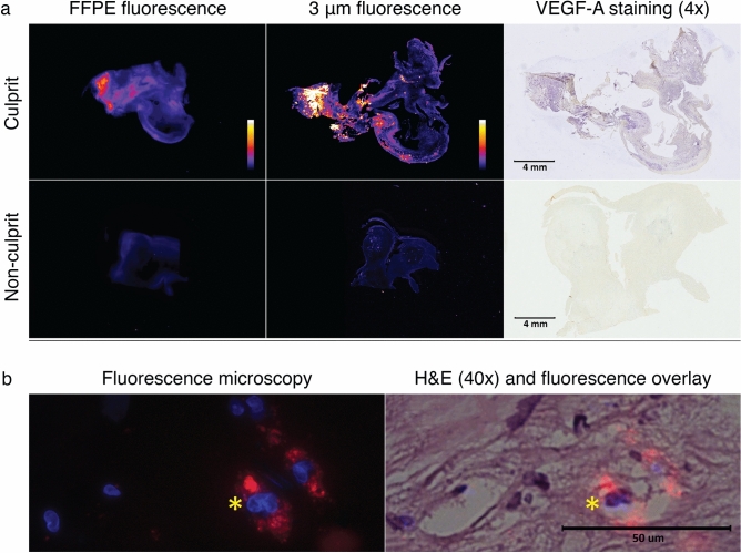
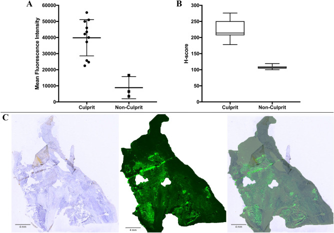
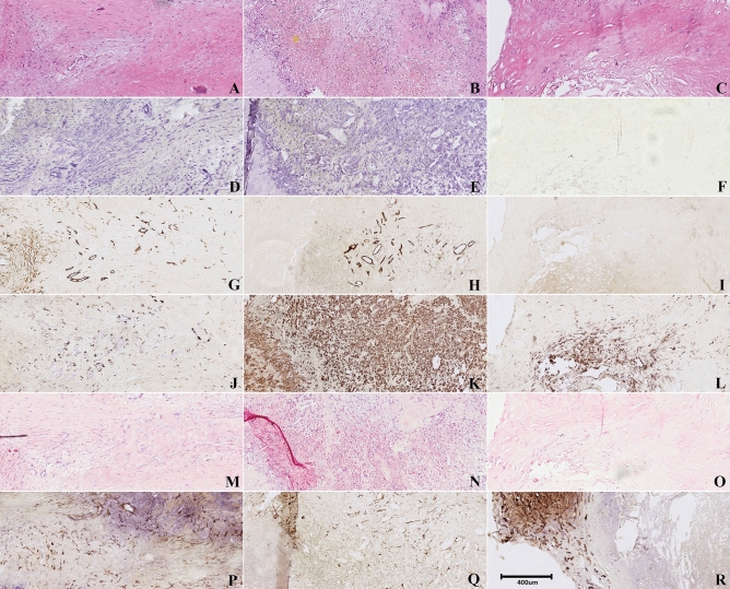
Vascular endothelial growth factor-A (VEGF-A) is assumed to play a crucial role in the development and rupture of vulnerable plaques in the atherosclerotic process. We used a VEGF-A targeted fluorescent antibody (bevacizumab-IRDye800CW [bevacizumab-800CW]) to image and visualize the distribution of VEGF-A in (non-)culprit carotid plaques ex vivo. Freshly endarterectomized human plaques (n = 15) were incubated in bevacizumab-800CW ex vivo. Subsequent NIRF imaging showed a more intense fluorescent signal in the culprit plaques (n = 11) than in the non-culprit plaques (n = 3). A plaque received from an asymptomatic patient showed pathologic features similar to the culprit plaques. Cross-correlation with VEGF-A immunohistochemistry showed co-localization of VEGF-A over-expression in 91% of the fluorescent culprit plaques, while no VEGF-A expression was found in the non-culprit plaques (p < 0.0001). VEGF-A expression was co-localized with CD34, a marker for angiogenesis (p < 0.001). Ex vivo near-infrared fluorescence (NIRF) imaging by incubation with bevacizumab-800CW shows promise for visualizing VEGF-A overexpression in culprit atherosclerotic plaques in vivo.
Conflict of interest statement
The authors declare no competing interests.
Figures
Similar articles
-
VEGF-Targeted Multispectral Optoacoustic Tomography and Fluorescence Molecular Imaging in Human Carotid Atherosclerotic Plaques.Diagnostics (Basel). 2021 Jul 7;11(7):1227. doi: 10.3390/diagnostics11071227. Diagnostics (Basel). 2021. PMID: 34359310 Free PMC article.
-
Imaging of the vulnerable carotid plaque: biological targeting of inflammation in atherosclerosis using iron oxide particles and MRI.Eur J Vasc Endovasc Surg. 2014 May;47(5):462-9. doi: 10.1016/j.ejvs.2014.01.017. Epub 2014 Mar 1. Eur J Vasc Endovasc Surg. 2014. PMID: 24594295
-
Increased vascularization of shoulder regions of carotid atherosclerotic plaques from patients with diabetes.J Vasc Surg. 2011 Nov;54(5):1324-1331.e5. doi: 10.1016/j.jvs.2011.04.061. Epub 2011 Jul 20. J Vasc Surg. 2011. PMID: 21764240
-
Angiogenesis in atherosclerotic plaque obtained from carotid endarterectomy: association between symptomatology and plaque morphology.Neurol Med Chir (Tokyo). 2010;50(12):1056-61. doi: 10.2176/nmc.50.1056. Neurol Med Chir (Tokyo). 2010. PMID: 21206178
-
Molecular imaging of carotid plaque vulnerability.Cerebrovasc Dis. 2015;39(1):5-12. doi: 10.1159/000369123. Epub 2014 Dec 24. Cerebrovasc Dis. 2015. PMID: 25547782 Review.
Cited by
-
Targeted optical fluorescence imaging: a meta-narrative review and future perspectives.Eur J Nucl Med Mol Imaging. 2021 Dec;48(13):4272-4292. doi: 10.1007/s00259-021-05504-y. Epub 2021 Oct 11. Eur J Nucl Med Mol Imaging. 2021. PMID: 34633509 Free PMC article. Review.
-
Research Advance of Chinese Medicine in Treating Atherosclerosis: Focus on Lipoprotein-Associated Phospholipase A2.Chin J Integr Med. 2024 Mar;30(3):277-288. doi: 10.1007/s11655-023-3611-6. Epub 2023 Dec 7. Chin J Integr Med. 2024. PMID: 38057549 Review.
-
Multi-Scale Imaging of Vascular Pathologies in Cardiovascular Disease.Front Med (Lausanne). 2022 Jan 5;8:754369. doi: 10.3389/fmed.2021.754369. eCollection 2021. Front Med (Lausanne). 2022. PMID: 35071257 Free PMC article. Review.
-
Novel treatment from a botanical formulation Si-Miao-Yong-an decoction inhibits vasa vasorum angiogenesis and stabilizes atherosclerosis plaques via the Wnt1/β-catenin signalling pathway.Pharm Biol. 2023 Dec;61(1):1364-1373. doi: 10.1080/13880209.2023.2249061. Pharm Biol. 2023. PMID: 37651108 Free PMC article.
-
Angiogenesis within atherosclerotic plaques: Mechanical regulation, molecular mechanism and clinical diagnosis.Mechanobiol Med. 2025 Feb 1;3(1):100114. doi: 10.1016/j.mbm.2025.100114. eCollection 2025 Mar. Mechanobiol Med. 2025. PMID: 40396135 Free PMC article. Review.
References
Publication types
MeSH terms
Substances
LinkOut - more resources
Full Text Sources
Other Literature Sources
