. 2021 Feb 3;11(1):2854.
doi: 10.1038/s41598-021-82499-4.

Biochemical and cellular characterisation of the Plasmodium falciparum M1 alanyl aminopeptidase (PfM1AAP) and M17 leucyl aminopeptidase (PfM17LAP)

Affiliations

Biochemical and cellular characterisation of the Plasmodium falciparum M1 alanyl aminopeptidase (PfM1AAP) and M17 leucyl aminopeptidase (PfM17LAP)

Rency Mathew et al. Sci Rep. .

Abstract

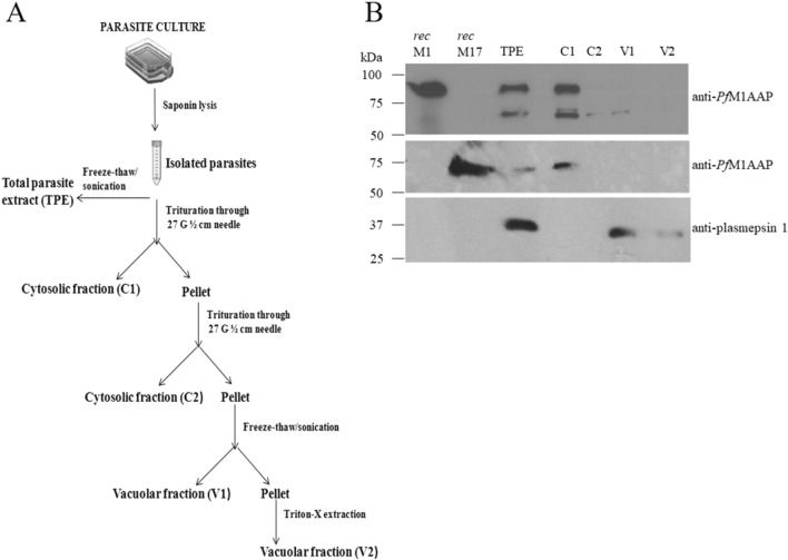
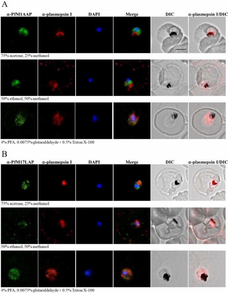
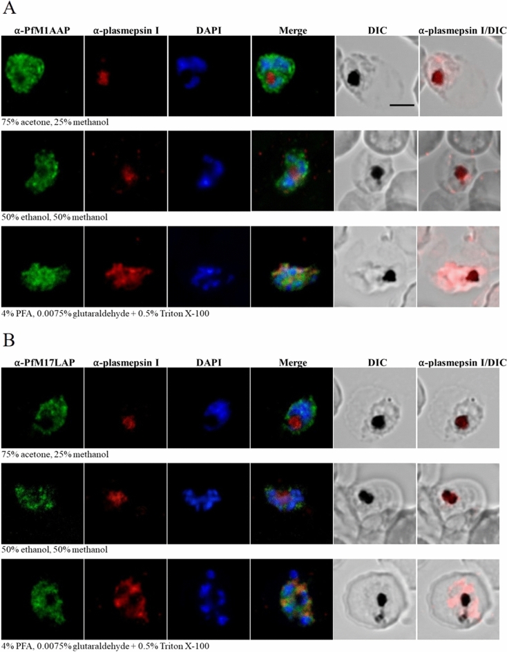
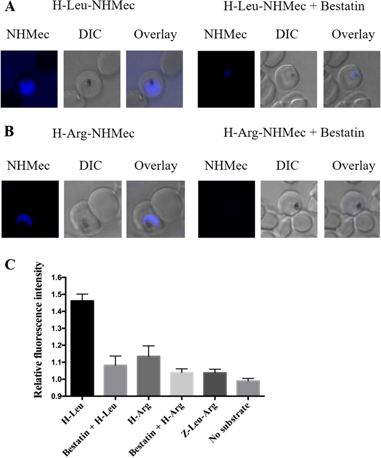
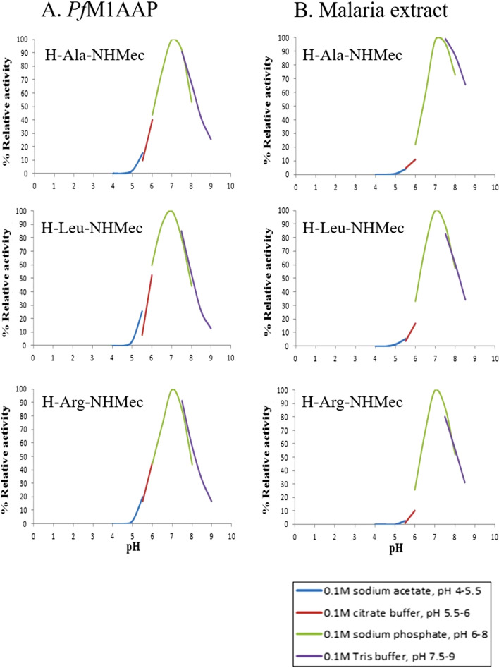
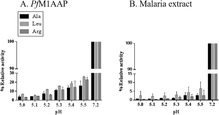
The Plasmodium falciparum M1 alanyl aminopeptidase and M17 leucyl aminopeptidase, PfM1AAP and PfM17LAP, are potential targets for novel anti-malarial drug development. Inhibitors of these aminopeptidases have been shown to kill malaria parasites in culture and reduce parasite growth in murine models. The two enzymes may function in the terminal stages of haemoglobin digestion, providing free amino acids for protein synthesis by the rapidly growing intra-erythrocytic parasites. Here we have performed a comparative cellular and biochemical characterisation of the two enzymes. Cell fractionation and immunolocalisation studies reveal that both enzymes are associated with the soluble cytosolic fraction of the parasite, with no evidence that they are present within other compartments, such as the digestive vacuole (DV). Enzyme kinetic studies show that the optimal pH of both enzymes is in the neutral range (pH 7.0-8.0), although PfM1AAP also possesses some activity (< 20%) at the lower pH range of 5.0-5.5. The data supports the proposal that PfM1AAP and PfM17LAP function in the cytoplasm of the parasite, likely in the degradation of haemoglobin-derived peptides generated in the DV and transported to the cytosol.


Conflict of interest statement

The authors declare no competing interests.

Figures

Figure 1
Figure 1
Distribution of PfM1AAP and PfM17LAP in the P. falciparum cellular compartments. (A) Flowchart depicting the isolation of P. falciparum parasites from host erythrocytes followed by fractionation of cell compartments. Parasites were isolated by saponin lysis of erythrocytes. Total parasite extracts (TPE) were prepared by freeze–thaw and sonication of the parasites in 10 mM Tris–HCl buffer, pH 7.2. Other samples were triturated four times through a syringe needle and centrifuged to obtain the first cytosolic fraction, C1, and a pellet. The pellet was resuspended in 10 mM Tris–HCl buffer, pH 7.2 and triturated/centrifuged to obtain the second cytosolic fraction, C2, and a pellet. This pellet was re-suspended in 10 mM Tris–HCl buffer, pH 7.2, and subjected to four rounds of freeze–thaw treatment followed by centrifugation to obtain a soluble vacuolar fraction, V1, and a pellet. The final detergent-soluble vacuolar fraction, V2, was obtained by incubating the pellet in 0.5% Triton X for 30 min on ice. (B) Representative immunoblots of three biological replicates showing the P. falciparum recombinant (rec) PfM1AAP, rec PfM17LAP, total parasite extract (TPE) and cellular fractions (C1, C2, V1 and V2) probed with anti-PfM1AAP (top panel), anti-PfM17LAP (middle panel), and anti-plasmepsin 1 antibodies (lower panel). Chemiluminescent molecular weight standards are shown on the left.
Figure 2
Figure 2
Detection of native PfM1AAP and PfM17LAP in malaria cytosolic extracts. Immunoblots of cytosolic fractions (C1) prepared either without (−) or with (+) a cocktail of protease inhibitors were probed with (A) anti-PfM1AAP, and (B) anti-PfM17LAP polyclonal antibodies prepared in rabbits and adsorbed against E. coli extracts (see “Materials and methods”). The chemiluminescent molecular size markers are shown on the left of each blot (lane 1).
Figure 3
Figure 3
Immunoblots with peptide antibodies. (A) Recombinant PfM1AAP was probed with antibodies prepared against 14-mer peptides derived from the N-terminal extension (PepA), spanning domain 1 (PepB) and domain 4 (PepC) (see Supplementary Fig. 1). Anti-PfM1AAP was used as a positive control. Anti-PepA does not react with rPfM1AAP, as this lacks the N-terminal extention, whereas both anti-PepB and anti-PepC do. Lane 1 was loaded with chemilluminescent molecular marker. Lanes 2–5 were loaded with 0.25 µg of recombinant M1. (B) Cytosolic fractions of malaria parasites prepared without (−) and with (+) a protease inhibitor cocktail were probed with anti-PepA, anti-PepB and anti-PepC antibodies. Extracts were separated on 4–15% SDS-PAGE gels and transferred to nitrocellulose membrane. Lanes 2 and 3 were probed with anti-PepA, lanes 4 and 5 with anti-PepB and lanes 6 and 7 with anti-PepC antibodies. The chemiluminescent molecular marker is shown in lane 1.
Figure 4
Figure 4
Localization of PfM1AAP and PfM17LAP in intra-erythrocytic P. falciparum 3D7 trophozoite-stage parasites. Immunofluorescence assays were carried out using air-dried blood smears fixed with 75% acetone and 25% methanol at − 20 °C for 5 min, or 50% ethanol and 50% methanol at − 20 °C for 2 min, or 4% PFA and 0.0075% glutaraldehyde for 20 min at room temperature. Fixed parasites were probed with polyclonal antibodies against (A) PfM1AAP and (B) PfM17LAP. Specific aminopeptidase staining (green, Alexa-Fluor 488) was observed in the cytosol of parasites. Parasite nuclei were visualized using DAPI (blue; 4,6-diamidino-2-phenylindole) and monoclonal antibodies against the DV marker plasmepsin I (α-plasmepsin I, red, Alexa-Fluor 594) were used as a control. Differential interference contrast (DIC) and α-plasmepsin I with DIC are shown for reference. Scale bar, 3 µm.
Figure 5
Figure 5
Localization of PfM1AAP and PfM17LAP in intra-erythrocytic P. falciparum 3D7 schizont-stage parasites. Immunofluorescence assays were carried out using air-dried blood smears fixed with 75% acetone and 25% methanol at − 20 °C for 5 min or 50% ethanol and 50% methanol at − 20 °C for 2 min or 4% PFA and 0.0075% glutaraldehyde for 20 min at room temperature. Fixed parasites were probed with polyclonal antibodies against (A) PfM1AAP and (B) PfM17LAP. Specific aminopeptidase staining (green, Alexa-Fluor 488) was observed in the cytosol of parasites. Parasite nuclei were visualized using DAPI (blue; 4,6-diamidino-2-phenylindole) and monoclonal antibodies against the DV marker plasmepsin I (α-plasmepsin I, red, Alexa-Fluor 594) were used as a control. Differential interference contrast (DIC) and α-plasmepsin I with DIC are shown for reference. Scale bar, 3 µm.
Figure 6
Figure 6
Proteolytic cleavage of fluorogenic substrates by PfM1AAP and PfM17LAP in the cytosol of live parasites. Erythrocytes infected with 3D7 P. falciparum parasites were incubated with either (A) 10 µM H-Leu-NHMec (substrate cleaved by PfM1AAP and PfM17LAP) or (B) 10 µM H-Arg-NHMec (substrate cleaved by PfM1AAP but not PfM17LAP) for 10 min. Proteolytic cleavage of substrates resulted in the release of the fluorescent free NHMec fluorophore in the parasite cytosol (blue staining). Previous incubation with 50 µM bestatin, an aminopeptidase inhibitor, resulted in inhibition of aminopeptidase activity, as revealed by the almost complete absence of fluorescence. Differential interference contrast (DIC) is shown for reference. (C) Quantification of NHMec fluorescence intensities in the cytosol relative to background (mean ± SEM). At least ten parasites were imaged per treatment under the same conditions on the same day. Z-Leu-Arg-NHMec (substrate not cleaved by PfM1AAP or PfM17LAP) was used for comparison.
Figure 7
Figure 7
pH profiles of PfM1AAP and soluble extracts of malaria parasites. Recombinant PfM1AAP enzyme (1 μg) (A) or soluble extract of malaria parasites (5 μg) (B) was incubated in a series of pH-controlled buffers (0.1 M sodium acetate pH 5.0–5.5, 0.1 M sodium citrate pH 5.5–6.0, 0.1 M sodium phosphate pH 6.0–8.0, 0.1 M Tris buffer pH 7.5–8.5) in the presence of the fluorogenic peptide substrates H-Ala-NHMec, H-Leu-NHMec or H-Arg-NHMec. The release of the fluorophore NHMec was monitored in a fluorimeter with excitation at 360 nm and emission at 460 nm. Reaction rates were used to plot pH-activity curves.
Figure 8
Figure 8
Activity profile PfM1AAP at low pH. Aminopeptidase activity of (A) recombinant PfM1AAP and (B) soluble extracts of P. falciparum malaria parasites in the acidic pH range of 5.0–5.5 (0.1 M sodium acetate buffer) compared with neutral pH 7.2 (PBS). The preparations were incubated in the buffers just prior to the addition of 10 μM substrates (H-Ala-NHMec, H-Leu-NHMec and H-Arg-NHMec).

References

    1. World malaria report 2019. Licence: CC BY-NC-SA 3.0 IGO (World Health Organization, Geneva, 2019).
    1. Khera A, Mukherjee R. Artemisinin resistance: Cause for worry? J. Mar. Med. Soc. 2019;21:4. doi: 10.4103/jmms.jmms_43_18. - DOI
    1. Vander Jagt DL, Baack BR, Hunsaker LA. Purification and characterization of an aminopeptidase from Plasmodium falciparum. Mol. Biochem. Parasitol. 1984;10:45–54. doi: 10.1016/0166-6851(84)90017-3. - DOI - PubMed
    1. Curley GP, et al. Aminopeptidases from Plasmodium falciparum, Plasmodium chabaudi chabaudi and Plasmodium berghei. J. Eukaryot. Microbiol. 1994;41:119–123. doi: 10.1111/j.1550-7408.1994.tb01483.x. - DOI - PubMed
    1. Skinner-Adams TS, et al. Plasmodium falciparum neutral aminopeptidases: New targets for anti-malarials. Trends. Biochem. Sci. 2010;35:53–61. doi: 10.1016/j.tibs.2009.08.004. - DOI - PubMed

Publication types

MeSH terms

Grants and funding