. 2021 Feb 3;11(1):2959.
doi: 10.1038/s41598-021-82619-0.

RasGRP2 inhibits glyceraldehyde-derived toxic advanced glycation end-products from inducing permeability in vascular endothelial cells

Affiliations

RasGRP2 inhibits glyceraldehyde-derived toxic advanced glycation end-products from inducing permeability in vascular endothelial cells

Jun-Ichi Takino et al. Sci Rep. .

Abstract

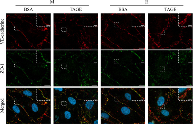
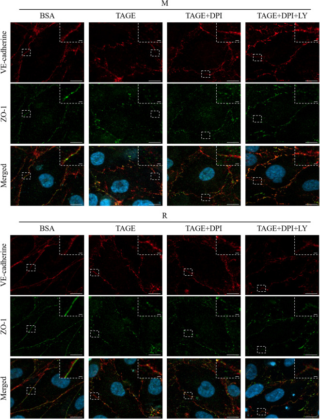
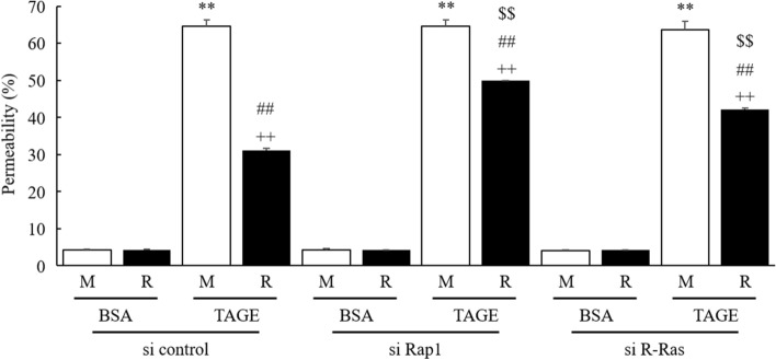
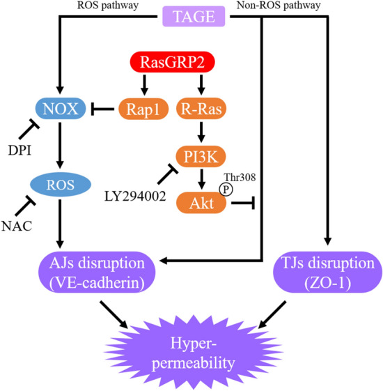
Advanced glycation end-products (AGEs) are formed by the non-enzymatic reaction of sugars and proteins. Among the AGEs, glyceraldehyde-derived toxic AGEs (TAGE) are associated with various diseases, including diabetic complications such as diabetic retinopathy (DR). The risk of developing DR is strongly associated with poor glycemic control, which causes AGE accumulation and increases AGE-induced vascular permeability. We previously reported that Ras guanyl nucleotide releasing protein 2 (RasGRP2), which activates small G proteins, may play an essential role in the cell response to toxicity when exposed to various factors. However, it is not known whether RasGRP2 prevents the adverse effects of TAGE in vascular endothelial cells. This study observed that TAGE enhanced vascular permeability by disrupting adherens junctions and tight junctions via complex signaling, such as ROS and non-ROS pathways. In particular, RasGRP2 protected adherens junction disruption, thereby suppressing vascular hyper-permeability. These results indicate that RasGRP2 is an essential protective factor of vascular permeability and may help develop novel therapeutic strategies for AGE-induced DR.


Conflict of interest statement

The authors declare no competing interests.

Figures

Figure 1
Figure 1
Barrier formation and suppression of TAGE-induced vascular hyper-permeability by RasGRP2. TEER value was measured using the Millicell-ERS. (a) Time-dependent change on barrier formation. The TEER value after 8 days for HUVEC was set to 100%. Square: HUVEC, circle: M cells, triangle: R cells. (b) Time-dependent change after BSA or TAGE treatment. The TEER value after 6 days for M cells was set to 100%. M mock cells, R RasGRP2-stable overexpression cells. Data shown as the mean ± SD (n = 3), **P < 0.01 compared with M cells of 0 h, ++P < 0.01 compared with R cells of 0 h, and #P < 0.05, ##P < 0.01 compared with each TAGE-treated M cells.
Figure 2
Figure 2
Reduction of TAGE-induced vascular hyper-permeability and ROS production by RasGRP2. (a) Permeability was measured using FITC-Dextran. (b) Cell viability was determined by Cell Counting Kit-8 assay. (c) Intracellular ROS was determined by CellROX Green. M mock cells, R RasGRP2-stable overexpression cells. Data shown as the mean ± SD (n = 3), **P < 0.01 compared with BSA-treated M cells, ++P < 0.01 compared with BSA-treated R cells, and ##P < 0.01 compared with TAGE-treated M cells.
Figure 3
Figure 3
Reduction of TAGE-induced vascular hyper-permeability via ROS and non-ROS pathways by RasGRP2. NAC, a ROS scavenger, DPI, an inhibitor of NOX, and LY, an inhibitor of the PI3K-Akt pathway, were used to inhibit TAGE-induced ROS production, TAGE-induced ROS production via NOX and RasGRP2-induced Akt activation, respectively. (a) Intracellular ROS was determined by CellROX Green. (b) Permeability was measured using FITC-Dextran. M mock cells, R RasGRP2-stable overexpression cells, NAC N-acetyl cysteine, DPI diphenyleneiodonium, LY LY294002. Data shown as the mean ± SD (n = 3), **P < 0.01 compared with BSA-treated M cells, ++P < 0.01 compared with BSA-treated R cells, ##P < 0.01 compared with each TAGE-treated M cells, and $$P < 0.01 compared with TAGE-treated cells alone.
Figure 4
Figure 4
Expression of VE-cadherin and ZO-1 proteins by TAGE. (a) VE-cadherin (approximately 130 kDa), ZO-1 (approximately 220 kDa) and β-actin (approximately 48 kDa) proteins were detected using western blotting. Size markers (kDa) are shown on the left. (b) Densitometry analysis for VE-cadherin protein. (c) Densitometry analysis for ZO-1 protein. M mock cells, R RasGRP2-stable overexpression cells. Data shown as the mean ± SD (n = 3).
Figure 5
Figure 5
Protective effect of RasGRP2 against TAGE-induced VE-cadherin perturbation. VE-cadherin and ZO-1 proteins were stained and visualized using confocal microscopy. M mock cells, R RasGRP2-stable overexpression cells. VE-cadherin (red), ZO-1 (green), and merged, including nucleus (blue), images are shown. The dashed box indicates the enlarged area, which is shown in the upper right corner. Scale bar, 10 μm. An enlarged area is shown for each image with a scale bar of 2 μm.
Figure 6
Figure 6
Protection of TAGE-induced VE-cadherin perturbation via ROS and non-ROS pathways by RasGRP2. DPI, an inhibitor of NOX, and LY, an inhibitor of the PI3K-Akt pathway, were used to inhibit TAGE-induced ROS production via NOX and RasGRP2-induced Akt activation, respectively. VE-cadherin and ZO-1 proteins were stained and visualized using confocal microscopy. M mock cells, R RasGRP2-stable overexpression cells, DPI diphenyleneiodonium, LY LY294002. VE-cadherin (red), ZO-1 (green), and merged, including nucleus (blue), images are shown. The dashed box indicates the enlarged area, which is shown in the upper right corner. Scale bar, 10 μm. An enlarged area is shown for each image with a scale bar of 2 μm.
Figure 7
Figure 7
RasGRP2 via Rap1 and R-Ras pathways protects against TAGE-induced vascular hyper-permeability. Cells were treated with siRNAs against Rap1, R-Ras or negative control siRNA. Permeability was measured using FITC-Dextran. M mock cells, R RasGRP2-stable overexpression cells. Data shown as the mean ± SD (n = 3), **P < 0.01 compared with BSA-treated M cells, ++P < 0.01 compared with BSA-treated R cells, ##P < 0.01 compared with each TAGE-treated M cells, and $$P < 0.01 compared with each si control-treated cells.
Figure 8
Figure 8
Proposed model for TAGE-induced vascular hyper-permeability suppression via the Rap1 and R-Ras pathways by RasGRP2. TAGE glyceraldehyde-derived toxic advanced glycation end-products, RasGRP2 ras guanyl nucleotide releasing protein 2, NOX NADPH oxidase, ROS reactive oxygen species, PI3K phosphoinositide 3-kinase, AJs adherens junctions, TJs tight junctions, VE-cadherin vascular endothelial-cadherin, ZO-1 zonula occludens-1, Thr threonine, P phosphorylation.

References

    1. Takeuchi M, Makita Z. Alternative routes for the formation of immunochemically distinct advanced glycation end-products in vivo. Curr. Mol. Med. 2001;1:305–315. doi: 10.2174/1566524013363735. - DOI - PubMed
    1. Takeuchi M, Yamagishi S. TAGE (toxic AGEs) hypothesis in various chronic diseases. Med. Hypotheses. 2004;63:449–452. doi: 10.1016/j.mehy.2004.02.042. - DOI - PubMed
    1. Sato T, et al. TAGE (toxic AGEs) theory in diabetic complications. Curr. Mol. Med. 2006;6:351–358. doi: 10.2174/156652406776894536. - DOI - PubMed
    1. Abe R, Yamagishi S. AGE-RAGE system and carcinogenesis. Curr. Pharm. Des. 2008;14:940–945. doi: 10.2174/138161208784139765. - DOI - PubMed
    1. Hyogo H, Yamagishi S. Advanced glycation end products (AGEs) and their involvement in liver disease. Curr. Pharm. Des. 2008;14:969–972. doi: 10.2174/138161208784139701. - DOI - PubMed

Publication types

MeSH terms

Substances

LinkOut - more resources