

Value of Intra-Perinodular Textural Transition Features from MRI in Distinguishing Between Benign and Malignant Testicular Lesions
- PMID: 33536790
- PMCID: PMC7850382
- DOI: 10.2147/CMAR.S288378
Value of Intra-Perinodular Textural Transition Features from MRI in Distinguishing Between Benign and Malignant Testicular Lesions
Abstract
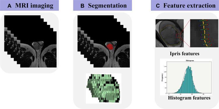
Purpose: To compare the performance of histogram analysis and intra-perinodular textural transition (Ipris) for distinguishing between benign and malignant testicular lesions.
Patients and methods: This retrospective study included 76 patients with 80 pathologically confirmed testicular lesions (55 malignant, 25 benign). All patients underwent preoperative T2-weighted imaging (T2WI) on a 3.0T MR scanner. All testicular lesions were manually segmented on axial T2WI, and histogram and Ipris features were extracted. Thirty enrolled patients were randomly selected to estimate the robustness of the features. We used intraclass correlation coefficients (ICCs) to evaluate intra- and interobserver agreement of features, independent t-test or Mann-Whitney U-test to compare features between benign and malignant lesions, and receiver operating characteristic curve analysis to evaluate the diagnostic performance of features.
Results: Eighteen histogram features and forty-eight Ipris features were extracted from T2WI of each lesion. Most (60/66) histogram and Ipris features had good robustness (ICC of both intra- and interobserver variabilities >0.6). Three histogram and nine Ipris features were significantly different between the benign and malignant groups. The area under the curve values for Energy, TotalEnergy, and Ipris_shell1_id_std were 0.807, 0.808, and 0.708, respectively, which were relatively higher than those of other features.
Conclusion: Ipris features may be useful for identifying benign and malignant testicular tumors but have no significant advantage over conventional histogram features.
Keywords: Ipris; magnetic resonance imaging; testicular disease; texture analysis.
© 2021 Zhang et al.
Conflict of interest statement
The authors report no conflicts of interest in this work.
Figures


References
-
- Cheng L, Albers P, Berney DM, et al. Testicular cancer. Nat Rev Dis Primers. 2018;4(1):29. - PubMed
LinkOut - more resources
Full Text Sources
Other Literature Sources
Research Materials
