. 2021 Jan 18:11:623190.
doi: 10.3389/fphys.2020.623190. eCollection 2020.

Transcriptomic Coupling of PKP2 With Inflammatory and Immune Pathways Endogenous to Adult Cardiac Myocytes

Affiliations

Transcriptomic Coupling of PKP2 With Inflammatory and Immune Pathways Endogenous to Adult Cardiac Myocytes

Marta Pérez-Hernández et al. Front Physiol. .

Abstract

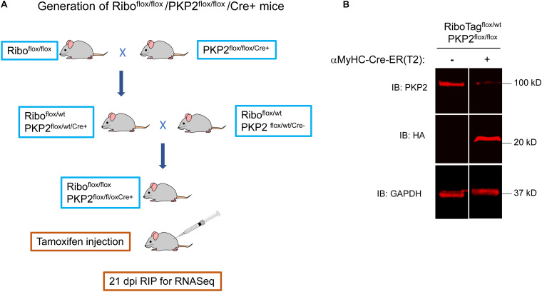
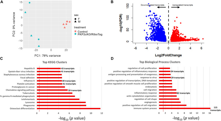
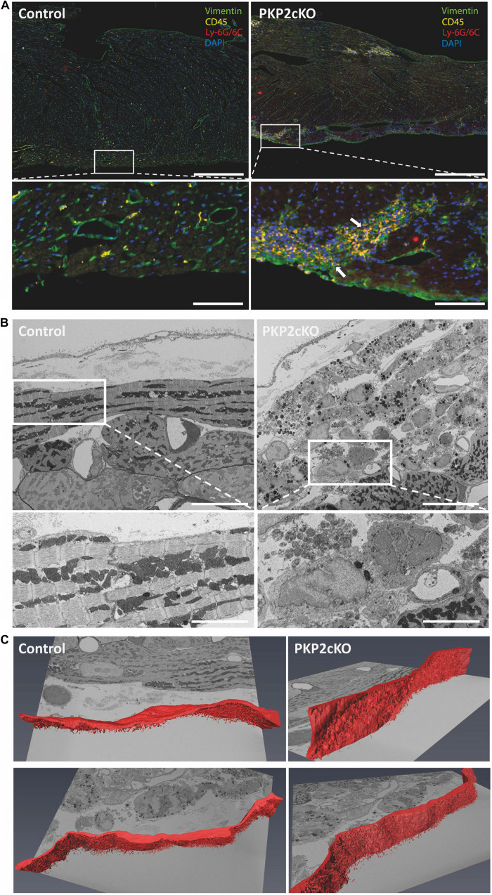
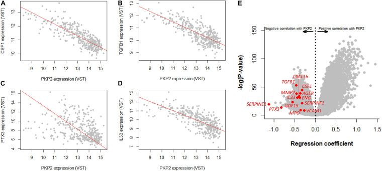
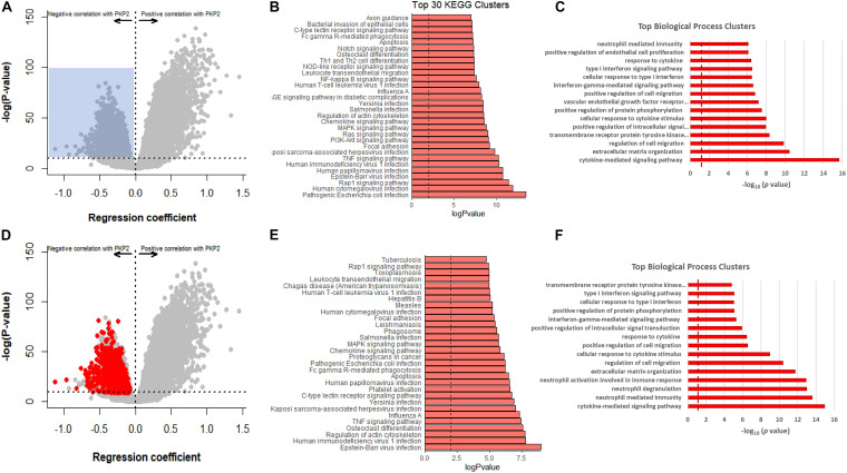
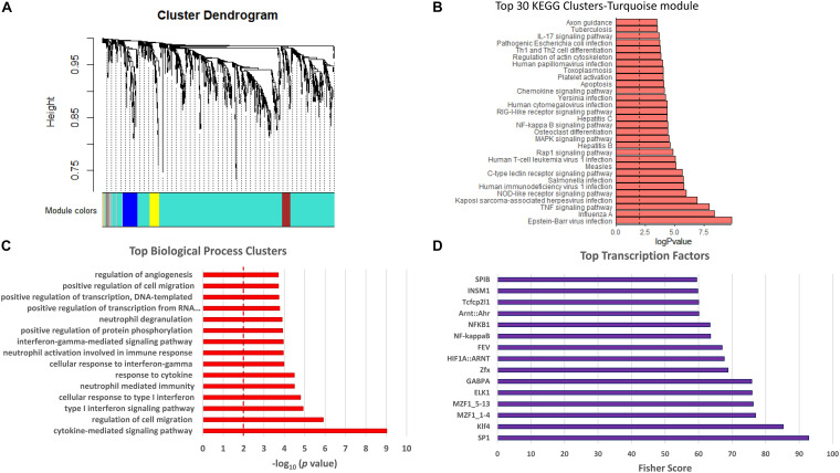
Plakophilin-2 (PKP2) is classically defined as a component of the desmosome. Besides its role in cell-cell adhesion, PKP2 can modulate transcription through intracellular signals initiated at the site of cell-cell contact. Mutations in PKP2 associate with arrhythmogenic right ventricular cardiomyopathy (ARVC). Recent data demonstrate that inflammation plays a key role in disease progression; other results show an abundance of anti-heart antibodies in patients with confirmed diagnosis of ARVC. Here, we test the hypothesis that, in adult cardiac myocytes, PKP2 transcript abundance is endogenously linked to the abundance of transcripts participating in the inflammatory/immune response. Cardiac-specific, tamoxifen (TAM)-activated PKP2-knockout mice (PKP2cKO) were crossed with a RiboTag line to allow characterization of the ribosome-resident transcriptome of cardiomyocytes after PKP2 knockdown. Data were combined with informatics analysis of human cardiac transcriptome using GTEx. Separately, the presence of non-myocyte cells at the time of analysis was assessed by imaging methods. We identified a large number of transcripts upregulated consequent to PKP2 deficiency in myocytes, inversely correlated with PKP2 abundance in human transcriptomes, and part of functional pathways associated with inflammatory/immune responses. Our data support the concept that PKP2 is transcriptionally linked, in cardiac myocytes, to genes coding for host-response molecules even in the absence of exogenous triggers. Targeted anti-inflammatory therapy may be effective in ARVC.

Keywords: GTEx; arrhythmogenic cardiomyopathy; inflammation/immune response; plakophilin-2; transcriptome.


Conflict of interest statement

The authors declare that the research was conducted in the absence of any commercial or financial relationships that could be construed as a potential conflict of interest.

Figures

FIGURE 1
FIGURE 1
Generation of Riboflox/flox/PKP2flox/flox/Cre+ mice. (A) RiboTagflox/flox/-PKP2flox/flox mice were obtained crossing RiboTag mice (JAX B6N.129-Rpl22/J 011029) with PKP2cKO/αMyHC-Cre-ER(T2). Cre + and Cre- mice were tamoxifen-injected and used 21 days post injection (dpi). (B) Mice hearts were analyzed by western blot. The 23-kDa 3HA epitope-tagged ribosomal protein RPL22 was detected by an HA antibody. A heterozygous RiboTagflox/wt was used. For experiments, we used homozygous RiboTagflox/flox to increase 3HA-RPL22 expression and facilitate breeding scheme.
FIGURE 2
FIGURE 2
Upregulated transcripts in hearts of PKP2cKO/RiboTag mice. (A) Principal component analysis (PCA) based on RNA-seq of myocyte control or PKP2cKO/RiboTag mice. The 78% of variance can be explained by treatment (PKP2 knockdown) whereas sex explained only 9% of variance, allowing data to be combined. (B) Volcano plot representing differential transcriptome of PKP2cKO/RiboTag myocytes vs control. “Upregulated” (red) or “downregulated” (blue) refers to abundance in PKP2 knocked-down myocytes (control as reference). Inclusion criteria are as follows: Log2 fold change ± 0.5 and FDR < 0.05. (C) Significantly enriched Kyoto Encyclopedia of Genes and Genomes (KEGG) pathways from upregulated genes. (D) Significantly enriched biological process clusters from upregulated genes in PKP2cKO.
FIGURE 3
FIGURE 3
Non-myocyte cell infiltration in ventricular tissue of wildtype and PKP2cKO mice. (A) Opal Multiplex immunohistochemistry staining for vimentin (green), CD45 (yellow), Ly-6G/Ly-6C (red), and DAPI (blue) in control and PKP2cKO ventricular tissue 21 days post-tamoxifen injection (dpi). Scale bar = 200 μm. White insets depict zoomed-in view; arrows highlight infiltration of CD45 and Ly-6G/6C-positive cells in PKP2cKO subepicardium. Scale bar = 50 μm. (B) Serial block face scanning electron microscopy (SBF-SEM) images of control (left) and PKP2cKO (right) subepicardial ventricular tissue, 14 dpi. Upper left panel (control), top to bottom: pericardial space; epicardial cell layer; basal membrane; first myocytes aligned parallel to epicardial surface. In right panel (PKP2cKO), see abundance of subepicardial non-myocyte cells and injured myocytes. A first layer of “healthy” myocytes is seen in bottom right corner. Scale bar = 10 μm. White frames indicate areas enlarged in bottom. Scale bar = 5 μm. (C) Single frames of complete z-stack of SBF-SEM images, with segmentation analysis of epicardial layer (red). Complete stack is in Supplementary Movies.
FIGURE 4
FIGURE 4
GTEx-based correlation analysis of human left ventricle transcriptome in relation to PKP2 expression. (A–D) Linear regression analysis of a transcript (CSF1, TGFB1, PTX3, and IL33, respectively) vs PKP2 expression. Notice negative slope for the four transcripts, all related with inflammation and immune response. Each gray point represents normalized level of PKP2 vs that of the given transcript for each individual in the 386 dataset. (E) Repeating this process for PKP2 against the entire transcriptome generates a volcano plot. Specific transcripts related to inflammation/immune system are highlighted in red. VST, variance stabilizing transformation.
FIGURE 5
FIGURE 5
GTEx-based PKP2 transcript correlation analysis, in comparison to RNA-seq PKP2cKO/RiboTag. (A) Correlation analysis of PKP2 normalized transcript abundance against other transcripts in the GTEx left ventricle database. Dots in gray are the same as in Figure 4E. Blue shadow indicates area selected for pathway analysis (regression coefficient < −0.5 and p-adjusted > 1E-10. (B,C) Top 30 Kyoto Encyclopedia of Genes and Genomes (KEGG) pathways and biological process analysis, respectively, of transcripts inversely correlated with PKP2. (D) Same plot as (A) but red circles indicate 912 transcripts significantly upregulated in RiboTag RNA-seq dataset. (E,F) Top 30 KEGG pathways and biological process analysis of red transcripts.
FIGURE 6
FIGURE 6
Weighted gene co-expression network analysis (WGCNA). Data included were the same as those in red in Figure 5D, i.e., significantly upregulated in RNA-seq dataset of PKP2cKO/RiboTag and negatively correlated with PKP2 expression in GTEx. (A) Cluster dendrogram of genes from WGCNA analysis, with dissimilarity based on topological overlap. Four modules detected and assigned to colors turquoise, blue, brown, and yellow, with number of genes ranging from 39 to 740. (B) Top 30 Kyoto Encyclopedia of Genes and Genomes (KEGG) pathways from the most abundant module (turquoise). (C) Top biological process associated with transcripts in the most abundant module (turquoise). (D) Transcription factor analysis for genes in turquoise module.

References

    1. Austin K. M., Trembley M. A., Chandler S. F., Sanders S. P., Saffitz J. E., Abrams D. J., et al. (2019). Molecular mechanisms of arrhythmogenic cardiomyopathy. Nat. Rev. Cardiol. 16 519–537. - PMC - PubMed
    1. Basso C., Corrado D., Marcus F. I., Nava A., Thiene G. (2009). Arrhythmogenic right ventricular cardiomyopathy. Lancet 373 1289–1300. - PubMed
    1. Bass-Zubek A. E., Hobbs R. P., Amargo E. V., Garcia N. J., Hsieh S. N., Chen X., et al. (2008). Plakophilin 2: a critical scaffold for PKC alpha that regulates intercellular junction assembly. J. Cell Biol. 181 605–613. 10.1083/jcb.200712133 - DOI - PMC - PubMed
    1. Bockstahler M., Fischer A., Goetzke C. C., Neumaier H. L., Sauter M., Kespohl M., et al. (2020). Heart-Specific Immune Responses in an Animal Model of AutoimmuneRelated Myocarditis Mitigated by an Immunoproteasome Inhibitor and Genetic Ablation. Circulation 141 1885–1902. 10.1161/circulationaha.119.043171 - DOI - PubMed
    1. Caforio A. L. P., Re F., Avella A., Marcolongo R., Baratta P., Seguso M., et al. (2020). Evidence From Family Studies for Autoimmunity in Arrhythmogenic Right Ventricular Cardiomyopathy: Associations of Circulating Anti-Heart and Anti-Intercalated Disk Autoantibodies With Disease Severity and Family History. Circulation 141 1238–1248. 10.1161/circulationaha.119.043931 - DOI - PubMed

LinkOut - more resources