. 2021 Jan 18:11:631444.
doi: 10.3389/fmicb.2020.631444. eCollection 2020.

PfAP2-G2 Is Associated to Production and Maturation of Gametocytes in Plasmodium falciparum via Regulating the Expression of PfMDV-1

Affiliations

PfAP2-G2 Is Associated to Production and Maturation of Gametocytes in Plasmodium falciparum via Regulating the Expression of PfMDV-1

Yaozheng Xu et al. Front Microbiol. .

Abstract

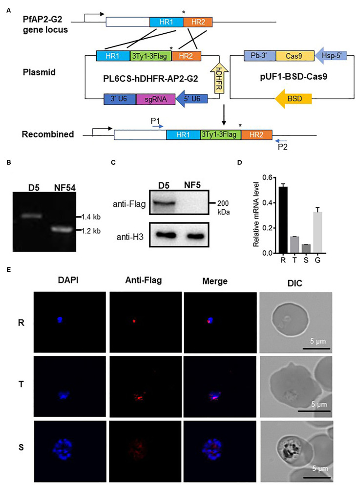
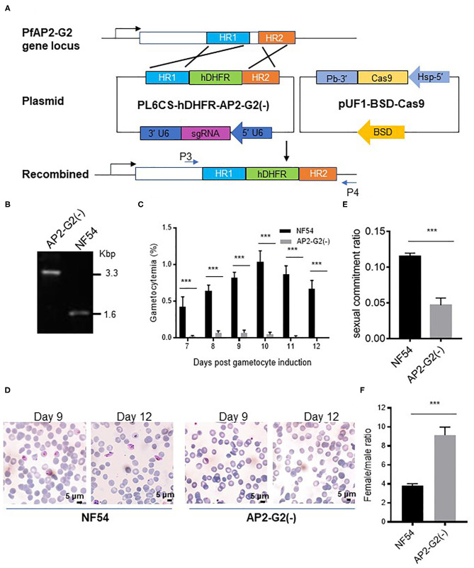
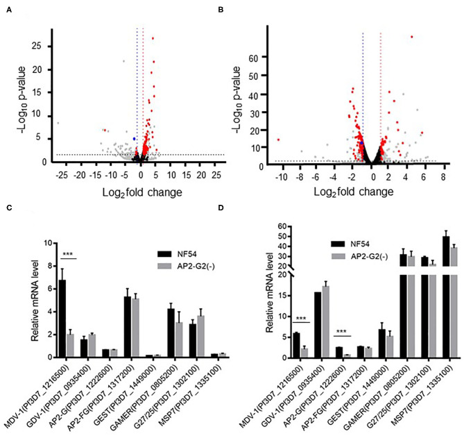
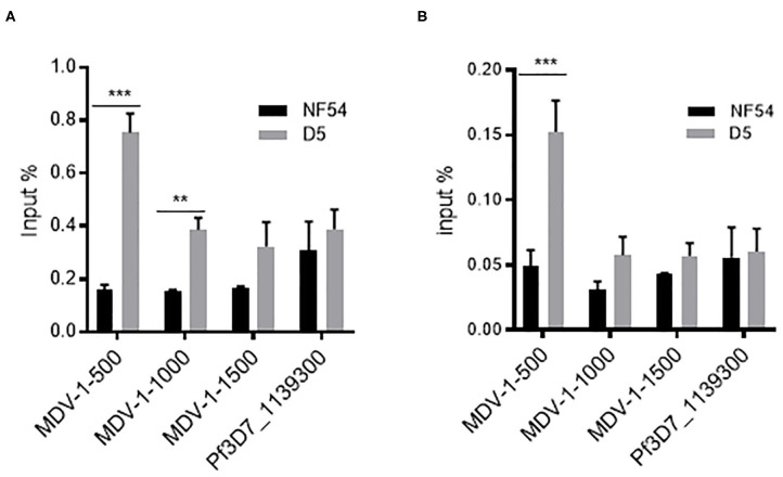
Gametocyte is the sole form of the Plasmodium falciparum which is transmissible to the mosquito vector. Here, we report that an Apicomplexan Apetala2 (ApiAP2) family transcription factor, PfAP2-G2 (Pf3D7_1408200), plays a role in the development of gametocytes in P. falciparum by regulating the expression of PfMDV-1 (Pf3D7_1216500). Reverse transcriptase-quantitative PCR (RT-qPCR) analysis showed that PfAP2-G2 was highly expressed in the ring stage. Indirect immunofluorescence assay showed nuclear localization of PfAP2-G2 in asexual stages. The knockout of PfAP2-G2 led to a ~95% decrease in the number of mature gametocytes with a more substantial influence on the production and maturation of the male gametocytes, resulting in a higher female/male gametocyte ratio. To test the mechanism of this phenotype, RNA-seq and RT-qPCR showed that disruption of PfAP2-G2 led to the down-regulation of male development gene-1 (PfMDV-1) in asexual stages. We further found that PfAP2-G2 was enriched at the transcriptional start site (TSS) of PfMDV-1 by chromatin immunoprecipitation and qPCR assay in both ring stage and schizont stage, which demonstrated that PfMDV-1 is one of the targets of PfAP2-G2. In addition, RT-qPCR also showed that PfAP2-G (Pf3D7_1222600), the master regulator for sexual commitment, was also down-regulated in the PfAP2-G2 knockout parasites in the schizont stage, but no change in the ring stage. This phenomenon suggested that PfAP2-G2 played a role at the asexual stage for the development of parasite gametocytes and warrants further investigations in regulatory pathways of PfAP2-G2.

Keywords: Plasmodium falciparum; gametocytes; malaria; sexual development; transcriptional factor.


Conflict of interest statement

The authors declare that the research was conducted in the absence of any commercial or financial relationships that could be construed as a potential conflict of interest.

Figures

Figure 1
Figure 1
Expression of PfAP2-G2 in P. falciparum development. (A) Schematic representation of the generation of transgenic PfAP2-G2-3Ty1-3Flag line. Co-transfection of the plasmid pUF1-BSD-Cas9 with PL6CS-hDHFR-AP2-G2 leads to the integration of the PfAP2-G2-3Ty1-3Flag containing cassette into the endogenous PfAP2-G2 locus. HR1 and HR2 are the two homologous arms used for recombination. The asterisk (*)indicates the stop codon. P1 and P2 are primers used to verify plasmid integration. (B) PCR analysis of genomic DNA from the NF54 parasites and the PfAP2-G2-3Ty1-3Flag clone D5. Predicted DNA fragment sizes with P1 and P2 are 1,423 bp from D5 parasites and 1,217 bp from the NF54 parasites. (C) Western blot analysis of D5 parasites. Proteins were separated by 6% SDS-PAGE and probed with the mouse anti-Flag antibody and against P. falciparum H3 as a protein loading control. (D) RT-qPCR analysis of PfAP2-G2 expression in the ring (14~16 h) (R), trophozoite (34~36h) (T), schizont (44~46 h) (S) stages and gametocyte stage IV~V(G). Primers for RT-qPCR are shown in Supplementary Table 1. The experiment was repeated three times. Error bars indicate standard deviation (SD). (E) IFA analysis of PfAP2-G2 during asexual parasite development. PfAP2-G2 localization was identified by using the anti-Flag antibody (red), while nuclei were counterstained with DAPI. Scale bars, 5 μm.
Figure 2
Figure 2
Phenotypic analysis of the PfAP2-G2(-) parasites. (A) Schematic representation of the generation of transgenic PfAP2-G2 knockout line. Co-transfection of the plasmid pUF1-BSD-Cas9 with PL6CS-hDHFR-AP2-G2(-) leads to knockout of PfAP2-G2 structural domain in the endogenous locus. HR1 and HR2 are the two homologous arms used for recombination. P3 and P4 are primers used to verify the deletion in PfAP2-G2. (B) PCR analysis of genomic DNA from the NF54 parasites and the PfAP2-G2(-) clone. Predicted DNA fragment sizes with P3 and P4 are 3,330 bp from the PfAP2-G2(-) parasites and 1,590 bp from the NF54 parasites. (C) Daily gametocytemia after day 6 post-gametocyte induction. Results are the mean of three biological replicates (10,000 RBCs counted per sample). (D) Giemsa-stained blood smears of NF54 (left) and the PfAP2-G2(-) parasites (right) on days 9 and 12 to illustrate the gross morphology of the gametocytes and their disappearance in day 12 culture in the PfAP2-G2(-) parasites. Scale bars, 5 μm. (E) Sexual commitment ratio on day 6 post-induction. Results are the mean of three biological replicates (1000 infected RBCs counted per sample). (F) Female/male gametocyte ratio on day 6 post-induction. Results are the mean of three biological replicates (1000 infected RBCs counted per sample). Sex was differentiated based on the staining of all gametocytes by the anti-Pfs16 antibodies and strong fluorescence of male gametocytes by the anti-α-tubulin II IFA. The experiment was repeated three times. All error bars indicate SD. ***P < 0.001.
Figure 3
Figure 3
Transcriptomic analysis of NF54 and the PfAP2-G2(-) parasites by RNA-seq. (A,B) Volcano plot of log2 fold change (x axis) in gene expression of PfAP2-G2(-) parasites vs. NF54 parasites in ring stage (A) and schizont stage (B) against the significance of change shown as the –log10 p-value (y axis). Red dots indicate genes with significant changes in expression level. Blue dots indicate the PfMDV-1 gene. Gray dots indicate genes whose TPM was <10. Black dots indicate genes whose p-value was more than 0.05 or | log2 fold change | was <1 (C,D) RT-qPCR validation of the RNA-seq data using selected genes essential for gametocyte development at the ring (C) and schizont (D) stages. A gene (MSP7), which is unrelated to gametocyte development, was used as a negative control. Primers for RT-qPCR are shown in Supplementary Table 1. The experiment was repeated three times. All error bars indicate SD. ***P < 0.001.
Figure 4
Figure 4
ChIP-qPCR analysis of PfAP2-G2 binding to the promoter region of PfMDV-1 at ring stage (A) and schizont stage (B). MDV1-500, MDV1-1000, and MDV1-1500 indicate primers that are located at about 500 bp, 1,000 bp, 1,500 bp before the start codon of PfMDV-1, respectively. TSS of PfMDV-1 is mapped to about 500 bp upstream of the start codon. Pf3D7_1139300 was used as a negative control. Primers for ChIP-qPCR are shown in Supplementary Table 1. The experiment was repeated three times. All error bars indicate SD. **P < 0.01; ***P < 0.001.

Similar articles

Cited by

References

    1. Amoah L. E., Acquah F. K., Nyarko P. B., Cudjoe E., Donu D., Ayanful-Torgby R., et al. (2020). Comparative analysis of asexual and sexual stage Plasmodium falciparum development in different red blood cell types. Malar. J. 19:200 10.1186/s12936-020-03275-9 - DOI - PMC - PubMed
    1. Balaji S., Babu M. M., Iyer L. M., Aravind L. (2005). Discovery of the principal specific transcription factors of Apicomplexa and their implication for the evolution of the AP2-integrase DNA binding domains. Nucleic Acids Res. 33, 3994–4006. 10.1093/nar/gki709 - DOI - PMC - PubMed
    1. Bancells C., Llorà-Batlle O., Poran A., Nötzel C., Rovira-Graells N., Elemento O., et al. . (2019). Revisiting the initial steps of sexual development in the malaria parasite Plasmodium falciparum. Nat. Microbiol. 4, 144–154. 10.1038/s41564-018-0291-7 - DOI - PMC - PubMed
    1. Bischoff E., Vaquero C. (2010). In silico and biological survey of transcription-associated proteins implicated in the transcriptional machinery during the erythrocytic development of Plasmodium falciparum. BMC Genomics 11:34. 10.1186/1471-2164-11-34 - DOI - PMC - PubMed
    1. Campbell T. L., De Silva E. K., Olszewski K. L., Elemento O., Llinas M. (2010). Identification and genome-wide prediction of DNA binding specificities for the ApiAP2 family of regulators from the malaria parasite. PLoS Pathog. 6:e1001165. 10.1371/journal.ppat.1001165 - DOI - PMC - PubMed

LinkOut - more resources