

The Dynamic Expression of Potential Mediators of Severe Acute Respiratory Syndrome Coronavirus 2 Cellular Entry in Fetal, Neonatal, and Adult Rhesus Monkeys
- PMID: 33537060
- PMCID: PMC7848180
- DOI: 10.3389/fgene.2020.607479
The Dynamic Expression of Potential Mediators of Severe Acute Respiratory Syndrome Coronavirus 2 Cellular Entry in Fetal, Neonatal, and Adult Rhesus Monkeys
Abstract
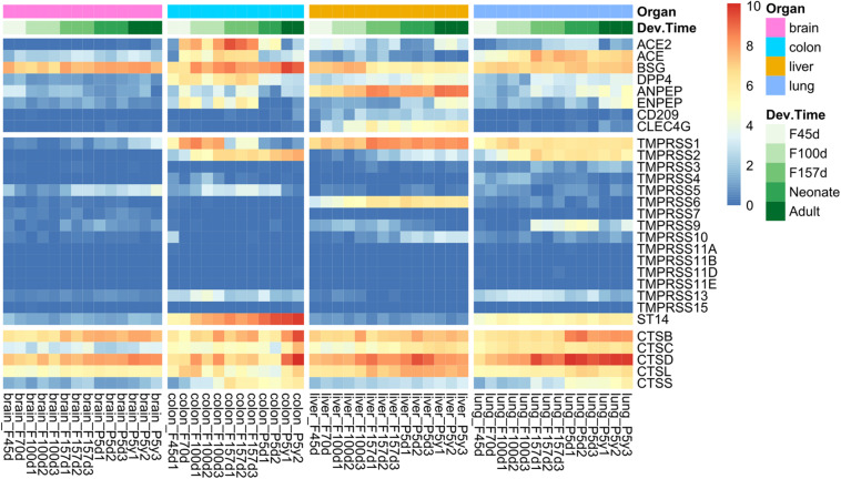
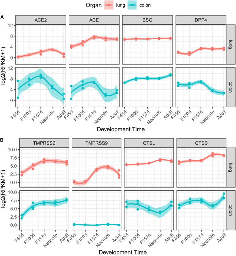
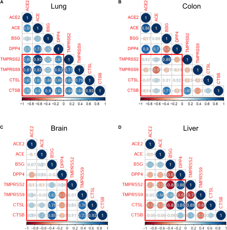
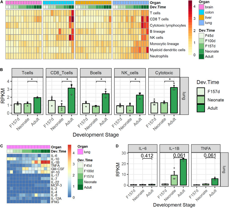
The coronavirus disease 2019 (COVID-19) pandemic, induced by the pathogenic severe acute respiratory syndrome coronavirus 2 (SARS-CoV-2), has spread rapidly all over the world. There is considerable variability among neonates, children, and adults in the incidence of infection and severe disease following exposure to SARS-CoV-2. In our study, we analyzed the transcriptome data of primate animal model of Rhesus monkeys to evaluate the expression levels of possible SARS-CoV-2 receptors and proteases and immunologic features in the lungs, colons, livers, and brains at different developmental stages. Our results revealed that ACE2 and TMPRSS2 were highly expressed in neonates compared with other populations, which imply the high incidence of infection. Other potential receptors and Type II transmembrane serine proteases (TTSPs) and cathepsin of endosomal proteases also exhibited dynamic and differential expression patterns. The expression of receptors (ACE2, BSG, and DPP4) and proteases (TMPRSS2, TMPRSS9, CTSL, and CTSB) were highly correlated during lung development, suggesting the high susceptibility of the lungs. TMPRSS9 was specifically highly expressed in the lungs and reached the highest level in neonates, similar to TMPRSS2. Moreover, the immune cell infiltration analysis revealed immunity immaturity in neonates, implying the association with the mild or moderate type of COVID-19. The results might help researchers design protective and therapeutic strategies for COVID-19 in populations at different ages.
Keywords: ACE2; Rhesus monkey; SARS-CoV-2; TMPRSS2; development.
Copyright © 2021 Cao, Zhang, Liu, Ma and Mi.
Conflict of interest statement
The authors declare that the research was conducted in the absence of any commercial or financial relationships that could be construed as a potential conflict of interest.
Figures
Similar articles
-
Single-cell RNA sequencing of SARS-CoV-2 cell entry factors in the preconceptional human endometrium.Hum Reprod. 2021 Sep 18;36(10):2709-2719. doi: 10.1093/humrep/deab183. Hum Reprod. 2021. PMID: 34329437 Free PMC article.
-
Single-cell analysis of SARS-CoV-2 receptor ACE2 and spike protein priming expression of proteases in the human heart.Cardiovasc Res. 2020 Aug 1;116(10):1733-1741. doi: 10.1093/cvr/cvaa191. Cardiovasc Res. 2020. PMID: 32638018 Free PMC article.
-
Integrated Bioinformatic Analysis of SARS-CoV-2 Infection Related Genes ACE2, BSG and TMPRSS2 in Aerodigestive Cancers.J Inflamm Res. 2021 Mar 10;14:791-802. doi: 10.2147/JIR.S300127. eCollection 2021. J Inflamm Res. 2021. PMID: 33732005 Free PMC article.
-
Targeting the viral-entry facilitators of SARS-CoV-2 as a therapeutic strategy in COVID-19.J Med Virol. 2021 Sep;93(9):5260-5276. doi: 10.1002/jmv.27019. Epub 2021 May 3. J Med Virol. 2021. PMID: 33851732 Free PMC article. Review.
-
SARS-CoV-2 pandemics: An update of CRISPR in diagnosis and host-virus interaction studies.Biomed J. 2023 Apr;46(2):100587. doi: 10.1016/j.bj.2023.02.007. Epub 2023 Feb 25. Biomed J. 2023. PMID: 36849044 Free PMC article. Review.
Cited by
-
A therapy for suppressing canonical and noncanonical SARS-CoV-2 viral entry and an intrinsic intrapulmonary inflammatory response.Proc Natl Acad Sci U S A. 2024 Jul 23;121(30):e2408109121. doi: 10.1073/pnas.2408109121. Epub 2024 Jul 19. Proc Natl Acad Sci U S A. 2024. PMID: 39028694 Free PMC article.
-
A genome-wide CRISPR screen identifies interactors of the autophagy pathway as conserved coronavirus targets.PLoS Biol. 2021 Dec 28;19(12):e3001490. doi: 10.1371/journal.pbio.3001490. eCollection 2021 Dec. PLoS Biol. 2021. PMID: 34962926 Free PMC article.
-
An Update on Animal Models for Severe Acute Respiratory Syndrome Coronavirus 2 Infection and Countermeasure Development.Front Microbiol. 2021 Nov 8;12:770935. doi: 10.3389/fmicb.2021.770935. eCollection 2021. Front Microbiol. 2021. PMID: 34819926 Free PMC article. Review.
-
Differential effects of age, sex and dexamethasone therapy on ACE2/TMPRSS2 expression and susceptibility to SARS-CoV-2 infection.Front Immunol. 2022 Nov 3;13:1021928. doi: 10.3389/fimmu.2022.1021928. eCollection 2022. Front Immunol. 2022. PMID: 36405732 Free PMC article.
-
Identification of Embryonic Chicken Proteases Activating Newcastle Disease Virus and Their Roles in the Pathogenicity of Virus Used as In Ovo Vaccine.J Virol. 2023 May 31;97(5):e0032423. doi: 10.1128/jvi.00324-23. Epub 2023 Apr 12. J Virol. 2023. PMID: 37042750 Free PMC article.
References
LinkOut - more resources
Full Text Sources
Other Literature Sources
Miscellaneous