Review
. 2021 Jan 7:8:608026.
doi: 10.3389/fcell.2020.608026. eCollection 2020.

Mitochondrial Dysfunction in Astrocytes: A Role in Parkinson's Disease?

Affiliations
Review

Mitochondrial Dysfunction in Astrocytes: A Role in Parkinson's Disease?

Collin M Bantle et al. Front Cell Dev Biol. .

Abstract

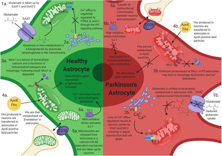
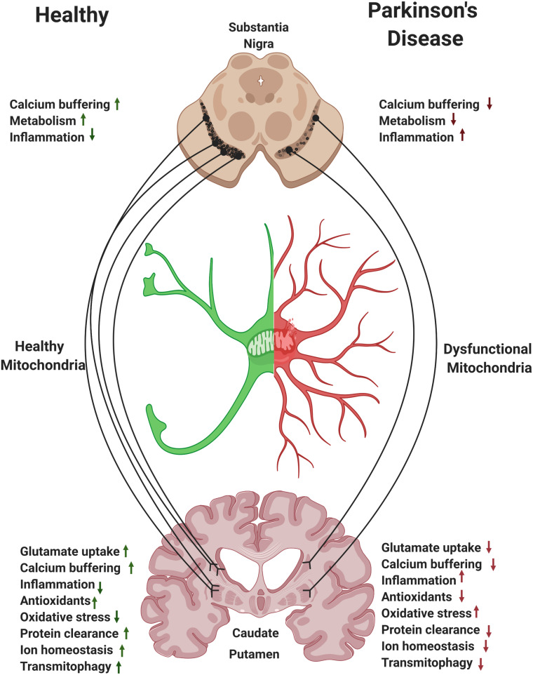
Mitochondrial dysfunction is a hallmark of Parkinson's disease (PD). Astrocytes are the most abundant glial cell type in the brain and are thought to play a pivotal role in the progression of PD. Emerging evidence suggests that many astrocytic functions, including glutamate metabolism, Ca2+ signaling, fatty acid metabolism, antioxidant production, and inflammation are dependent on healthy mitochondria. Here, we review how mitochondrial dysfunction impacts astrocytes, highlighting translational gaps and opening new questions for therapeutic development.

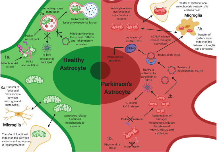
Keywords: NLRP3; PINK1/Parkin pathway; Parkinson’s disease; astrocyte; cGAS/STING pathway; inflammation; mitochondria.


Conflict of interest statement

All authors are employees and shareholders of Biogen.

Figures

FIGURE 1
FIGURE 1
How mitochondrial dysfunction in astrocytes can contribute to Parkinson’s disease (PD) progression. Mitochondrial dysfunction in astrocytes may elicit neuronal toxicity through multiple mechanisms. (1a) Functional astrocyte mitochondria are needed for glutamate regulation and metabolism. (1b) Dysfunctional astrocyte mitochondria likely have reduced glutamate uptake and metabolism, resulting in excitatory neurotoxicity in neurons. (2a) Astrocytes house large stores of intracellular calcium and other ions, which is negatively regulated by ITPKA, B, and C. (2b) Perturbations of these calcium reservoirs results in an increase in mitochondrial intracellular calcium through VDAC1 and MCU, resulting in signal transduction and astrocytic cell death. (3a) Miro1 is a sensor of extracellular calcium and a facilitator of mitochondrial transport and mitophagy. Following mitochondrial stress, Miro1 is degraded, which facilitates the clearance of damaged mitochondria. (3b) Defective proteostasis of Miro1 in PD astrocytes may lead to mitophagy dysfunction and Ca2+ disbalance. (4a) Toxic fatty acids (FAs) are produced in neurons and are transferred to astrocytic lipid droplets by ApoE-positive lipid particles. Astrocytes then consume the FAs stored in lipid droplets via mitochondrial β-oxidation in response to neuronal activity and turn on a detoxification gene expression program. (4b) Loss of mitochondrial function in astrocytes prevents proper metabolism of these FAs and results in FA induced toxicity (4b). (5a,b) Astrocytes serve as the primary cell type responsible for the clearance and transfer of damaged and healthy mitochondria to and from neurons. The transfer is completed in a calcium-dependent manner, regulated by CD38. During transmitophagy, mitophagy is thought to begin in neurons and be completed in astrocytes. (6a) Mild mtROS induces DJ-1 activation, which results in antioxidant gene transcription. (6b) High levels of mtROS inactives DJ-1 and prevents antioxidant gene transcription.
FIGURE 2
FIGURE 2
Mitochondrial dysfunction in astrocytes contributes to neuroinflammatory activation of astrocytes. Mutations in PINK1 and Parkin likely elicit neuroinflammatory activation in astrocytes. (1a) After mitochondrial stress, PINK1 protein accumulates on the outer mitochondrial membrane. Phosphorylation of polyubiquitin and Parkin induces autophagosome maturation and eventually delivery to the lysosome. (1b) Loss-of-function (LOF) mutations in PINK1 and Parkin prevent PINK1 accumulation on the mitochondrial membrane and the activation of Parkin during mitochondrial stress, resulting in the accumulation of damaged mitochondria within astrocytes. This likely facilitates chronic inflammatory activation through multiple inflammatory pathways, including NLRP3 activation. (2a) Functional mitophagy prevents the release of damage-associated molecular patterns (DAMPs)/pathogen-associated molecular patterns (PAMPs) and activation of the NLR family, pyrin domain containing 3 (NLRP3) inflammasome in astrocytes. (2b) The accumulation of damaged mitochondria likely results in the release of intracellular DAMPs/PAMPs, cardiolipin, and mtROS, which activates NLRP3 and the cyclic guanosine monophosphate–adenosine monophosphate cGAMP synthase (cGAS)/stimulator of interferon genes (STING) pathway, resulting in inflammatory activation and release of interleukin (IL)-1β and IL-18, and potentially inducing the activation of microglia via extracellular release of cGAMP. (3a) Transfer of functional mitochondria from astrocytes to neurons is thought to be neuroprotective; however, whether microglia release functional mitochondria to astrocytes is elusive. (3b) Joshi et al. have shown that activated microglia can release dysfunctional and fragmented mitochondria to astrocytes. Astrocytes then transfer dysfunctional mitochondria to neurons, resulting in neuronal death. However, whether this occurs in vivo is unknown.
FIGURE 3
FIGURE 3
Astrocytes and the nigrostriatal pathway. Within the nigrostriatal pathway, astrocyte numbers and functions are most abundant in the striatum/caudate putamen, where dopaminergic neurons support is the highest and where loss of astrocytic function is likely to have the most detrimental effect in Parkinson’s disease (PD). Reversing mitochondrial dysfunction in astrocytes in the striatum may induce significant therapeutic benefit in PD.

References

    1. Ablasser A., Chen Z. J. (2019). cGAS in action: expanding roles in immunity and inflammation. Science 363:eaat8657. 10.1126/science.aat8657 - DOI - PubMed
    1. Agarwal A., Wu P. H., Hughes E. G., Fukaya M., Tischfield M. A., Langseth A. J., et al. (2017). Transient opening of the mitochondrial permeability transition pore induces microdomain calcium transients in astrocyte processes. Neuron 93 587.e7–605.e7. - PMC - PubMed
    1. Ariga H., Takahashi-Niki K., Kato I., Maita H., Niki T., Iguchi-Ariga S. M. (2013). Neuroprotective function of DJ-1 in Parkinson’s disease. Oxid. Med. Cell Longev. 2013:683920. - PMC - PubMed
    1. Armada-Moreira A., Gomes J. I., Pina C. C., Savchak O. K., Goncalves-Ribeiro J., Rei N., et al. (2020). Going the extra (Synaptic) mile: excitotoxicity as the road toward neurodegenerative diseases. Front. Cell Neurosci. 14:90. 10.3389/fncel.2020.00090 - DOI - PMC - PubMed
    1. Bagur R., Hajnoczky G. (2017). Intracellular Ca(2+) sensing: its role in calcium homeostasis and signaling. Mol. Cell. 66 780–788. 10.1016/j.molcel.2017.05.028 - DOI - PMC - PubMed