

Optogenetic brain-stimulation reward: A new procedure to re-evaluate the rewarding versus aversive effects of cannabinoids in dopamine transporter-Cre mice
- PMID: 33538103
- PMCID: PMC9308103
- DOI: 10.1111/adb.13005
Optogenetic brain-stimulation reward: A new procedure to re-evaluate the rewarding versus aversive effects of cannabinoids in dopamine transporter-Cre mice
Abstract
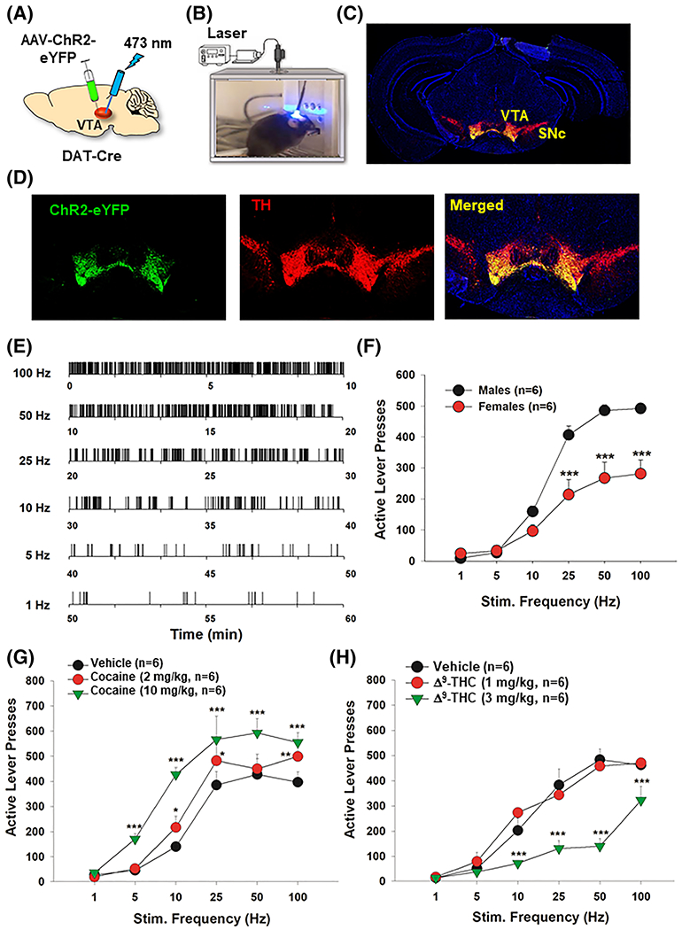
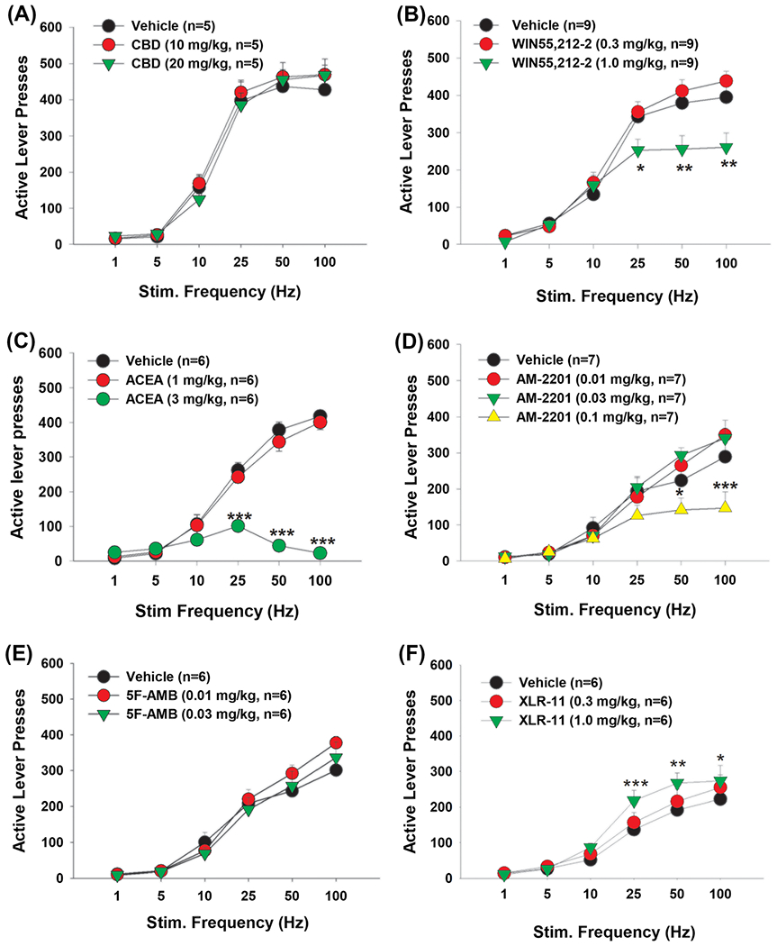
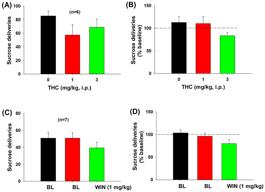
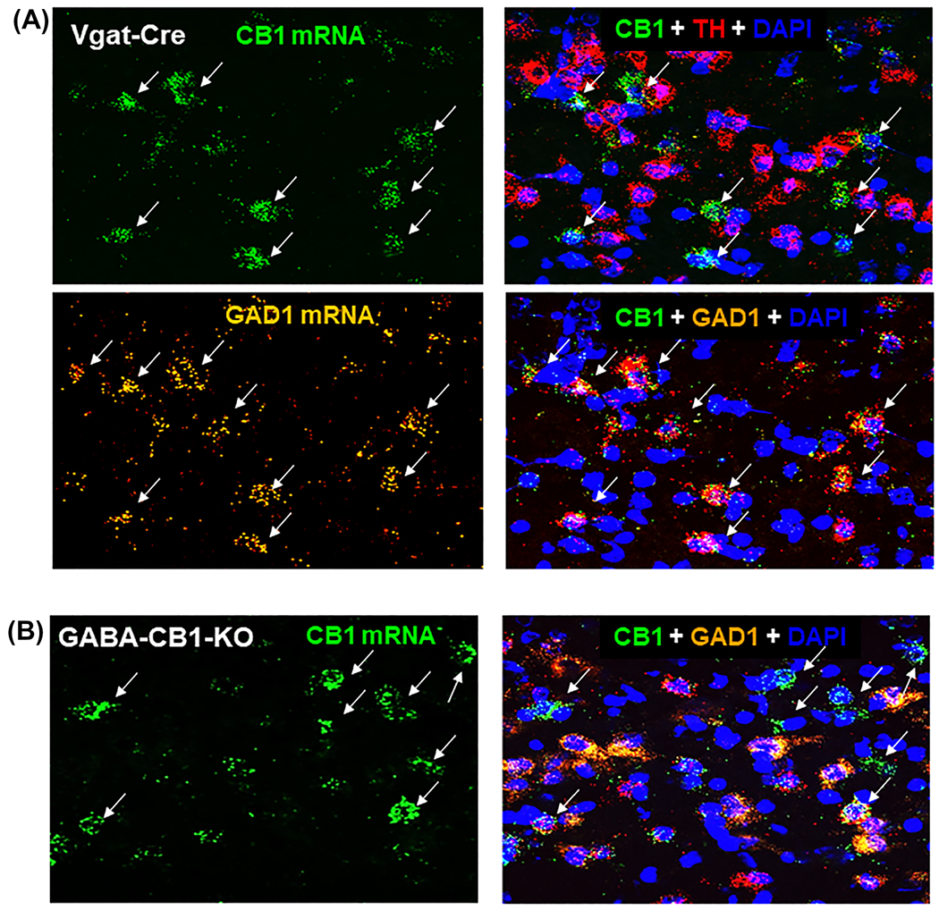
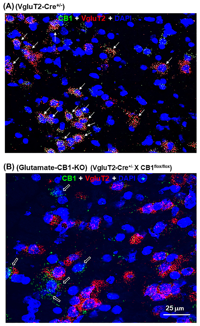
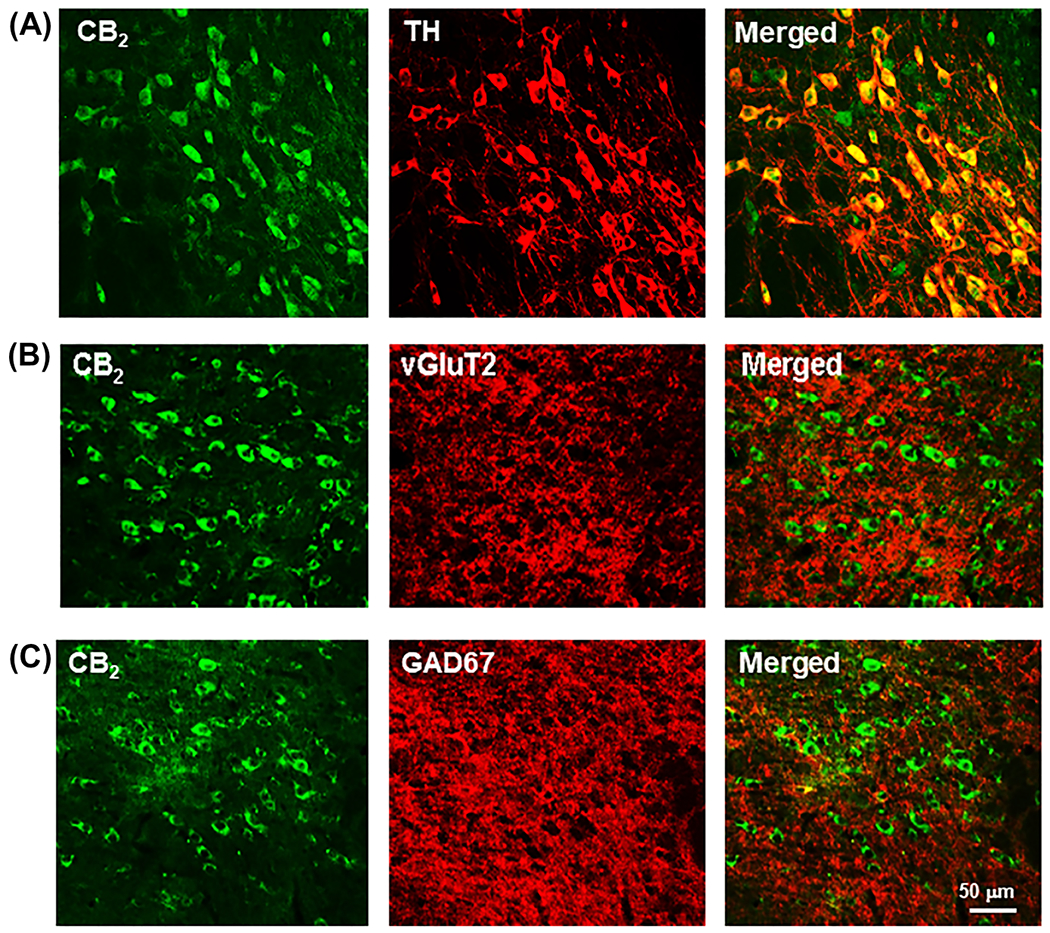
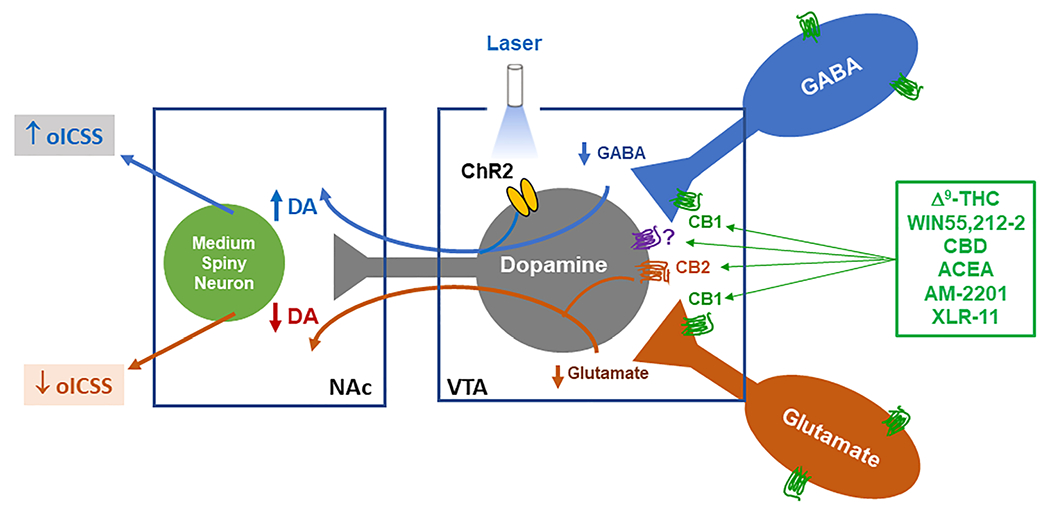
Despite extensive research, the rewarding effects of cannabinoids are still debated. Here, we used a newly established animal procedure called optogenetic intracranial self-stimulation (ICSS) (oICSS) to re-examine the abuse potential of cannabinoids in mice. A specific adeno-associated viral vector carrying a channelrhodopsin gene was microinjected into the ventral tegmental area (VTA) to express light-sensitive channelrhodopsin in dopamine (DA) neurons of transgenic dopamine transporter (DAT)-Cre mice. Optogenetic stimulation of VTA DA neurons was highly reinforcing and produced a classical "sigmoidal"-shaped stimulation-response curve dependent upon the laser pulse frequency. Systemic administration of cocaine dose-dependently enhanced oICSS and shifted stimulation-response curves upward, in a way similar to previously observed effects of cocaine on electrical ICSS. In contrast, Δ9 -tetrahydrocannabinol (Δ9 -THC), but not cannabidiol, dose-dependently decreased oICSS responding and shifted oICSS curves downward. WIN55,212-2 and ACEA, two synthetic cannabinoids often used in laboratory settings, also produced dose-dependent reductions in oICSS. We then examined several new synthetic cannabinoids, which are used recreationally. XLR-11 produced a cocaine-like increase, AM-2201 produced a Δ9 -THC-like reduction, while 5F-AMB had no effect on oICSS responding. Immunohistochemistry and RNAscope in situ hybridization assays indicated that CB1 Rs are expressed mainly in VTA GABA and glutamate neurons, while CB2 Rs are expressed mainly in VTA DA neurons. Together, these findings suggest that most cannabinoids are not reward enhancing, but rather reward attenuating or aversive in mice. Activation of CB1 R and/or CB2 R in different populations of neurons in the brain may underlie the observed actions.
Keywords: WIN55,212-2; brain-stimulation reward; cannabinoids; cocaine; dopamine; intracranial self-stimulation; optogenetics; ventral tegmental area; Δ9-THC.
Published 2021. This article is a U.S. Government work and is in the public domain in the USA.
Conflict of interest statement
CONFLICT OF INTEREST
None of the authors have any conflicts of interest.
Figures
Similar articles
-
Re-examining the role of ventral tegmental area dopaminergic neurons in motor activity and reinforcement by chemogenetic and optogenetic manipulation in mice.Metab Brain Dis. 2019 Oct;34(5):1421-1430. doi: 10.1007/s11011-019-00442-z. Epub 2019 Jul 16. Metab Brain Dis. 2019. PMID: 31313126
-
CB1 Receptor Activation on VgluT2-Expressing Glutamatergic Neurons Underlies Δ9-Tetrahydrocannabinol (Δ9-THC)-Induced Aversive Effects in Mice.Sci Rep. 2017 Sep 26;7(1):12315. doi: 10.1038/s41598-017-12399-z. Sci Rep. 2017. PMID: 28951549 Free PMC article.
-
Cannabinoid CB1 Receptors Are Expressed in a Subset of Dopamine Neurons and Underlie Cannabinoid-Induced Aversion, Hypoactivity, and Anxiolytic Effects in Mice.J Neurosci. 2023 Jan 18;43(3):373-385. doi: 10.1523/JNEUROSCI.1493-22.2022. Epub 2022 Dec 14. J Neurosci. 2023. PMID: 36517243 Free PMC article.
-
Optical Intracranial Self-Stimulation (oICSS): A New Behavioral Model for Studying Drug Reward and Aversion in Rodents.Int J Mol Sci. 2024 Mar 19;25(6):3455. doi: 10.3390/ijms25063455. Int J Mol Sci. 2024. PMID: 38542425 Free PMC article. Review.
-
Reward and aversion in a heterogeneous midbrain dopamine system.Neuropharmacology. 2014 Jan;76 Pt B(0 0):351-9. doi: 10.1016/j.neuropharm.2013.03.019. Epub 2013 Apr 8. Neuropharmacology. 2014. PMID: 23578393 Free PMC article. Review.
Cited by
-
Elevating levels of the endocannabinoid 2-arachidonoylglycerol blunts opioid reward but not analgesia.bioRxiv [Preprint]. 2024 Apr 2:2024.04.02.585967. doi: 10.1101/2024.04.02.585967. bioRxiv. 2024. Update in: Sci Adv. 2024 Nov 29;10(48):eadq4779. doi: 10.1126/sciadv.adq4779. PMID: 38766079 Free PMC article. Updated. Preprint.
-
Receptor mechanisms underlying the CNS effects of cannabinoids: CB1 receptor and beyond.Adv Pharmacol. 2022;93:275-333. doi: 10.1016/bs.apha.2021.10.006. Epub 2021 Dec 13. Adv Pharmacol. 2022. PMID: 35341569 Free PMC article.
-
RDS-04-010: a novel atypical DAT inhibitor that inhibits cocaine taking and seeking and itself has low abuse potential in experimental animals.Transl Psychiatry. 2025 May 24;15(1):182. doi: 10.1038/s41398-025-03391-7. Transl Psychiatry. 2025. PMID: 40413193 Free PMC article.
-
Targeting Endocannabinoid Signaling in the Lateral Habenula as an Intervention to Prevent Mental Illnesses Following Early Life Stress: A Perspective.Front Synaptic Neurosci. 2021 May 28;13:689518. doi: 10.3389/fnsyn.2021.689518. eCollection 2021. Front Synaptic Neurosci. 2021. PMID: 34122037 Free PMC article.
-
Therapeutic potential of PIMSR, a novel CB1 receptor neutral antagonist, for cocaine use disorder: evidence from preclinical research.Transl Psychiatry. 2022 Jul 18;12(1):286. doi: 10.1038/s41398-022-02059-w. Transl Psychiatry. 2022. PMID: 35851573 Free PMC article.
References
-
- Grigsby TM, Hoffmann LM, Moss MJ. Marijuana use and potential implications of marijuana legalization. Pediatr Rev. 2020;41(2):61–72. - PubMed
-
- National Institute on Drug Abuse. What is the scope of marijuana use in the United States? https://www.drugabuse.gov/publications/research-reports/marijuana/what-s.... Published 2020. Accessed May 30, 2020.
-
- National Institute on Drug Abuse. Is marijuana addictive? https://www.drugabuse.gov/publications/research-reports/marijuana/mariju.... Published 2020. Accessed May 30, 2020.
-
- Fattore L, Fadda P, Spano MS, Pistis M, Fratta W. Neurobiological mechanisms of cannabinoid addiction. Mol Cell Endocrinol. 2008;286(1–2 Suppl 1):S97–S107. - PubMed
Publication types
MeSH terms
Substances
Grants and funding
LinkOut - more resources
Full Text Sources
Other Literature Sources
Molecular Biology Databases
