. 2021 Feb 14;13(6):3644-3653.
doi: 10.1039/d0nr06587k. Epub 2021 Feb 4.

Laser nanobubbles induce immunogenic cell death in breast cancer

Affiliations

Laser nanobubbles induce immunogenic cell death in breast cancer

Hieu T M Nguyen et al. Nanoscale. .

Abstract

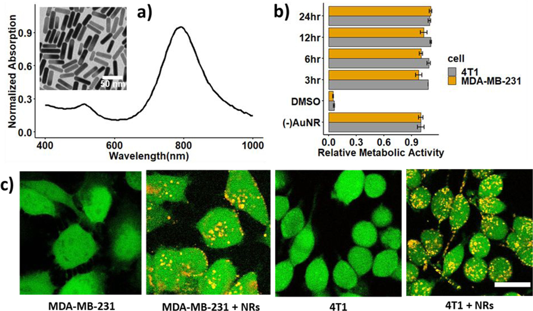
Recent advances in immunotherapy have highlighted a need for therapeutics that initiate immunogenic cell death in tumors to stimulate the body's immune response to cancer. This study examines whether laser-generated bubbles surrounding nanoparticles ("nanobubbles") induce an immunogenic response for cancer treatment. A single nanosecond laser pulse at 1064 nm generates micron-sized bubbles surrounding gold nanorods in the cytoplasm of breast cancer cells. Cell death occurred in cells treated with nanorods and irradiated, but not in cells with irradiation treatment alone. Cells treated with nanorods and irradiation had increased damage-associated molecular patterns (DAMPs), including increased expression of chaperone proteins human high mobility group box 1 (HMGB1), adenosine triphosphate (ATP), and heat shock protein 70 (HSP70). This enhanced expression of DAMPs led to the activation of dendritic cells. Overall, this treatment approach is a rapid and highly specific method to eradicate tumor cells with simultaneous immunogenic cell death signaling, showing potential as a combination strategy for immunotherapy.


Conflict of interest statement

Conflicts of interest

There are no conflicts to declare.

Figures

Figure 1
Figure 1
Gold nanorods (AuNRs) internalization in breast cancer cells: a) absorption spectrum of Mutab-coated AuNRs and TEM image of the nanorods (insert); b) relative metabolic activity of 4T1 and MDA-MB-231 cells incubated with AuNRs; c) two-photon images of MDA-MB-231 and 4T1 cells without and with AuNRs incubated for 6 hours. Scale bar: 20 μm
Figure 2
Figure 2
Cell death resulting from laser (1064 nm) irradiation of AuNRs-incubated MDA-MBA-231 cells: a) cell death with varying fluence from 0.7–5 J/cm2; b-d) cell death specific to irradiation of AuNRs-embedded MDA-MBA-231 cells at 3 J/cm2: without AuNRs incubation (left), with AuNRs incubation (middle) and with a mix of these two populations in 2:1 ratio (right). A two-photon microscope acquires the images with live cells stained in green (calcein-AM) and dead cells stained in red (ethidium homodimer-1). Beamwidth is highlighted with a white dashed line. Scale bar: 0.5 mm
Figure 3
Figure 3
Imaging bubbles following rapid heating and water vaporization around AuNRs. (a) Assembly of the optical system to image bubbles, (b) montage of bubbles formed in two examples of 4T1 cells embedded with AuNRs after one pulse of laser irradiation at 3 J/cm2, (c) mean intensity of bubble over its time course with polynomial fitting (red line), the red dash line highlights the bubble pixels being monitored, (d) bubble’s lifetime vs. diameter with polynomial fitting (red line). Scale bar: 10 μm
Figure 4
Figure 4
Extracellular release of damage associated patterns from laser irradiation of MDA-MB-231 and 4T1 cells at 3 J/cm2 and 2 cm2 of cells area per sample (50% of the well area), a) HSP70, HMGB1, and ATP; b) Time-dependent release of DAMPs after laser irradiation. Groups: CTR: cells without any treatment, NR: cells incubated with AuNRs, L: cells irradiated with a laser, NR+L: cell incubated with AuNRs and irradiated with a laser, DOX: cell treated with doxorubicin for 24 hours as the positive control. The number of samples per group n = 3
Figure 5
Figure 5
Activation of dendritic cells from irradiation of AuNRs-embedded 4T1 cells a) experimental layout describing 4T1 co-cultured with DCs in a transwell setting, b) Dot plot of DCs expressing MHC II and CD86, c) Percentage of mature dendritic cells as CD11c+ MHCII+ and CD86+, and median intensity of DCs that express MHCII and CD86. Six groups of dendritic cells: CTR: DCs without treatment; 4T1cc: DCs co-cultured with 4T1 cells; 4T1cc+L: DCs cultured with irradiated 4T1 cells; 4T1cc+NR+L: DCs co-cultured with irradiated AuNRs-embedded 4T1 cells; 4T1cc+NR+L x 2: DCs co-cultured with twice irradiated AuNRs-embedded 4T1 cells, the second irradiation is 12 hours after the first irradiation; LPS: DCs treated with LPS at 1 μg/ml for 12 hours. Number of samples per group n = 3. Statistical analysis was performed with analysis variance (ANOVA) in combination with Tukey test, * means p-value < 0.05

References

    1. Maio M, Grob J-J, Aamdal S, Bondarenko I, Robert C, Thomas L, Garbe C, Chiarion-Sileni V, Testori A, Chen T-T, Tschaika M. and Wolchok JD, J. Clin. Oncol, 2015, 33, 1191–1196. - PMC - PubMed
    1. Hodi FS, O’Day SJ, McDermott DF, Weber RW, Sosman JA, Haanen JB, Gonzalez R, Robert C, Schadendorf D, Hassel JC, Akerley W, van den Eertwegh AJM, Lutzky J, Lorigan P, Vaubel JM, Linette GP, Hogg D, Ottensmeier CH, Lebbé C, Peschel C, Quirt I, Clark JI, Wolchok JD, Weber JS, Tian J, Yellin MJ, Nichol GM, Hoos A. and Urba WJ, N. Engl. J. Med, 2010, 363, 711–723. - PMC - PubMed
    1. Wei SC, Duffy CR and Allison JP, Cancer Discovery, 2018, 8, 1069–1086. - PubMed
    1. Nanda R, Chow LQM, Dees EC, Berger R, Gupta S, Geva R, Pusztai L, Pathiraja K, Aktan G, Cheng JD, Karantza V. and Buisseret L, J. Clin. Oncol, 2016, 34, 2460–2467. - PMC - PubMed
    1. Sharma P. and Allison JP, Science, 2015, 348, 56–61. - PubMed