Review
. 2021 Feb 5;128(3):433-450.
doi: 10.1161/CIRCRESAHA.120.318003. Epub 2021 Feb 4.

Endothelial Cell Receptors in Tissue Lipid Uptake and Metabolism

Affiliations
Review

Endothelial Cell Receptors in Tissue Lipid Uptake and Metabolism

Nada A Abumrad et al. Circ Res. .

Abstract

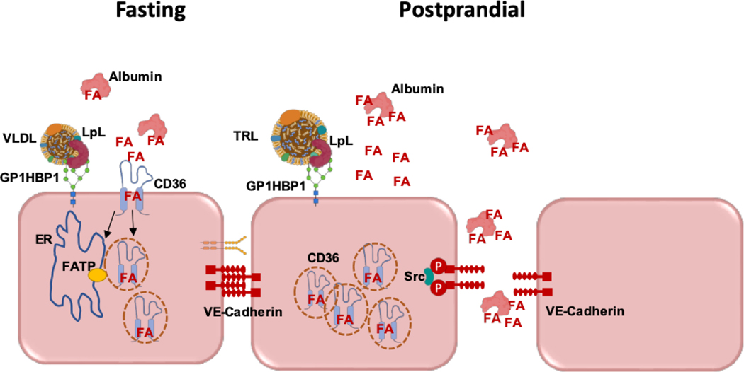
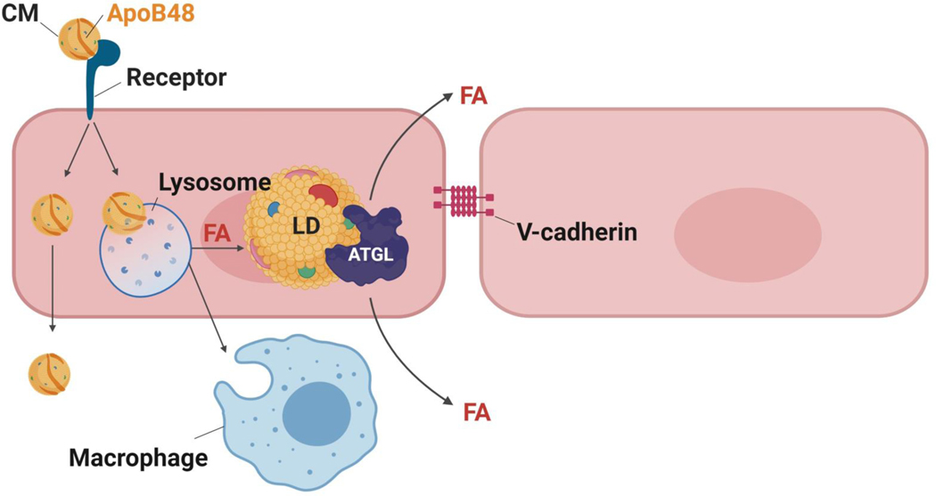
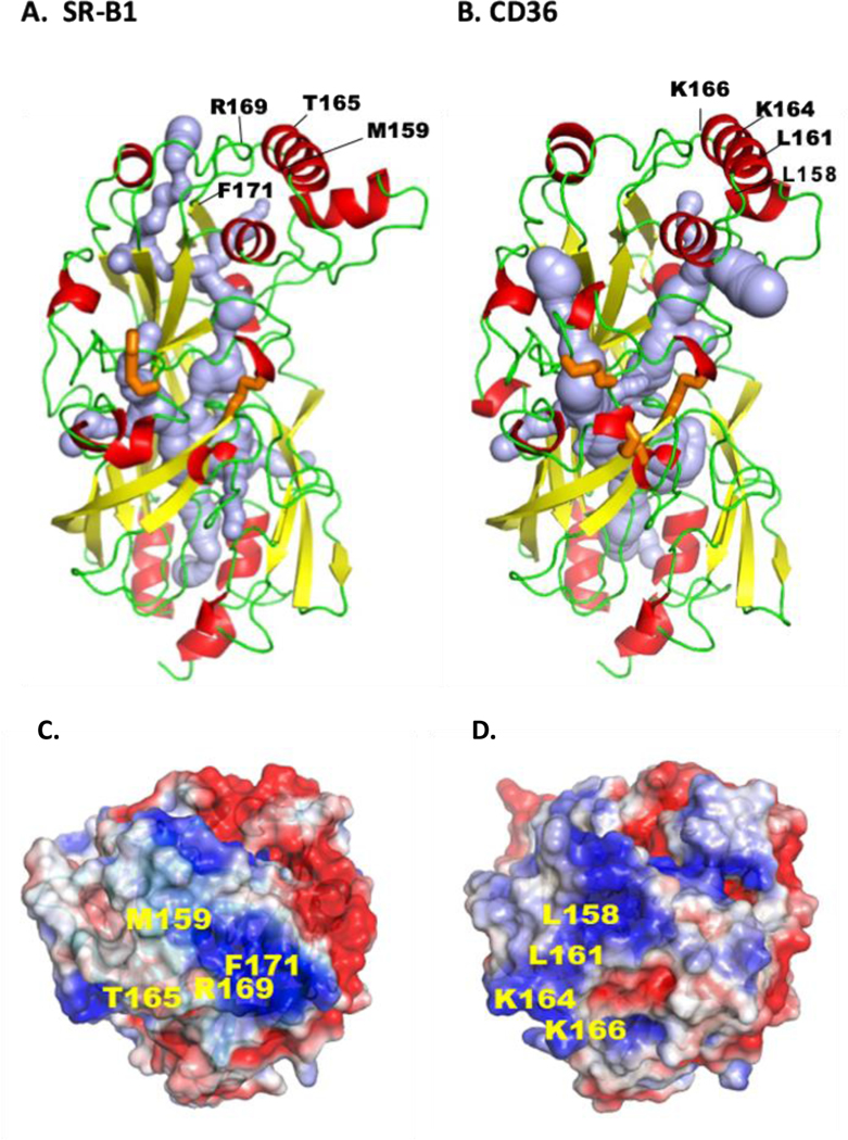
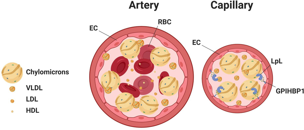
Lipid uptake and metabolism are central to the function of organs such as heart, skeletal muscle, and adipose tissue. Although most heart energy derives from fatty acids (FAs), excess lipid accumulation can cause cardiomyopathy. Similarly, high delivery of cholesterol can initiate coronary artery atherosclerosis. Hearts and arteries-unlike liver and adrenals-have nonfenestrated capillaries and lipid accumulation in both health and disease requires lipid movement from the circulation across the endothelial barrier. This review summarizes recent in vitro and in vivo findings on the importance of endothelial cell receptors and uptake pathways in regulating FAs and cholesterol uptake in normal physiology and cardiovascular disease. We highlight clinical and experimental data on the roles of ECs in lipid supply to tissues, heart, and arterial wall in particular, and how this affects organ metabolism and function. Models of FA uptake into ECs suggest that receptor-mediated uptake predominates at low FA concentrations, such as during fasting, whereas FA uptake during lipolysis of chylomicrons may involve paracellular movement. Similarly, in the setting of an intact arterial endothelial layer, recent and historic data support a role for receptor-mediated processes in the movement of lipoproteins into the subarterial space. We conclude with thoughts on the need to better understand endothelial lipid transfer for fuller comprehension of the pathophysiology of hyperlipidemia, and lipotoxic diseases such as some forms of cardiomyopathy and atherosclerosis.

Keywords: cardiomyopathy; chylomicrons; fatty acids; lipoprotein; triglyceride.


Figures

Figure 1.
Figure 1.. Pathways of FA transfer across ECs.
Left: The concentration of FAs is likely to modulate the pathway mediating their transit from the circulation to subendothelial cells. At low FA levels, as found during fasting, transfer is receptor-mediated involving cell surface CD36. FAs are internalized together with CD36, most likely in vesicles that might deliver the FA to the ER for activation by FATP3 or FATP4, for oxidation or incorporation into cell lipids. How and in what form the FA exits the basolateral EC surface to the subendothelial space is not known and is under investigation. Right: At higher FA concentrations such as locally occurs during lipolysis of triglyceride-rich lipoproteins (TRLs), EC CD36 will be saturated, being mostly internalized and cellular FA uptake would mainly occur via another pathway that we hypothesize involves paracellular flux between ECs. Activation of paracellular flux might occur by promoting loosening of EC junctions through phosphorylation of VE-cadherin by Src, protein kinase C or other kinases influenced by FAs. These processes are likely restricted to capillaries, as ECs of large arteries mostly do not express CD36, GPIHBP1 and do not have associated LpL.
Figure 2.
Figure 2.. Non-LpL mediated uptake of CMs and VLDL by ECs.
In capillaries, most FAs from TG are liberated by LpL, but with LpL deficiency or in arteries a novel pathway for CM uptake is likely to be present that does not involve CD36. In arteries, this additional pathway leads to uptake of lipids or chylomicrons to form intracellular lipid droplets.
Figure 3.
Figure 3.. Tertiary structures of CD36 and SR-B1.
Both receptors are members of the CD36 family that also includes LIMP2, and both play important roles in endothelial lipid transport. The comparison is based on the crystal structures. The ectodomain of human CD36 (A) is derived from its crystallography and homology modeling of human SR-BI (B). For both proteins the α-helices are in red, the β-sheets in yellow and the loops in green. Crystal structure identified a hydrophobic internal lipid transport tunnel (light blue) that traverses the length of these proteins ending at the bilayer proximity. This tunnel can be accessed from the apex region and residues identified to be important for ligand binding are highlighted. C and D show the apex regions’ electrostatic surface potential for SR-B1 (C) and CD36 (D) and residues implicated in binding of LDL and HDL (SR-B1), and FA and oxidized-LDL (CD36) are indicated. The tunnel is surrounded by short α-helices and the disulfide bridges (orange) would stabilize structure and dimensions of the cavity for it to accommodate the lipid ligands. E shows linear sequence of the C-terminal domains for SR-B1 and CD36 highlighting the PDZ binding sequence (red) of SR-B1 and the palmitoylated (green) and ubiquitinated (orange) residues of CD36 important for signaling. The tertiary structures were rendered in PyMol211, cavity prediction used CaverWeb 1.0212 and the electrostatic surface potential map used APBS212 with color gradient of −2 (red) to +2 (blue) kT/e.
Figure 4.
Figure 4.. Lipoprotein interaction with endothelial cells differs in arteries and capillaries.
A. In an artery, the rapid blood flow likely causes margination of larger particles, leading to a gradient with more chylomicrons in proximity of the arterial wall. B. This gradient is less likely in capillaries, still chylomicrons would still interact more with lipoprotein lipase (LpL) than the smaller VLDL.

References

    1. Duncan JG, Bharadwaj KG, Fong JL, Mitra R, Sambandam N, Courtois MR, Lavine KJ, Goldberg IJ and Kelly DP. Rescue of cardiomyopathy in peroxisome proliferator-activated receptor-alpha transgenic mice by deletion of lipoprotein lipase identifies sources of cardiac lipids and peroxisome proliferator-activated receptor-alpha activators. Circulation. 2010;121:426–35. - PMC - PubMed
    1. Yang J, Sambandam N, Han X, Gross RW, Courtois M, Kovacs A, Febbraio M, Finck BN and Kelly DP. CD36 deficiency rescues lipotoxic cardiomyopathy. Circ Res. 2007;100:1208–17. - PubMed
    1. Sletten AC, Peterson LR and Schaffer JE. Manifestations and mechanisms of myocardial lipotoxicity in obesity. J Intern Med. 2018;284:478–491. - PMC - PubMed
    1. Glatz JFC, Zuurbier CJ and Larsen TS. Targeting metabolic pathways to treat cardiovascular diseases. Biochim Biophys Acta Mol Basis Dis. 2020;1866:165879. - PubMed
    1. Goldberg IJ, Eckel RH and Abumrad NA. Regulation of fatty acid uptake into tissues: lipoprotein lipase- and CD36-mediated pathways. J Lipid Res. 2009;50 Suppl:S86–90. - PMC - PubMed

Publication types