. 2021 Feb 5;128(3):335-357.
doi: 10.1161/CIRCRESAHA.120.316738. Epub 2020 Dec 2.

KLF5 Is Induced by FOXO1 and Causes Oxidative Stress and Diabetic Cardiomyopathy

Affiliations

KLF5 Is Induced by FOXO1 and Causes Oxidative Stress and Diabetic Cardiomyopathy

Ioannis D Kyriazis et al. Circ Res. .

Abstract

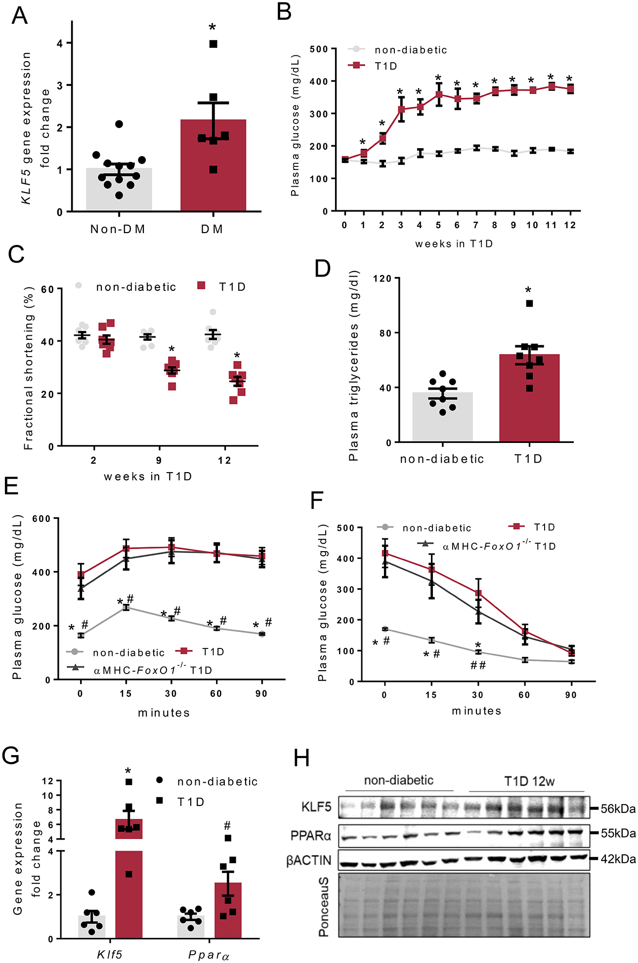
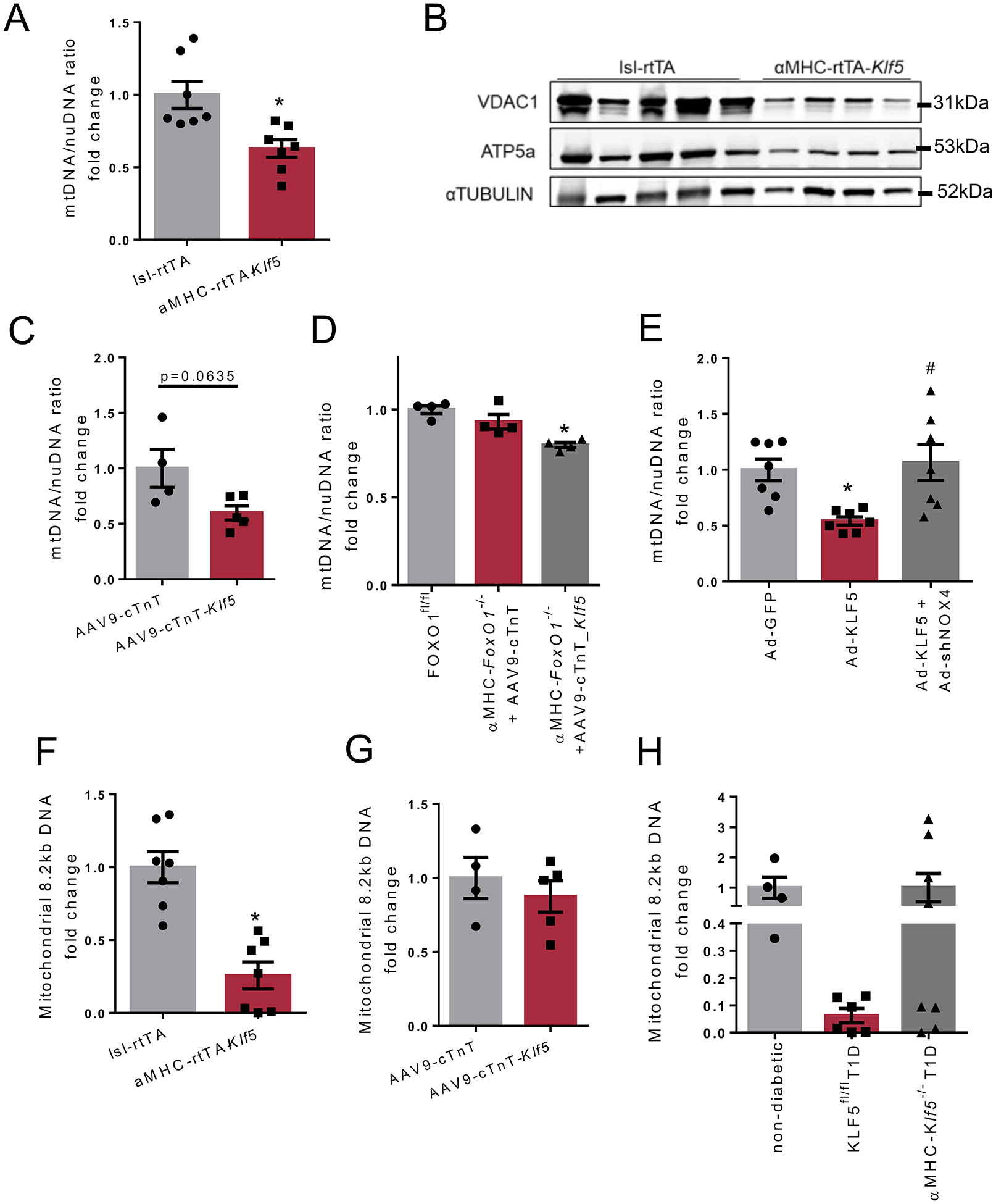
Rationale: Diabetic cardiomyopathy (DbCM) is a major complication in type-1 diabetes, accompanied by altered cardiac energetics, impaired mitochondrial function, and oxidative stress. Previous studies indicate that type-1 diabetes is associated with increased cardiac expression of KLF5 (Krüppel-like factor-5) and PPARα (peroxisome proliferator-activated receptor) that regulate cardiac lipid metabolism.

Objective: In this study, we investigated the involvement of KLF5 in DbCM and its transcriptional regulation.

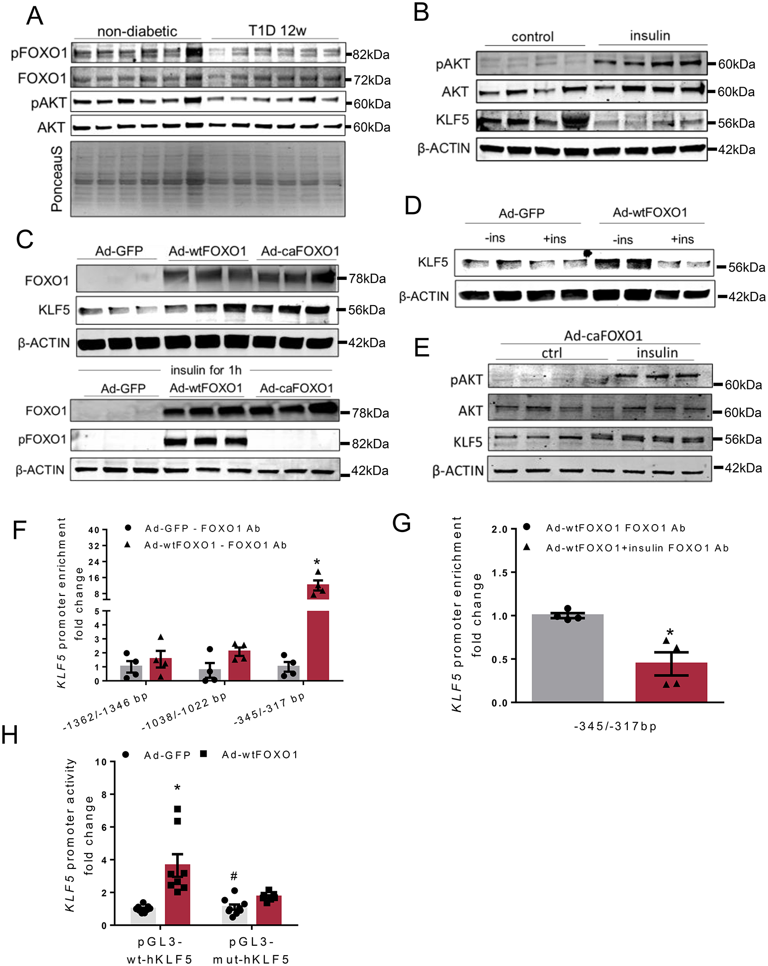
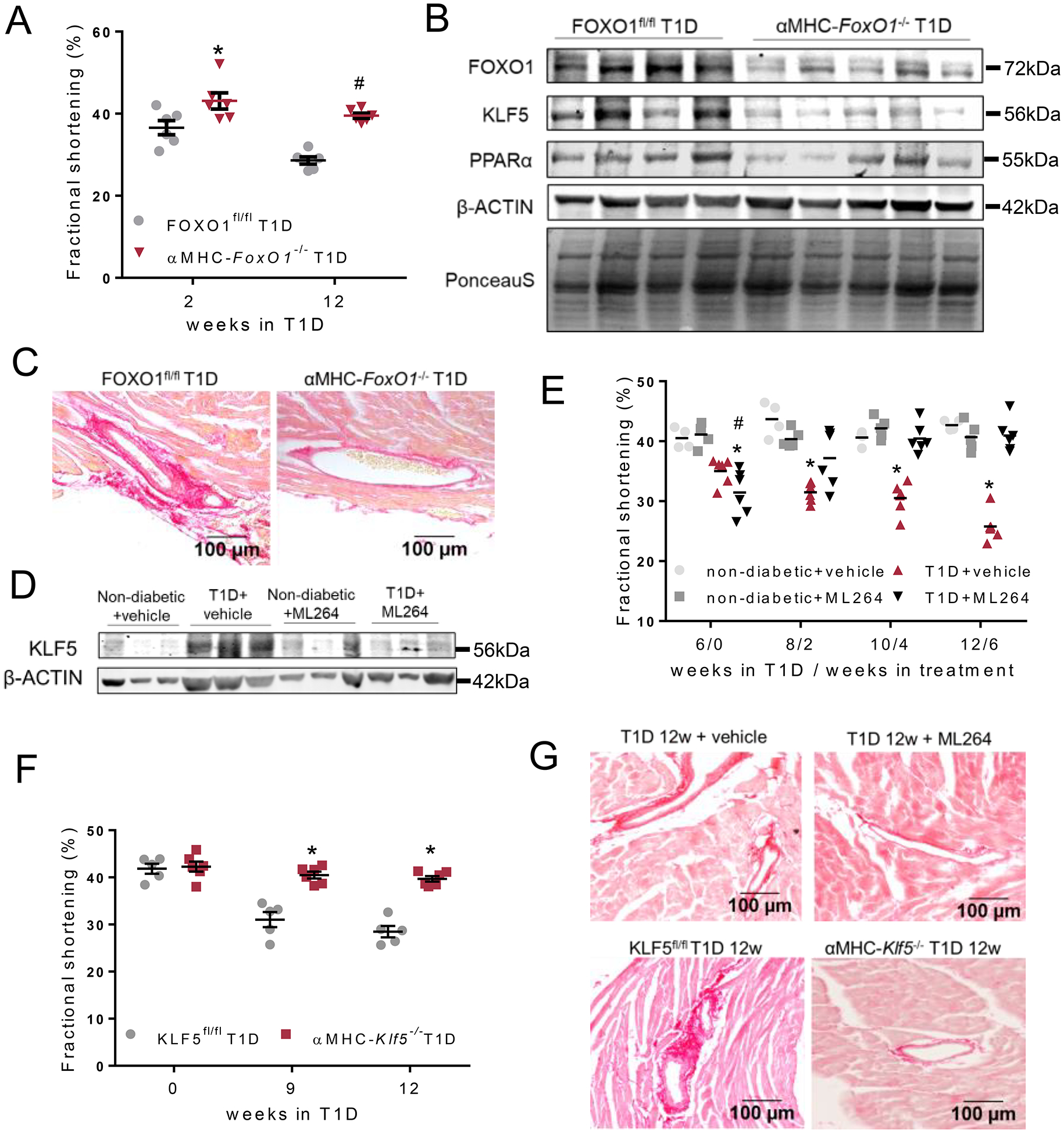
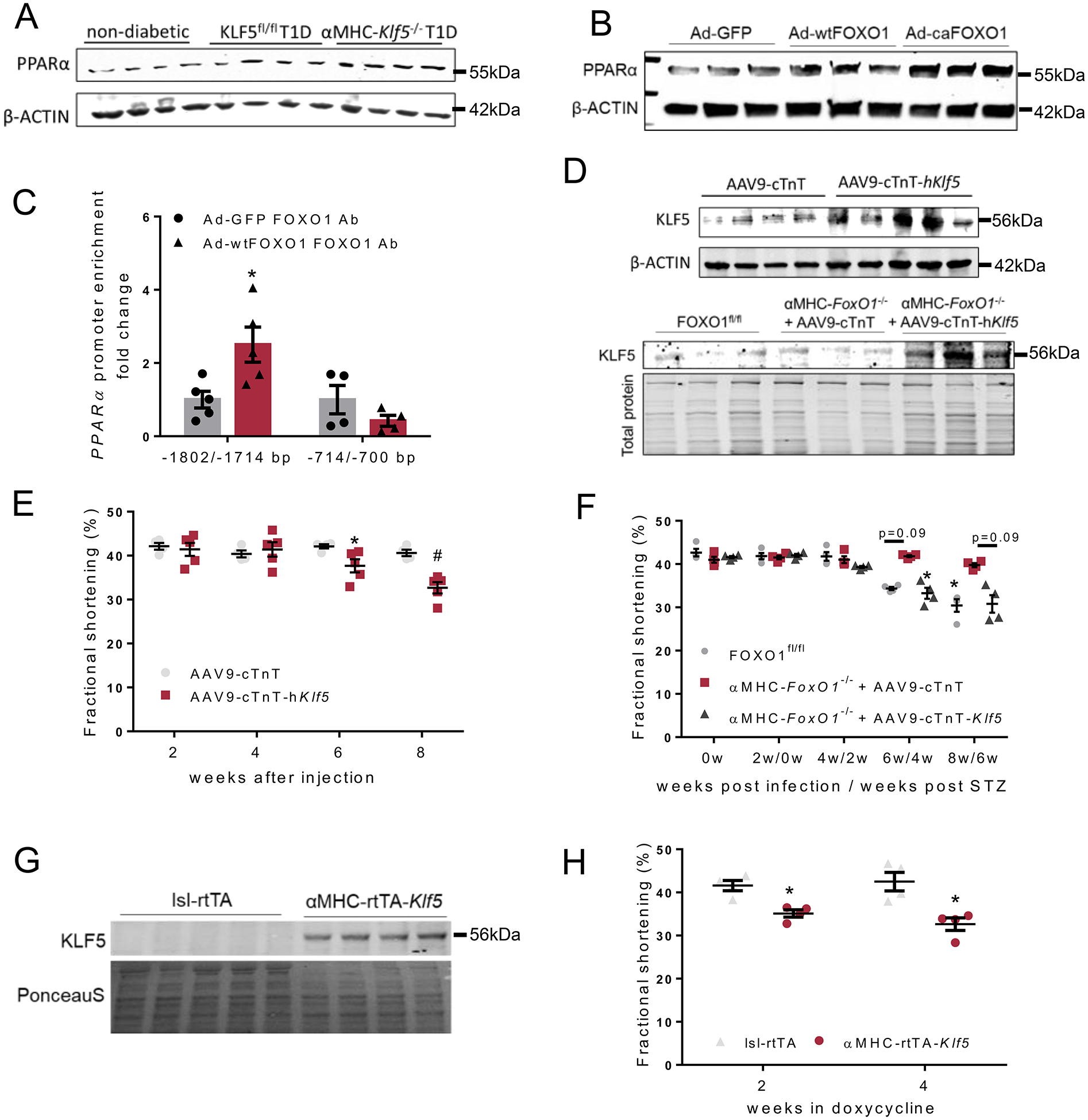
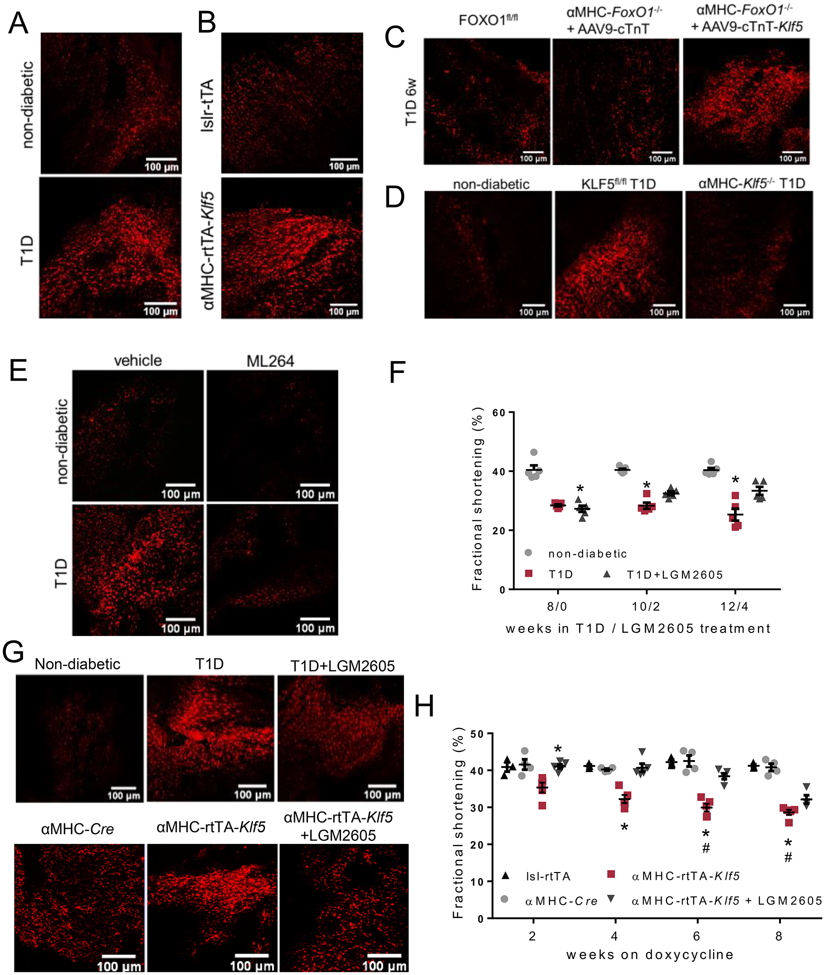
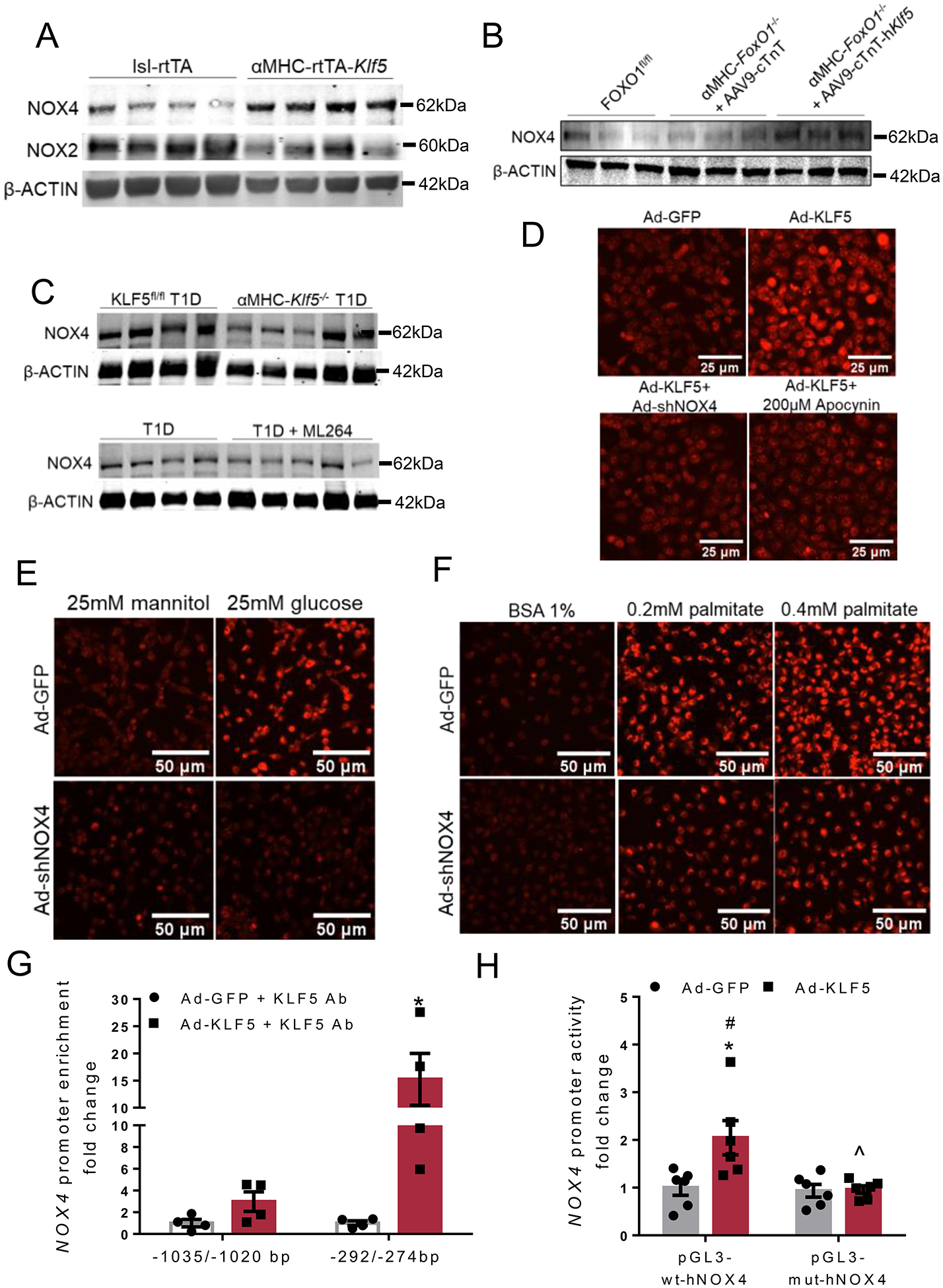
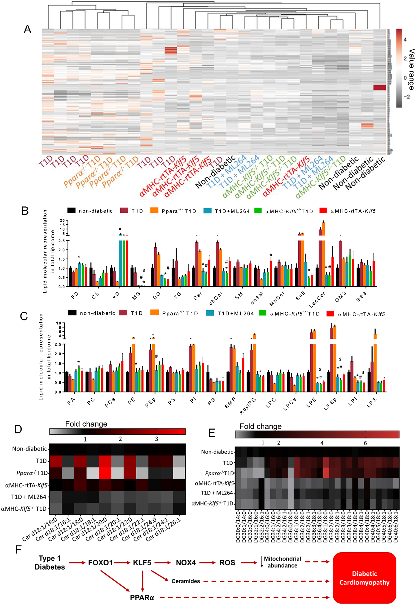
Methods and results: KLF5 mRNA levels were assessed in isolated cardiomyocytes from cardiovascular patients with diabetes and were higher compared with nondiabetic individuals. Analyses in human cells and diabetic mice with cardiomyocyte-specific FOXO1 (Forkhead box protein O1) deletion showed that FOXO1 bound directly on the KLF5 promoter and increased KLF5 expression. Diabetic mice with cardiomyocyte-specific FOXO1 deletion had lower cardiac KLF5 expression and were protected from DbCM. Genetic, pharmacological gain and loss of KLF5 function approaches and AAV (adeno-associated virus)-mediated Klf5 delivery in mice showed that KLF5 induces DbCM. Accordingly, the protective effect of cardiomyocyte FOXO1 ablation in DbCM was abolished when KLF5 expression was rescued. Similarly, constitutive cardiomyocyte-specific KLF5 overexpression caused cardiac dysfunction. KLF5 caused oxidative stress via direct binding on NADPH oxidase (NOX)4 promoter and induction of NOX4 (NADPH oxidase 4) expression. This was accompanied by accumulation of cardiac ceramides. Pharmacological or genetic KLF5 inhibition alleviated superoxide formation, prevented ceramide accumulation, and improved cardiac function in diabetic mice.

Conclusions: Diabetes-mediated activation of cardiomyocyte FOXO1 increases KLF5 expression, which stimulates NOX4 expression, ceramide accumulation, and causes DbCM.

Keywords: NADPH oxidases; ceramides; diabetic cardiomyopathy; forkhead box protein O1; oxidative stress; peroxisome proliferator-activated receptors.


Conflict of interest statement

DISCLOSURES

MCS has patents No. US 10,045,951 B2, No. US 10,030,040 B2, and No. US 9,987,321 B2 issued, patents No. PCT/US2016/049780, No. PCT/US17/35960, and No. PCT/US2008/006694 pending, and has a founder’s equity position in LignaMed, LLC. All other authors have no competing interests or conflicts to report.

Figures

Fig. 1:
Fig. 1:
A: KLF5 gene expression in cardiomyocytes of aortic stenosis patients with diabetes or without diabetes. B-C: Plasma glucose levels (B) and fractional shortening (C) in non-diabetic and type-1 diabetic C57Bl/6 mice. D: Plasma triglyceride levels in non-diabetic and diabetic C57BL/6 mice 12 weeks post-STZ administration. E, F: Plasma glucose levels in non-diabetic and diabetic FOXO1fl/fl and diabetic αMHC-FoxO1−/− mice 12 weeks post-STZ administration that were subjected to glucose (E) or insulin (F) tolerance test. G: Cardiac KLF5 and PPARα mRNA levels in non-diabetic and diabetic C57BL/6 mice, 12 weeks post-STZ administration. H: Immunoblots of KLF5, PPARα, β-ACTIN and PonceauS staining (analysis shown in Online Figure I-F), in cardiac protein lysates from non-diabetic and diabetic C57BL/6 mice, 12 weeks post-STZ injections.
Fig. 2.
Fig. 2.
A: Immunoblots of cardiac pSer256-FOXO1, total FOXO1, pSer473-AKT and total AKT and PonceauS staining in non-diabetic and diabetic C57BL/6 mice, 12 weeks post-STZ administration (analysis shown in Online Figure III-A). B: Immunoblots of pSer473-AKT, total AKT, KLF5 and β-ACTIN proteins from control and insulin-stimulated AC16 cells (analysis shown in Online Figure IV-A). C: Immunoblots of total FOXO1, pSer256-FOXO1 and β-ACTIN proteins from AC16 cells treated with adenoviruses expressing GFP, wtFOXO1 or caFOXO1 without (analysis shown in Online Figure IV-C) or with insulin treatment. D: Immunoblots of KLF5 and β-ACTIN proteins from AC16 cells treated with adenoviruses expressing GFP and wtFOXO1 without or with insulin stimulation (analysis shown in Online Figure IV-F). E: Immunoblots of pSer473-AKT, total AKT, KLF5 and β-ACTIN proteins from AC16 cells treated with adenovirus expressing caFOXO1 (analysis shown in Online Figure IV-G) without or with insulin treatment. F: FOXO1 enrichment in KLF5 promoter sequence fragments precipitated with FOXO1 antibody from AC16 cells expressing GFP or wtFOXO1. G:FOXO1 enrichment in KLF5 promoter sequence fragments precipitated with FOXO1 antibody from AC16 cells expressing wtFOXO1 and treated with insulin. H: Luciferase activity normalized to firefly from AC16 cells transfected with plasmids containing the wild type KLF5 −1757/−263bp promoter fragment (pGL3BV-wt-hKLF5) or the mutant [-1757/-263bp](-342/-317bp)N→A (pGL3BV-mut-hKLF5) and infected with adenoviruses expressing GFP or wtFOXO1.
Fig. 3.
Fig. 3.
A: Left ventricular fractional shortening of diabetic FOXO1fl/fl and αMHC-FoxO1−/− mice 2 and 12 weeks post-STZ. B: Immunoblots of FOXO1, KLF5, PPARα, β-ACTIN protein and PonceauS staining in hearts obtained from diabetic FOXO1fl/fl and αMHC-FoxO1−/− mice 12 weeks post-STZ administration (analysis shown in Online Figure VII-K). C: Representative images of picrosirius red staining (20X magnification) of cardiac tissue sections of diabetic FOXO1fl/fl and αMHC-FoxO1−/− mice. Additional images and quantification in Online Figures IX-A,B. D: Immunoblot of cardiac KLF5 and β-ACTIN proteins in non-diabetic treated with 6 weeks with vehicle or ML264 or diabetic C57BL/6 mice treated with vehicle or ML264 starting 6 weeks post-STZ administration (analysis shown in Online Figure X-A). E: Left ventricular fractional shortening of diabetic or control non-diabetic mice both treated with either vehicle or ML264 for 6 weeks starting 6 weeks post-STZ administration. F: Left ventricular fractional shortening of diabetic KLF5fl/fl and αMHC-Klf5−/− mice 9 weeks and 12 weeks post-STZ administration. G: Picrosirius red staining and perivascular fibrosis analysis of cardiac tissue from diabetic C57BL/6 mice treated with ML264 or control vehicle and diabetic KLF5fl/fl or αMHC-Klf5−/− mice 12 weeks post-STZ administration. Additional images and quantification analysis in Online Figures XII-A,B or Online Figures XIII–A,B.
Fig. 4.
Fig. 4.
A: Immunoblots of cardiac PPARα and β-ACTIN proteins in non-diabetic, diabetic KLF5fl/fl and diabetic αMHC-Klf5−/− mice 12 weeks post-STZ administration (analysis shown in Online Figure XVII). B: Immunoblots of PPARα and β-ACTIN proteins in AC16 cells infected with adenoviruses expressing GFP, wtFOXO1 or caFOXO1 (analysis shown in Online Figure XIX–B). C: FOXO1 enrichment in PPARα promoter sequence fragments precipitated with FOXO1 antibody from AC16 cells expressing GFP or wtFOXO1. D: Immunoblot of cardiac KLF5 and β-ACTIN proteins in C57BL/6 mice infected with AAV9-cTnT or AAV9-cTnT-hKlf5 (analysis shown in Online Figure XXIV–A), and immunoblot of cardiac KLF5 and total protein stain of diabetic FOXO1fl/fl mice and diabetic αMHC-FoxO1−/− mice infected with control AAV9-cTnT or AAV9-cTnT-hKlf5 (analysis shown in Online Figure XXIV–B). E: Left ventricular fractional shortening of C57BL/6 mice infected with AAV9-cTnT or AAV9-cTnT-hKlf5 2, 4, 6 and 8 weeks post-AAV9 administration. F: Left ventricular fractional shortening of diabetic FOXO1fl/fl mice and diabetic αMHC-FoxO1−/− mice infected with AAV9-cTnT or AAV9-cTnT-hKlf5. G: Immunoblot of cardiac KLF5 and PonceauS staining of lsl-rtTA-TRE-Klf5 and αMHC-rtTA-Klf5 mice fed on doxycycline enriched diet for 2 weeks (analysis shown in Online Figure XXV–D). H: Left ventricular fractional shortening of lsl-rtTA-TRE-Klf5 and αMHC-rtTA-Klf5 mice fed on doxycycline enriched diet for 2 and 4 weeks.
Fig. 5.
Fig. 5.
A-E: Representative DHE fluorescence images in fresh cardiac tissue of non-diabetic C57BL/6 and diabetic C57BL/6 (A) mice 12 weeks post-STZ, lsl-rtTA-TRE-Klf5 and αMHC-rtTA-Klf5 mice fed on doxycycline-enriched diet for 10 days (B), diabetic FOXO1fl/fl mice and diabetic αMHC-FoxO1−/− mice infected with AAV9-cTnT or AAV9-cTnT-hKlf5 (C), non-diabetic C57BL/6, diabetic KLF5fl/fl and diabetic αMHC-Klf5−/− mice (D), non-diabetic and diabetic mice treated with ML264 or vehicle (E). F: Left ventricular fractional shortening of non-diabetic C57BL/6, diabetic C57BL/6 and diabetic C57BL/6 mice treated with LGM2605. G: Representative DHE fluorescence images in fresh cardiac tissue of non-diabetic C57BL/6, diabetic C57BL/6 and diabetic C57BL/6 mice treated with LGM2605 and doxycycline-fed αMHC-Cre, αMHC-rtTA-Klf5 mice treated with LGM2605 or control saline. H: Left ventricular fractional shortening of doxycycline-fed lsl-rtTA mice, αMHC-Cre, αMHC-rtTA-Klf5 mice treated with LGM2605 or control saline.
Fig. 6.
Fig. 6.
A-C: Immunoblots of cardiac NOX4 (A, B, C), NOX2 (A) and β-ACTIN (A, B, C) proteins of lsl-rtTA-TRE-Klf5 and αMHC-rtTA-Klf5 mice fed on doxycycline-enriched diet for 10 days (A; analysis shown in Online Figures XXXIII–F,G), diabetic FOXO1fl/fl mice and diabetic αMHC-FoxO1−/− mice infected with AAV9-cTnT or AAV9-cTnT-hKlf5 (B; analysis shown in Online Figure XXXIII–H), diabetic KLF5fl/fl or αMHC-Klf5−/− mice (C; analysis shown in Online Figure XXXIII–I), and diabetic mice treated with ML264 or control vehicle (C; analysis shown in Online Figure XXXIII–J). D-F: Representative DHE fluorescence images (D, E, F) of AC16 cells infected with adenovirus expressing GFP, KLF5, KLF5+shNOX4 or KLF5 followed by treatment with apocynin (D), AC16 cells infected with adenovirus expressing GFP and shNOX4 treated with 25mM mannitol or 25mM glucose (E), and AC16 cells infected with adenovirus expressing GFP and shNOX4 and treated with 0.2mM or 0.4mM palmitate (PA) dissolved in 1% BSA fraction V (F). G:KLF5 enrichment in NOX4 promoter sequence fragments precipitated with KLF5 antibody from AC16 cells expressing GFP or KLF5. H: Luciferase activity normalized to firefly in AC16 cells transfected with reporter plasmids containing the wild type NOX4 −1546/+54bp promoter fragment (pGL3-wt-hNOX4) or the mutant [−1547/+54bp](-282/-276bp)N→A (pGL3-mut-hNOX4) and infected with adenoviruses expressing GFP or KLF5.
Fig. 7.
Fig. 7.
A: Mitochondrial DNA/nuclear DNA ratio in cardiac tissue of lsl-rtTA-TRE-Klf5 and αMHC-rtTA-Klf5 mice fed on doxycycline-enriched diet for 10 days. B: Immunoblots of cardiac VDAC1, ATP5a and α-TUBULIN in lsl-rtTA-TRE-Klf5 and αMHC-rtTA-Klf5 mice fed on doxycycline-enriched diet for 10 days (analysis shown in Online Figures XXXVII–H,I). C-E: Mitochondrial DNA/nuclear DNA ratio in cardiac tissue of C57BL/6 mice infected with AAV9-cTnT or AAV9-cTnT-hKlf5 (C), diabetic FOXO1fl/fl mice and diabetic αMHC-FoxO1−/− mice infected with AAV9-cTnT or AAV9-cTnT-hKlf5 (D), AC16 cells infected with adenovirus expressing GFP, KLF5 or KLF5+shNOX4 (E). F-H: Amplification of mitochondrial 8.2kb DNA sequence normalized to mitochondrial abundance of lsl-rtTA-TRE-Klf5 and αMHC-rtTA-Klf5 mice fed on doxycycline-enriched diet for 10 days (F), C57BL/6 mice infected with AAV9-cTnT or AAV9-cTnT-hKlf5 (G), and non-diabetic C57BL/6, diabetic KLF5fl/fl and diabetic αMHC-Klf5−/− mice (H).
Fig. 8.
Fig. 8.
A-C: Heatmap (A) with Euclidean hierarchical clustering of lipid species (lipid species that are listed on the right side of the heat map are described in Online Table XVII). Cardiac lipid species representation to total lipidome (presented as fold-change compared to non-diabetic wt mice) in non-diabetic C57BL/6 mice, diabetic C57BL/6 mice, diabetic Pparα−/− mice, diabetic C57BL/6 mice treated with ML264, diabetic aMHC-Klf5−/− mice and aMHC-rtTA-Klf5 mice fed on doxycycline-enriched diet for 10 days; FC; free cholesterol, CE; cholesterol ester, AC; acylcarnitines, MG; monoglycerides, DG; diacylglycerols, TG; triglycerides, Cer; ceramides, dhCer; dihydroceramides, SM; sphingomyelins, dhSM; dihydrosphingomyelins, MhCer; monohexosylceramides, Sulf; sulfatides, LacCer; lactosylceramides, GM3; gangliosides, GB3; globotrioseacylceramides) (B) and PA; phosphatidic acids, PC; phosphatidylcholines, PCe; phosphatidylcholine ethers, PE; phosphatidylethanolamines, PEp; plasmalogen phosphatidylethanolamines, PS; phosphatidylserines, PI; phosphatidylinositols, PG; phosphatidylglycerols, BMP; bis(monoacylglycero)phosphates, AcylPG; acylphosphatidylglycerols, LPC; lysophosphosphatidylcholines, LPCe; lysophosphosphatidylcholine ethers, LPE; lysophosphatidylethanolamines, LPEp; plasmalogen lysophosphatidylethanolamines, LPI; lysophosphatidylinositols and LPS; lysophosphatidylserines (C). D-E; Heatmaps of ceramides family (D) and diacylglycerol family (E) in non-diabetic C57BL/6 mice, diabetic C57BL/6 mice, diabetic Pparα−/− mice, diabetic C57BL/6 mice treated with ML264, diabetic αMHC-Klf5−/− mice, and αMHC-rtTA-Klf5 mice fed on doxycycline-enriched diet for 10 days. F: Schematic representation of the proposed model about pathways that are involved in diabetic cardiomyopathy.

Comment in

References

    1. Ogurtsova K, da Rocha Fernandes JD, Huang Y, Linnenkamp U, Guariguata L, Cho NH, Cavan D, Shaw JE, Makaroff LE. Idf diabetes atlas: Global estimates for the prevalence of diabetes for 2015 and 2040. Diabetes Res Clin Pract. 2017;128:40–50 - PubMed
    1. Lee MMY, McMurray JJV, Lorenzo-Almoros A, Kristensen SL, Sattar N, Jhund PS, Petrie MC. Diabetic cardiomyopathy. Heart. 2018 - PubMed
    1. Zinman B, Wanner C, Lachin JM, Fitchett D, Bluhmki E, Hantel S, Mattheus M, Devins T, Johansen OE, Woerle HJ, Broedl UC, Inzucchi SE, Investigators E-RO. Empagliflozin, cardiovascular outcomes, and mortality in type 2 diabetes. N Engl J Med. 2015;373:2117–2128 - PubMed
    1. Nolan CJ, Ruderman NB, Kahn SE, Pedersen O, Prentki M. Insulin resistance as a physiological defense against metabolic stress: Implications for the management of subsets of type 2 diabetes. Diabetes. 2015;64:673–686 - PMC - PubMed
    1. Bayeva M, Sawicki KT, Ardehali H. Taking diabetes to heart--deregulation of myocardial lipid metabolism in diabetic cardiomyopathy. J Am Heart Assoc. 2013;2:e000433. - PMC - PubMed

Publication types

MeSH terms