. 2021 Feb 2;22(3):1477.
doi: 10.3390/ijms22031477.

CLytA-DAAO Chimeric Enzyme Bound to Magnetic Nanoparticles. A New Therapeutical Approach for Cancer Patients?

Affiliations

CLytA-DAAO Chimeric Enzyme Bound to Magnetic Nanoparticles. A New Therapeutical Approach for Cancer Patients?

María Fuentes-Baile et al. Int J Mol Sci. .

Abstract

D-amino acid oxidase (DAAO) is an enzyme that catalyzes the oxidation of D-amino acids generating H2O2. The enzymatic chimera formed by DAAO bound to the choline-binding domain of N-acetylmuramoyl-L-alanine amidase (CLytA) induces cytotoxicity in several pancreatic and colorectal carcinoma and glioblastoma cell models. In the current work, we determined whether the effect of CLytA-DAAO immobilized in magnetic nanoparticles, gold nanoparticles, and alginate capsules offered some advantages as compared to the free CLytA-DAAO. Results indicate that the immobilization of CLytA-DAAO in magnetic nanoparticles increases the stability of the enzyme, extending its time of action. Besides, we compared the effect induced by CLytA-DAAO with the direct addition of hydrogen peroxide, demonstrating that the progressive generation of reactive oxygen species by CLytA-DAAO is more effective in inducing cytotoxicity than the direct addition of H2O2. Furthermore, a pilot study has been initiated in biopsies obtained from pancreatic and colorectal carcinoma and glioblastoma patients to evaluate the expression of the main genes involved in resistance to CLytA-DAAO cytotoxicity. Based on our findings, we propose that CLytA-DAAO immobilized in magnetic nanoparticles could be effective in a high percentage of patients and, therefore, be used as an anti-cancer therapy for pancreatic and colorectal carcinoma and glioblastoma.

Keywords: alginate capsules; cytotoxicity; enzymatic therapy; gold nanoparticle; hydrogen peroxide; magnetic nanoparticle; oxidative stress; reactive oxygen species.


Conflict of interest statement

The authors declare no conflict of interest.

Figures

Figure 1
Figure 1
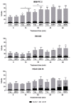
Cell death induced by CLytA-DAAO free, immobilized in magnetic nanoparticles (MNPs), in gold nanoparticles (GNPs), and immobilized in alginate capsules. (A) IMIM-PC-2 pancreatic carcinoma cell line was treated with 2 U/mL CLytA-DAAO, free or immobilized in MNPs, GNPs or alginate capsules, and 1 mM D-Ala for 24 h. (B) SW-480 colorectal carcinoma cell line and HGUE-GB-18 and HGUE-GB-37 glioblastoma cell lines were treated with 2 U/mL CLytA-DAAO, free or immobilized in MNPs, and 1 mM D-Ala for 24 h. Cell viability was determined using Muse cell analyzer. Graphs represent cell death percentage (mean ± SD) after subtracting cell death in the control untreated (n ≥ 3). (C) IMIM-PC-2 cell line was treated with CLytA-DAAO bound to MNPs in a concentration range between 0.05–5 U/mL for 72 h. Graph shows the proliferation percentage ± SD normalized with respect to the control untreated (n ≥ 6). (D) IMIM-PC-2 cell line was treated with 5 or 10 U/mL CLytA-DAAO immobilized in magnetic nanoparticles for 24 h and the cell distribution in each phase of cell cycle was determined by flow cytometry. Graph shows the cell percentage in each phase of cell cycle (mean ± SD) (n ≥ 3). ** Indicates a p-value < 0.01 and *** p-value < 0.001.
Figure 2
Figure 2
Anti-proliferative effect of CLytA-DAAO, free and bound to MNPs, on pancreatic carcinoma, colorectal carcinoma, and glioblastoma cell lines. Cells were treated with CLytA-DAAO, free or immobilized, in a concentration range between 0.025-2 U/mL and 1 mM D-Ala for 72 h and cell proliferation was determined by MTT assay. Graph shows the proliferation percentage ± SD respect to control untreated versus the logarithm of the concentration (n ≥ 6).
Figure 3
Figure 3
Anti-proliferative effect of CLytA-DAAO, free and bound to MNPs, on pancreatic and colorectal carcinoma cell lines. Cells were treated with CLytA-DAAO, free or immobilized, in a concentration range between 0.025-2 U/mL and 1 mM D-Ala for 24 h and cell proliferation was determined by MTT assay. Graph shows the proliferation percentage ± SD respect to control untreated versus the logarithm of the concentration (n ≥ 6).
Figure 4
Figure 4
Intracellular reactive oxygen species (ROS) increase after CLytA-DAAO, free and bound to MNPs, and D-Ala treatment in IMIM-PC-2 pancreatic carcinoma cell line, SW-480 colorectal carcinoma cell line, and HGUE-GB-37 glioblastoma cell line. Cells were treated with 2 U/mL CLytA-DAAO and 1 mM D-Ala for 20–120 min. Free radical production was determined through DCFH2-DA probe and each treatment time had a control untreated that only contained the probe. Graph shows the fold change (FC) ± SD of fluorescent intensity with respect to the control (n ≥ 6). * Indicates a p-value < 0.05 and *** < 0.001.
Figure 5
Figure 5
Cells accumulation in subG1 and G2/M phases after a CLytA-DAAO, free or bound to MNPs, and D-Ala treatment in IMIM-PC-2 pancreatic carcinoma cell line, SW-480 colorectal carcinoma cell line, and HGUE-GB-18 glioblastoma cell line. Cells were treated with 2 U/mL CLytA-DAAO and 1 mM D-Ala for a short time (15–60 min) and then, treatment was removed, replacing the medium. Cells were incubated until 24 h from the treatment addition were completed. Graph shows the percentage of cells ± SD in subG1 and G2/M phases (n ≥ 3). * Indicates a p-value < 0.05, ** < 0.01 and *** < 0.001.
Figure 6
Figure 6
Cell death induced by CLytA-DAAO, free and bound to MNPs, and D-Ala after incubation of the enzyme at 37 °C in IMIM-PC-2 pancreatic carcinoma cell line, SW-480 colorectal carcinoma cell line, and HGUE-GB-37 glioblastoma cell line. Cells were treated with 2 U/mL CLytA-DAAO and 1 mM D-Ala for 24 h. The CLytA-DAAO used was pre-incubated at 37 °C for 30 min, 1, 2, and 3 h before adding it to the cells. Graph shows the percentage of cell death normalizing the treatment with CLytA-DAAO without pre-incubation as 100% ± SD (n ≥ 3). * Indicates a p-value < 0.05, ** < 0.01 and *** < 0.001.
Figure 7
Figure 7
CLytA-DAAO release from MNPs through the choline addition. (A) SW-480 cell line was treated with 1–100 mM choline for 72 h and cell proliferation was determined by MTT assay. (B) SW-480 cell line was treated with 2 U/mL CLytA-DAAO, 1 mM D-Ala and increasing concentrations of choline (0.1–50 mM) for 72 h and cell proliferation was determined by MTT assay. The CLytA-DAAO enzyme was used: free and bound to MNPs in combination with choline and bound to MNPs after a pre-incubation with choline for 10 min. Graphs show the proliferation percentage ± SD respect to control untreated (n ≥ 6). ** Indicates a p-value < 0.01 and *** < 0.001.
Figure 8
Figure 8
H2O2 effect in pancreatic carcinoma, colorectal carcinoma, and glioblastoma cell models. H2O2 anti-proliferative effect induced in pancreatic carcinoma (A), colorectal carcinoma (B) and glioblastoma (C) cell lines. Cells were treated with H2O2, in a concentration range between 50–600 μM for 72 h and cell proliferation was determined by MTT assay. Graph shows the proliferation percentage ± SD respect to control untreated versus the logarithm of the concentration (n ≥ 6). (D) Variations in cell cycle distribution after H2O2 treatment in Hs766T, IMIM-PC-2, and RWP-1 pancreatic carcinoma cell lines, SW-480 and SW-620 colorectal carcinoma cell lines and HGUE-GB-18, HGUE-GB-37, and HGUE-GB-39 glioblastoma cell lines. Cells were treated with 600 μM H2O2 for 24 h and cell cycle distribution was determined by flow cytometry. Graph shows the cells percentage ± SD in each phase of cell cycle after subtracting the cells percentage in the control untreated (n ≥ 3). * Indicates a p-value < 0.05, ** < 0.01 and *** < 0.001.
Figure 9
Figure 9
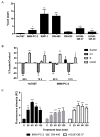
Differential effect of H2O2 with respect to CLytA-DAAO and D-Ala treatment. (A) Plasmatic membrane rupture induced by H2O2 in Hs766T, IMIM-PC-2, and RWP-1 cell lines from pancreatic carcinoma, SW-480 and SW-620 cell lines from colorectal carcinoma and HGUE-GB-37 and HGUE-GB-39 cell lines from glioblastoma. Cells were treated with 600 μM H2O2 for 24 h and cell viability was determined using Muse cell analyzer. Graph represents cell death percentage (mean ± SD) after subtracting cell death in the control untreated (n ≥ 3). (B) Variations in cell cycle distribution after H2O2 treatment for 48 and 72 h. Hs766T and IMIM-PC-2 pancreatic carcinoma cell lines were treated with 600 μM H2O2 for 48 and 72 h and cell cycle distribution was determined by flow cytometry. Graph shows the cells percentage ± SD in each phase of cell cycle after subtracting the cells percentage in the control untreated (n ≥ 3). (C) Intracellular ROS increase after H2O2 treatment in IMIM-PC-2 pancreatic carcinoma cell line, SW-480 colorectal carcinoma cell line and HGUE-GB-37 glioblastoma cell line. Cells were treated with 600 μM H2O2 for 20–120 min. Free radical production was determined through DCFH2-DA probe and each treatment time had a control untreated that only contained the probe. Graph shows the fold change (FC) ± SD of fluorescent intensity with respect to the control (n ≥ 6). * Indicates a p-value < 0.05, ** p-value < 0.01 and *** p-value < 0.001.
Figure 10
Figure 10
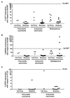
Gene expression in cell lines and patient biopsies from pancreatic carcinoma, colorectal carcinoma, and glioblastoma. Graphs show the expression percentage of CAT (A), NFE2L2 (B), and GPX2 (C). CAT and NFE2L2 expression are normalized with respect to the expression observed in Hs766T pancreatic carcinoma cell line and GPX2 is normalized with the expression observed in HT-29 colorectal carcinoma cell line (n ≥ 3).

References

    1. Illanes A., Cauerhff A., Wilson L., Castro G.R. Recent trends in biocatalysis engineering. Bioresour. Thechnol. 2012;115:48–57. doi: 10.1016/j.biortech.2011.12.050. - DOI - PubMed
    1. Ali Khan A., Alzohairy M.A. Recent Advances and Applications of Immobilized Enzyme Technologies: A Review. Res. J. Biol. Sci. 2010;5:565–575. doi: 10.3923/rjbsci.2010.565.575. - DOI
    1. Ansari S.A., Husain Q. Potential applications of enzymes immobilized on/in nano materials: A review. Biotechnol. Adv. 2012;30:512–523. doi: 10.1016/j.biotechadv.2011.09.005. - DOI - PubMed
    1. De la Rica R., Aili D., Stevens M.M. Enzyme-responsive nanoparticles for drug release and diagnostics. Adv. Drug Deliv. Rev. 2012;64:967–978. doi: 10.1016/j.addr.2012.01.002. - DOI - PubMed
    1. Koutsopoulos S. Molecular fabrications of smart nanobiomaterials and applications in personalized medicine. Adv. Drug Deliv. Rev. 2012;64:1459–1476. doi: 10.1016/j.addr.2012.08.002. - DOI - PubMed

MeSH terms