Review
. 2021 Feb 2;22(3):1499.
doi: 10.3390/ijms22031499.

Silk Fibroin as a Functional Biomaterial for Tissue Engineering

Affiliations
Review

Silk Fibroin as a Functional Biomaterial for Tissue Engineering

Weizhen Sun et al. Int J Mol Sci. .

Abstract

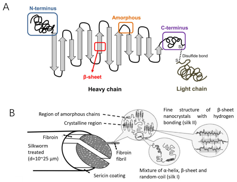
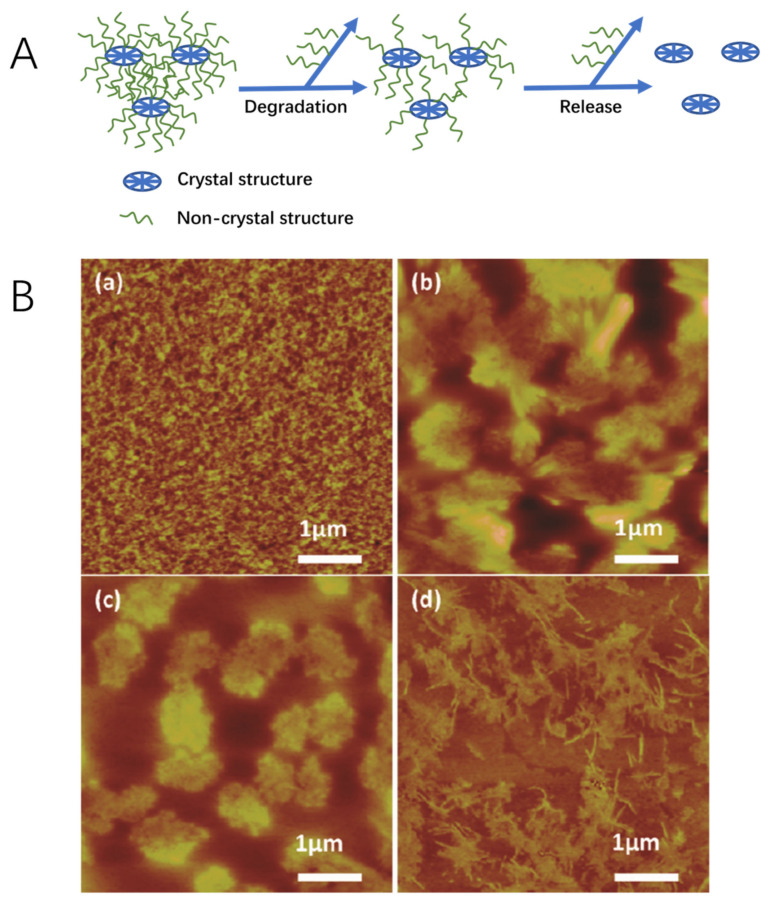
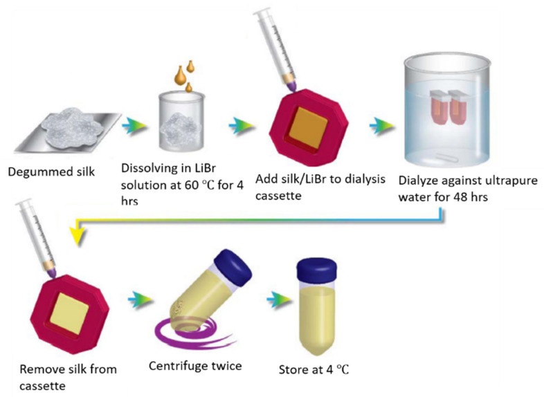
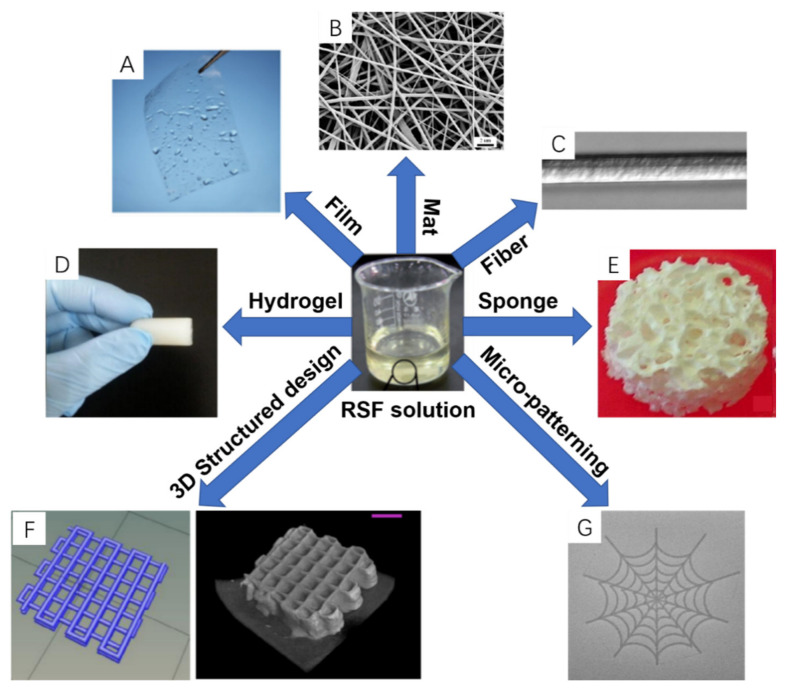
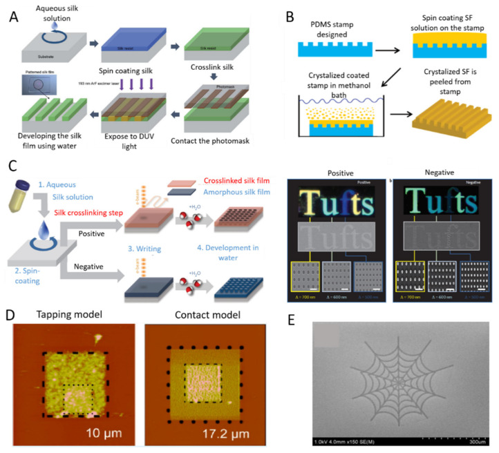
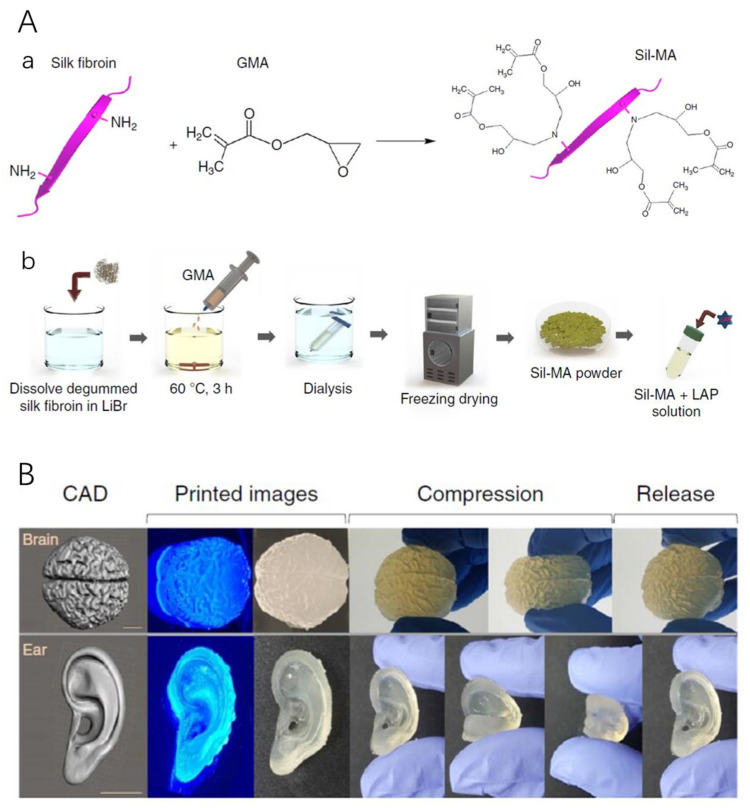
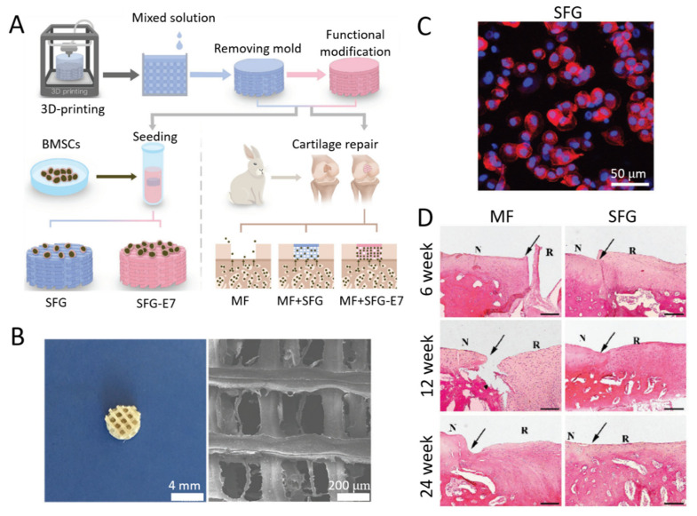
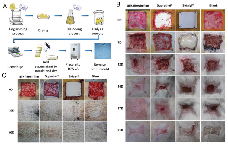
Tissue engineering (TE) is the approach to combine cells with scaffold materials and appropriate growth factors to regenerate or replace damaged or degenerated tissue or organs. The scaffold material as a template for tissue formation plays the most important role in TE. Among scaffold materials, silk fibroin (SF), a natural protein with outstanding mechanical properties, biodegradability, biocompatibility, and bioresorbability has attracted significant attention for TE applications. SF is commonly dissolved into an aqueous solution and can be easily reconstructed into different material formats, including films, mats, hydrogels, and sponges via various fabrication techniques. These include spin coating, electrospinning, freeze drying, physical, and chemical crosslinking techniques. Furthermore, to facilitate fabrication of more complex SF-based scaffolds with high precision techniques including micro-patterning and bio-printing have recently been explored. This review introduces the physicochemical and mechanical properties of SF and looks into a range of SF-based scaffolds that have been recently developed. The typical TE applications of SF-based scaffolds including bone, cartilage, ligament, tendon, skin, wound healing, and tympanic membrane, will be highlighted and discussed, followed by future prospects and challenges needing to be addressed.

Keywords: biomaterial; scaffold; silk fibroin; tissue engineering.


Conflict of interest statement

The authors declare no conflict of interest.

Figures

Figure 1
Figure 1
Schematic diagram of the silk structure. (A) heavy chain (i.e., N-terminus, β-sheets, Amorphous and C-terminus) and light chain which linked via disulphide bonds. Reproduced with permission from [40] (B) silkworm thread, fibril overall structure and silk fibroin polypeptide chains. reproduced with permission from [39].
Figure 2
Figure 2
(A) Schematic illustrating the SF degradation process mechanism. (B) Representative AFM images of (a) pure protease XIV solution and differently fabricated SF films: (b) slow drying process, (c) water annealing treatment, and (d) stretching treatment, after 12 h of exposure to protease XIV solution. The degraded SF particles that dissolved in protease XIV can be seen in (b), (c), and (d). Reproduced with permission from [82].
Figure 3
Figure 3
A schematical representation of the LiBr dissolution process to obtain RSF solution. The degummed silk is dissolved in 9.3 M LiBr solution at 60 °C for 4 h. The obtained solutions are dialyzed against ultrapure water to remove salt. Until a conductivity of < 5 µS is reached, RSF solutions are centrifuged twice and stored at 4 °C. Reprinted with permission from [21].
Figure 4
Figure 4
SF-based scaffolds with different representative structures: (A) Film; (B) Mat; (C) artificial fiber; (D) Hydrogel; (E) Sponge; (F) 3D structure design and printed scaffold; (G) Inkjet-printed silk pattern. Reprinted with permission from [33,97,98,99,100,101,102].
Figure 5
Figure 5
Micro-patterning of silk-based biomaterials. (A) Schematic diagram of ultraviolet lithography process which can form high-resolution silk fibroin micro-patterns by ArF (argon fluoride) excimer laser. (B) Schematic diagram of soft lithography of fabricating patterned silk films. (C) Schematic diagram of water-based electron-bean patterning on a silk film. Dark-field and electron microscopy images of silk nanostructures generated on positive and negative resist. (D) Atomic force microscopy (AFM) images of patterned silk films fabricated by AFM patterning in tapping mode and contact mode. (E) SEM images of micro-spider web fabricated by inkjet printing. Reprinted with permissions from [101,143,144,145,146].
Figure 6
Figure 6
(A) Regenerated silk fibroin (RSF) was chemically modified with glycidyl methacrylate (GMA) to form (Sil-MA) as a pre-hydrogel. (a) RSF covalently immobilized with GMA, generating a vinyl double bond as a UV-crosslinking site. (b) Schematic diagram of the methacrylation process of SF; LAP represents Lithium phenyl(2,4,6-trimethylbenzoyl) phosphinate which is a photoinitiator. (B) Representative 3D printed models (brain and ear) via a digital light processing (DLP) printer using Sil-MA as a bioink, showing complex structure reflecting their CAD images. Reprinted with permission from [154].
Figure 7
Figure 7
(A) Schematic diagram illustrating the fabrication of a 3D of scaffold made via Bioprinting up to the final in vivo implantation. (B) Microscopy and SEM images of the RSF-Gelatin scaffold (mixture of SF solution and gelatin solution at a mass ratio 1:2). (C) Phalloidin/Hoechst assay of chondrogenic morphology on the SFG scaffold after 21 days incubation. (D) Hematoxylin-eosin staining of repaired cartilage at 6, 12, and 24 weeks. (MF represents the microfracture control group; N represents normal cartilage; R represents repaired cartilage; the margins between repaired and normal cartilage are indicated by black arrows; scale bar: 200 μm). Reprinted with permission from [188].
Figure 8
Figure 8
(A) Schematic diagram of silk fibroin films via Temperature Controlled Water Vapor annealing (TCWVA). (B) RSF films implanted into full-thickness skin defects in rabbit models compared to Suprathel, Sidaiyi, and untreated tissue at 0, 7, 12, 14, 17, and 21 days. (C) RSF films implanted into full-thickness skin defects in a porcine model and compared to Suprathel, Sidaiyi, or untreated at 0, 30, and 90 days. Reprinted with permission from [97].

References

    1. Ma D., Wang Y., Dai W. Silk fibroin-based biomaterials for musculoskeletal tissue engineering. Mater. Sci. Eng. C. 2018;89:456–469. doi: 10.1016/j.msec.2018.04.062. - DOI - PubMed
    1. Singh D., Harding A.J., Albadawi E., Boissonade F.M., Haycock J.W., Claeyssens F. Additive manufactured biodegradable poly(glycerol sebacate methacrylate) nerve guidance conduits. Acta Biomater. 2018;78:48–63. doi: 10.1016/j.actbio.2018.07.055. - DOI - PubMed
    1. Cheng G., Davoudi Z., Xing X., Yu X., Cheng X., Li Z., Deng H., Wang Q. Advanced Silk Fibroin Biomaterials for Cartilage Regeneration. ACS Biomater. Sci. Eng. 2018;4:2704–2715. doi: 10.1021/acsbiomaterials.8b00150. - DOI - PubMed
    1. Yodmuang S., McNamara S.L., Nover A.B., Mandal B.B., Agarwal M., Kelly T.-A.N., Chao P.-H.G., Hung C., Kaplan D.L., Vunjak-Novakovic G. Silk microfiber-reinforced silk hydrogel composites for functional cartilage tissue repair. Acta Biomater. 2015;11:27–36. doi: 10.1016/j.actbio.2014.09.032. - DOI - PMC - PubMed
    1. Gui L., Niklason L.E. Vascular Tissue Engineering: Building Perfusable Vasculature for Implantation. Curr. Opin. Chem. Eng. 2014;3:68–74. doi: 10.1016/j.coche.2013.11.004. - DOI - PMC - PubMed

MeSH terms

LinkOut - more resources