. 2021 Feb 2;22(3):1501.
doi: 10.3390/ijms22031501.

The Effect of Cannabidiol on UV-Induced Changes in Intracellular Signaling of 3D-Cultured Skin Keratinocytes

Affiliations

The Effect of Cannabidiol on UV-Induced Changes in Intracellular Signaling of 3D-Cultured Skin Keratinocytes

Agnieszka Gęgotek et al. Int J Mol Sci. .

Abstract

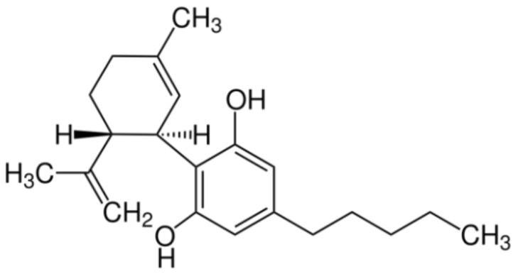
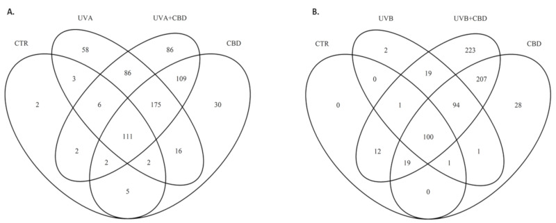
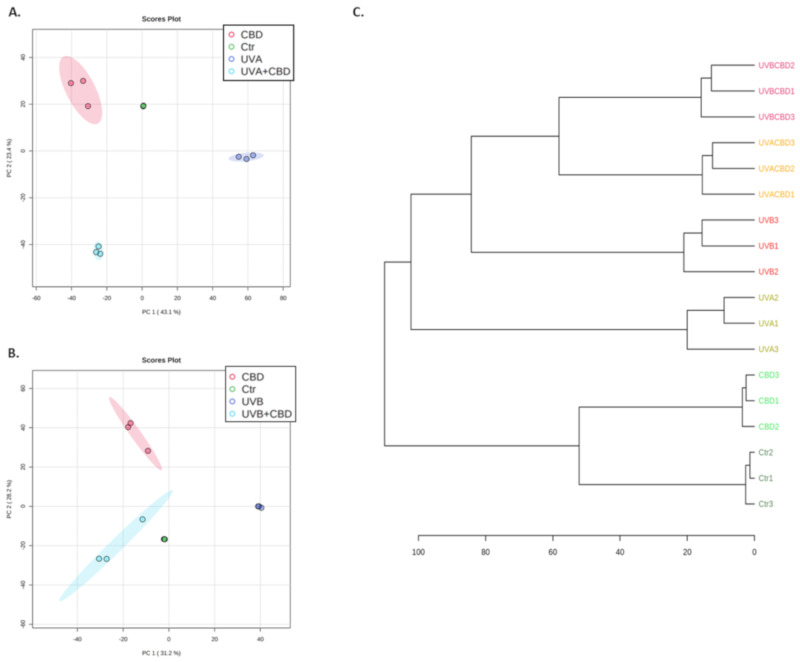
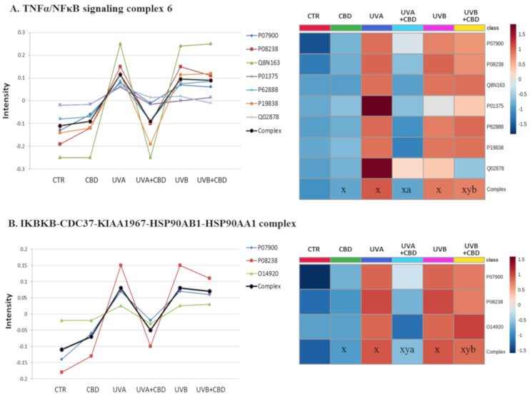
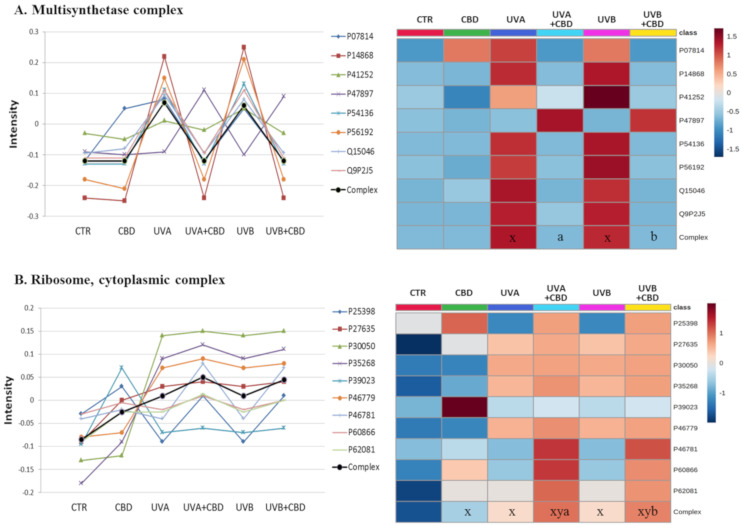
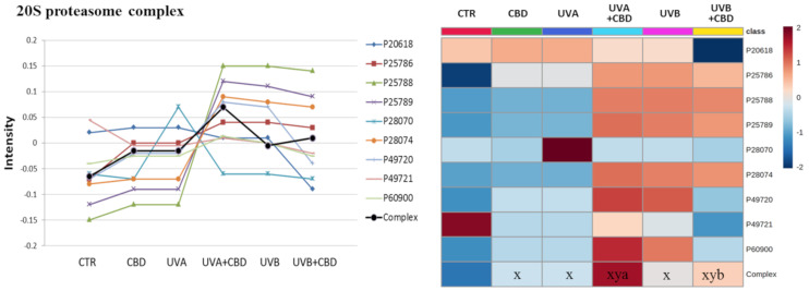
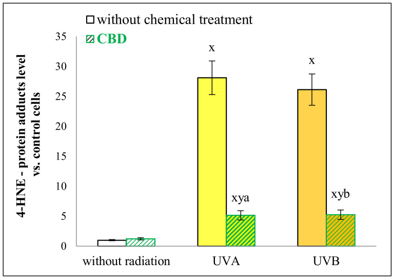
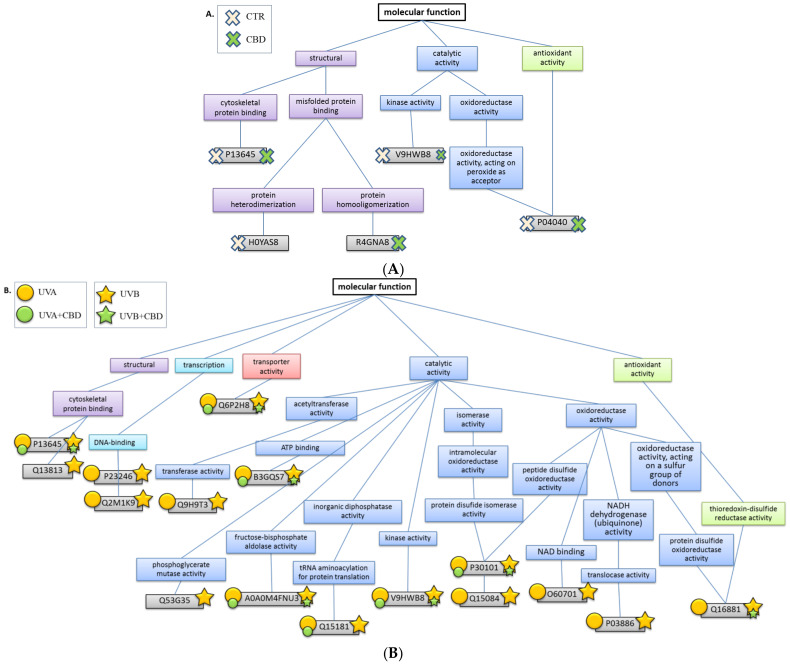
Human epidermal keratinocytes are constantly exposed to UV radiation. As a result, there is a significant need for safe and effective compounds to protect skin cells against this environmental damage. This study aimed to analyze the effect of phytocannabinoid-cannabinoid (CBD)-on the proteome of UVA/B irradiated keratinocytes. The keratinocytes were cultured in a three-dimensional (3D) system, designed to mimic epidermal conditions closely. The obtained results indicate that CBD protected against the harmful effects of UVA/B radiation. CBD decreased the expression of proinflammatory proteins, including TNFα/NFκB and IκBKB complex and decreased the expression of proteins involved in de novo protein biosynthesis, which are increased in UVA/B-irradiated cells. Additionally, CBD enhanced the UV-induced expression of 20S proteasome subunits. CBD also protected protein structures from 4-hydroxynonenal (HNE)-binding induced by UV radiation, which primarily affects antioxidant enzymes. CBD-through its antioxidant/anti-inflammatory activity and regulation of protein biosynthesis and degradation-protects skin cells against UVA/B-induced changes. In the future, its long-term use in epidermal cells should be investigated.

Keywords: UV irradiated skin; cannabidiol; keratinocytes; lipid peroxidation products adducts formation; proteomics; three-dimensional in vitro culture.


Conflict of interest statement

The authors declare no conflict of interest.

Figures

Figure 1
Figure 1
Chemical structure of cannabidiol (CBD).
Figure 2
Figure 2
Venn diagram showing protein distribution in the keratinocytes cultured in a three-dimensional culture model and treated with cannabidiol (4 μM) following UVA (30 J/cm2) (A) or UVB (60 mJ/cm2) (B) radiation. Data analyzed using RStudio (R version 3.6.2). The names and abundance of proteins are shown in Supplementary Table S1. Abbreviations: Ctr, control; CBD, cannabidiol.
Figure 3
Figure 3
Principal component analysis (PCA) of keratinocytes cultured in a three-dimensional culture model and treated with cannabidiol (4 μM) following UVA (30 J/cm2) (A) or UVB (60 mJ/cm2) (B) radiation, as well as a hierarchical dendrogram (C) of these samples. Abbreviations: Ctr, control; CBD, cannabidiol; PC, principal component.
Figure 4
Figure 4
The point graph and heatmap showing the average intensity of proteins creating TNFα/NFκB (A) and IκBKB/HSP90 (B) complexes in keratinocytes following UVA (30 J/cm2) or UVB (60 mJ/cm2) radiation and treated with cannabidiol (CBD, 4 μM) in a three-dimensional culture model. Proteins IDs: O14920, inhibitor of nuclear factor κ-B kinase subunit B; P01375, tumor necrosis factor α; P07900, heat shock protein HSP 90A; P08238, heat shock protein HSP 90B; P19838, nuclear factor NFκB p105; P62888, 60S ribosomal protein L30; Q02878, 60S ribosomal protein L6; Q8N163, cell cycle and apoptosis regulator protein 2. Statistical significances showed only for the whole complexes. X: statistically significant differences vs. non-treated cells, p < 0.05; y: statistically significant differences vs. CBD-treated cells, p < 0.05; a: statistically significant differences vs. UVA-irradiated cells, p < 0.05; b: statistically significant differences vs. UVB-irradiated cells, p < 0.05.
Figure 5
Figure 5
The point graph and heatmap showing the average intensity of proteins creating multisynthetase (A) and ribosome (B) complexes in keratinocytes following UVA (30 J/cm2) or UVB (60 mJ/cm2) radiation and treated with cannabidiol (CBD, 4 μM) in a three-dimensional culture model. Proteins IDs: P07814, glutamate/proline-tRNA ligase; P14868, aspartate-tRNA ligase; P41252, isoleucine-tRNA ligase; P47897, glutamine-tRNA ligase; P54136, arginine-tRNA ligase; P56192, methionine-tRNA ligase; Q15046, lysine-tRNA ligase; Q9P2J5, leucine-tRNA ligase; P62081, P46781, P25398, P60866, 40S ribosomal proteins S7, S9, S12, S20; P39023, P27635, P30050, P35268, P46779, 60S ribosomal protein L3, L10, L12, L22, L28. Statistical significances showed only for the whole complexes. x: statistically significant differences vs. non-treated cells, p < 0.05; y: statistically significant differences vs. CBD-treated cells, p < 0.05; a: statistically significant differences vs. UVA-irradiated cells, p < 0.05; b: statistically significant differences vs. UVB-irradiated cells, p < 0.05.
Figure 6
Figure 6
The point graph and heatmap showing the average intensity of proteins creating 20S proteasome complex in keratinocytes following UVA (30 J/cm2) or UVB (60 mJ/cm2) radiation and treated with cannabidiol (CBD, 4 μM) in a three-dimensional culture model. Proteins IDs: P25786, P25788, P25789, P60900, proteasome subunit α type-1, -3, -4, -6; P20618, P49721, P49720, P28070, P28074, proteasome subunit β type-1, -2, -3, -4, -5. Statistical significances showed only for the whole complexes. x: statistically significant differences vs. non-treated cells, p < 0.05; y: statistically significant differences vs. CBD-treated cells, p < 0.05; a: statistically significant differences vs. UVA-irradiated cells, p < 0.05; b: statistically significant differences vs. UVB-irradiated cells, p < 0.05.
Figure 7
Figure 7
The level of 4-hydroxynonenal (4-HNE)-protein adducts in keratinocytes following UVA (30 J/cm2) or UVB (60 mJ/cm2) radiation and treated with cannabidiol (CBD, 4 μM) in a three-dimensional culture model. x: statistically significant differences vs. non-treated cells, p < 0.05; y: statistically significant differences vs. CBD-treated cells, p < 0.05; a: statistically significant differences vs. UVA-irradiated cells, p < 0.05; b: statistically significant differences vs. UVB-irradiated cells, p < 0.05.
Figure 8
Figure 8
The biological function of proteins forming adducts with 4-hydroxynonenal (4-HNE) in keratinocytes treated in a three-dimensional culture model with cannabidiol (CBD, 4 μM) (A) and following UVA (30 J/cm2) or UVB (60 mJ/cm2) (B) radiation. The size of the stamps indicates the statistically significant differences between the estimated amount of the 4-HNE-protein adducts in the samples non-treated or treated with CBD.
Figure 9
Figure 9
SDS–PAGE separation and staining with Coomassie brilliant blue R-250 of proteins from control keratinocytes and irradiated with UVA (30 J/cm2), UVB (60 mJ/cm2) or/and treated with cannabidiol (CBD, 4 μM) in a three-dimensional (3D) culture model. The grid indicates the borders of the protein migration zones. Original photo of the gel is added in Supplementary Figure S1.

Similar articles

Cited by

References

    1. Cork M.J., Robinson D.A., Vasilopoulos Y., Ferguson A., Moustafa M., MacGowan A., Duff G.W., Ward S.J., Tazi-Ahnini R. New perspectives on epidermal barrier dysfunction in atopic dermatitis: Gene-environment interactions. J. Allergy Clin. Immunol. 2006;118:3–21. doi: 10.1016/j.jaci.2006.04.042. - DOI - PubMed
    1. D’Orazio J., Jarrett S., Amaro-Ortiz A., Scott T. UV radiation and the skin. Int. J. Mol. Sci. 2013;14:12222–12248. doi: 10.3390/ijms140612222. - DOI - PMC - PubMed
    1. Gegotek A., Biernacki M., Ambrozewicz E., Surazyński A., Wroński A., Skrzydlewska E. The cross-talk between electrophiles, antioxidant defence and the endocannabinoid system in fibroblasts and keratinocytes after UVA and UVB irradiation. J. Dermatol. Sci. 2016;81:107–117. doi: 10.1016/j.jdermsci.2015.11.005. - DOI - PubMed
    1. Łuczaj W., Gęgotek A., Skrzydlewska E. Antioxidants and HNE in redox homeostasis. Free Radic. Biol. Med. 2017;111:87–101. doi: 10.1016/j.freeradbiomed.2016.11.033. - DOI - PubMed
    1. Gęgotek A., Skrzydlewska E. Biological effect of protein modifications by lipid peroxidation products. Chem. Phys. Lipids. 2019;221:46–52. doi: 10.1016/j.chemphyslip.2019.03.011. - DOI - PubMed

MeSH terms

LinkOut - more resources