

Transcriptomic Analysis of Right Ventricular Remodeling in Two Rat Models of Pulmonary Hypertension: Identification and Validation of Epithelial-to-Mesenchymal Transition in Human Right Ventricular Failure
- PMID: 33541093
- PMCID: PMC7887079
- DOI: 10.1161/CIRCHEARTFAILURE.120.007058
Transcriptomic Analysis of Right Ventricular Remodeling in Two Rat Models of Pulmonary Hypertension: Identification and Validation of Epithelial-to-Mesenchymal Transition in Human Right Ventricular Failure
Abstract
Background: Right ventricular (RV) dysfunction is a significant prognostic determinant of morbidity and mortality in pulmonary arterial hypertension (PAH). Despite the importance of RV function in PAH, the underlying molecular mechanisms of RV dysfunction secondary to PAH remain unclear. We aim to identify and compare molecular determinants of RV failure using RNA sequencing of RV tissue from 2 clinically relevant animal models of PAH.
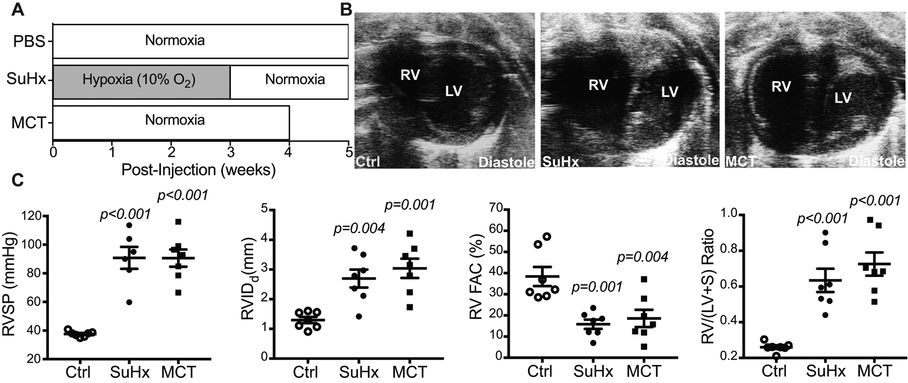
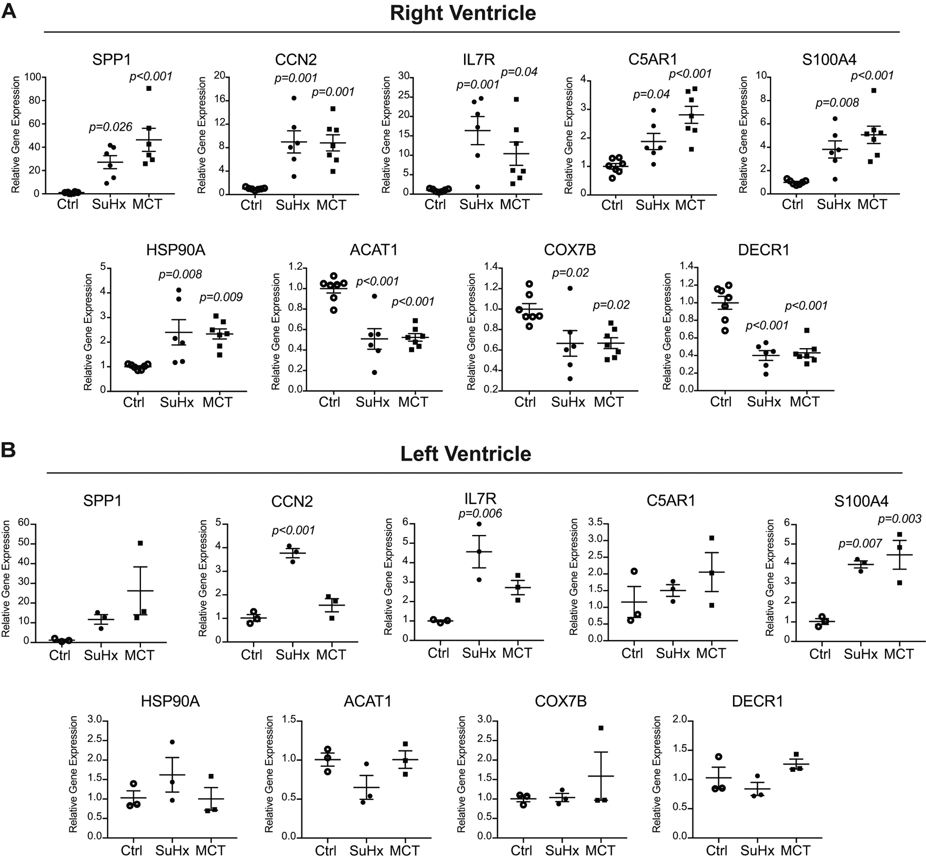
Methods: We performed RNA sequencing on RV from rats treated with monocrotaline or Sugen with hypoxia/normoxia. PAH and RV failure were confirmed by catheterization and echocardiography. We validated the RV transcriptome results using quantitative real-time polymerase chain reaction, immunofluorescence, and Western blot. Immunohistochemistry and immunofluorescence were performed on human RV tissue from control (n=3) and PAH-induced RV failure patients (n=5).
Results: We identified similar transcriptomic profiles of RV from monocrotaline- and Sugen with hypoxia-induced RV failure. Pathway analysis showed genes enriched in epithelial-to-mesenchymal transition, inflammation, and metabolism. Histological staining of human RV tissue from patients with RV failure secondary to PAH revealed significant RV fibrosis and endothelial-to-mesenchymal transition, as well as elevated cellular communication network factor 2 (top gene implicated in epithelial-to-mesenchymal transition/endothelial-to-mesenchymal transition) expression in perivascular areas compared with normal RV.
Conclusions: Transcriptomic signature of RV failure in monocrotaline and Sugen with hypoxia models showed similar gene expressions and biological pathways. We provide translational relevance of this transcriptomic signature using RV from patients with PAH to demonstrate evidence of epithelial-to-mesenchymal transition/endothelial-to-mesenchymal transition and protein expression of cellular communication network factor 2 (CTGF [connective tissue growth factor]). Targeting specific molecular mechanisms responsible for RV failure in monocrotaline and Sugen with hypoxia models may identify novel therapeutic strategies for PAH-associated RV failure.
Keywords: fibrosis; monocrotaline; pulmonary hypertension; right ventricular failure; transcriptome.
Figures


Comment in
-
Letter by Legchenko et al Regarding Article, "Transcriptomic Analysis of Right Ventricular Remodeling in Two Rat Models of Pulmonary Hypertension: Identification and Validation of Epithelial-to-Mesenchymal Transition in Human Right Ventricular Failure".Circ Heart Fail. 2021 Dec;14(12):e008503. doi: 10.1161/CIRCHEARTFAILURE.121.008503. Epub 2021 Dec 6. Circ Heart Fail. 2021. PMID: 34932370 No abstract available.
-
Response by Park et al to Letter Regarding Article, "Transcriptomic Analysis of Right Ventricular Remodeling in Two Rat Models of Pulmonary Hypertension: Identification and Validation of Epithelial-to-Mesenchymal Transition in Human Right Ventricular Failure".Circ Heart Fail. 2022 Jul;15(7):e008632. doi: 10.1161/CIRCHEARTFAILURE.122.008632. Epub 2022 Jun 29. Circ Heart Fail. 2022. PMID: 35766030 Free PMC article. No abstract available.
References
-
- Zelt JGE, Chaudhary KR, Cadete VJ, Mielniczuk LM, Stewart DJ. Medical Therapy for Heart Failure Associated With Pulmonary Hypertension. Circ Res. 2019;124:1551–1567. - PubMed
-
- Lahm T, Douglas IS, Archer SL, Bogaard HJ, Chesler NC, Haddad F, Hemnes AR, Kawut SM, Kline JA, Kolb TM, et al., American Thoracic Society Assembly on Pulmonary Circulation. Assessment of Right Ventricular Function in the Research Setting: Knowledge Gaps and Pathways Forward. An Official American Thoracic Society Research Statement. Am J Respir Crit Care Med. 2018;198:e15–e43. - PMC - PubMed
-
- Rain S, Andersen S, Najafi A, Gammelgaard Schultz J, da Silva Gonçalves Bós D, Handoko ML, Bogaard H-J, Vonk-Noordegraaf A, Andersen A, van der Velden J, et al. Right Ventricular Myocardial Stiffness in Experimental Pulmonary Arterial Hypertension: Relative Contribution of Fibrosis and Myofibril Stiffness. Circ Heart Fail. 2016. July;9(7):e002636. - PMC - PubMed
-
- Thiery JP, Acloque H, Huang RYJ, Nieto MA. Epithelial-mesenchymal transitions in development and disease. Cell. 2009;139:871–890. - PubMed
Publication types
MeSH terms
Substances
Grants and funding
LinkOut - more resources
Full Text Sources
Other Literature Sources
Medical
Miscellaneous
