. 2021 Feb 4;12(1):107.
doi: 10.1186/s13287-021-02175-2.

Local anesthetics impair the growth and self-renewal of glioblastoma stem cells by inhibiting ZDHHC15-mediated GP130 palmitoylation

Affiliations

Local anesthetics impair the growth and self-renewal of glioblastoma stem cells by inhibiting ZDHHC15-mediated GP130 palmitoylation

Xiaoqing Fan et al. Stem Cell Res Ther. .

Abstract

Background: A large number of preclinical studies have shown that local anesthetics have a direct inhibitory effect on tumor biological activities, including cell survival, proliferation, migration, and invasion. There are few studies on the role of local anesthetics in cancer stem cells. This study aimed to determine the possible role of local anesthetics in glioblastoma stem cell (GSC) self-renewal and the underlying molecular mechanisms.

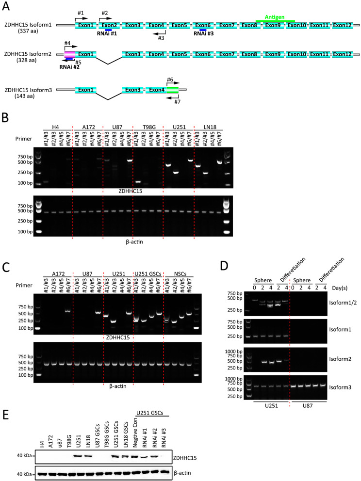
Methods: The effects of local anesthetics in GSCs were investigated through in vitro and in vivo assays (i.e., Cell Counting Kit 8, spheroidal formation assay, double immunofluorescence, western blot, and xenograft model). The acyl-biotin exchange method (ABE) assay was identified proteins that are S-acylated by zinc finger Asp-His-His-Cys-type palmitoyltransferase 15 (ZDHHC15). Western blot, co-immunoprecipitation, and liquid chromatograph mass spectrometer-mass spectrometry assays were used to explore the mechanisms of ZDHHC15 in effects of local anesthetics in GSCs.

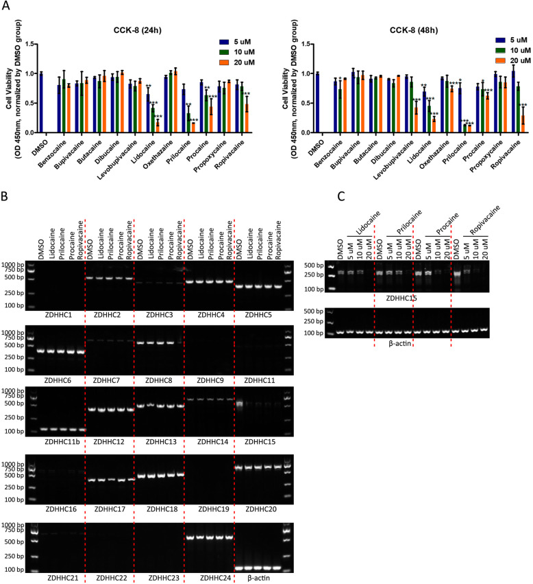
Results: In this study, we identified a novel mechanism through which local anesthetics can damage the malignant phenotype of glioma. We found that local anesthetics prilocaine, lidocaine, procaine, and ropivacaine can impair the survival and self-renewal of GSCs, especially the classic glioblastoma subtype. These findings suggest that local anesthetics may weaken ZDHHC15 transcripts and decrease GP130 palmitoylation levels and membrane localization, thus inhibiting the activation of IL-6/STAT3 signaling.

Conclusions: In conclusion, our work emphasizes that ZDHHC15 is a candidate therapeutic target, and local anesthetics are potential therapeutic options for glioblastoma.

Keywords: GP130; Glioblastoma stem cells; Local anesthetics; STAT3; Self-renewal; ZDHHC15.


Conflict of interest statement

The authors declare no conflicts of interest.

Figures

Fig. 1
Fig. 1
Local anesthetics prilocaine, lidocaine, procaine, and ropivacaine impaired cell survival through inhibition of ZDHHC15 expression. Histogram showing the viability of GSCs cultured in CSC medium with or without local anesthetics (procaine, benzocaine, procaine, dibucaine, tetracaine, oxadiazine, lidocaine, propoxycaine, levobupivacaine, bupivacaine, and ropivacaine) for 24 h (left) or 48 h (right) at different concentrations (5, 10, and 20 μM, respectively). Local anesthetics, such as prilocaine, lidocaine, procaine, and ropivacaine, killed GSCs in a concentration- and time-dependent manner. a ART-PCR analysis of mRNA levels of 24 known pats in GSC treated with prilocaine, lidocaine, procaine, and ropivacaine (20 μm each). β-Actin was used as a loading control. b RT-PCR analysis of ZDHHC15 mRNA levels in GSCs treated with different concentrations (5, 10, and 20 μM) of prilocaine, lidocaine, procaine, and ropivacaine. β-Actin was used as a loading control
Fig. 2
Fig. 2
ZDHHC15 isoform expression in GBM cells and GSCs. a Diagram of the ZDHHC15 splicing isoforms. The isoform 1 coding sequence contains 12 exons. Compared to isoform 1, variant 2 lacks coding for exon 2, and variant 3 lacks an in-frame coding exon and differs at the 3′ end. The locations of the primers and the sites targeted with stealth siRNAs are indicated on a diagram of the ZDHHC15 splicing isoforms. b RT-PCR analysis of the mRNA levels of ZDHHC15 splicing isoforms in six GBM cells (H4, A172, U87, T98G, U251, and LN18). Sequences encoding isoforms 1 and 2 using primers #1 and #3 (compared to isoform 1, variant 2 lacks 27 bp). Sequences encoding isoform 1 used primers #2 and #3. Sequences encoding isoform 2 used primers #4 and #5. Sequences encoding isoform 3 used primers #6 and #7. β-Actin served as the loading control. c RT-PCR analysis of the mRNA levels of ZDHHC15 splicing isoforms in GSCs and NSCs. β-Actin served as the loading control. d RT-PCR analysis of the mRNA levels of ZDHHC15 splicing isoforms during GSC self-renewal and the differentiation stage. β-Actin served as the loading control. e Western blot analysis of ZDHHC15 in six GBM cell lines (H4, A172, U87, T98G, U251, and LN18); four GSCs derived from U87, T98G, U251, and LN18; and U251 GSCs after transfection with ZDHHC15 stealth siRNAs. β-Actin served as the loading control
Fig. 3
Fig. 3
ZDHHC15 silencing or treatment with local anesthetics strongly induces differentiation of GSCs. a Glioma tissue sections (n = 60) were stained with an antibody against ZDHHC15. Scale bar, 200 μm. b Representative images showing U251 GSCs maintained under neurosphere conditions for 7 days after transfection with ZDHHC15 shRNA or treatment with prilocaine, lidocaine, procaine, or ropivacaine (20 μM). c The capacity of the U251 GSCs transfected with ZDHHC5 shRNA and treated with prilocaine, lidocaine, procaine, and ropivacaine (20 μM) to generate neurospheres was estimated by a serial dilution assay. d GSC neurospheres of all categories were stained for stem and differentiated cell markers as indicated. Cells were stained with antibodies against nestin and SOX2 for neural stem cell markers, and antibody staining of GFAP and MAP 2 were used as markers of differentiated cells. DAPI (4′,6-diamidino-2-phenylindole) was used as a nuclear stain. Scale bar, 100 μm
Fig. 4
Fig. 4
Identity of palmitoylated proteins mediated by ZDHHC15. a Principles of the ABE capture methods. U251 GSC was dissolved and incubated with ZDHHC15 antibody. In addition, it was cleaved with palmitate in the HAM+ group. The HAM− condition was used as a negative control. After the ABE reaction, streptavidin beads were used to enrich biotinylated proteins. The proteins enriched under HAM+ and HAM− conditions were identified by mass spectrometry (MS). In the HAM+ sample, proteins with at least 2-fold higher abundance compared to the control were considered as candidate proteins. Probability: 0–19% (2–3 times; n = 37), 20–49% (3–4 times; n = 6), and > 50% (> 4 times; n = 31). b Venn diagram showing the relationship between the expression patterns of different DHHCs in glioma using the Human Protein Atlas (HPA), previously validated S-acylated proteins, predicted S-acylated proteins using the CSS-Palm version 4.0 software, and function previously reported in glioma. c Lysates from U251 GSCs were subjected to IP with the ZDHHC15 antibody, followed by immunoblotting (IB) with anti-GP130, anti-LRP12, and anti-RIF1 antibodies
Fig. 5
Fig. 5
Palmitoylation inhibition mediated by the local anesthetics resulted in the disappearance of GP130 in the membrane fractions. a ABE was performed on proteins from U251 GSCs transfected with ZDHHC15 shRNA or treated with 2BP (50 μM) or PalmB (1 μM) for 48 h. The presence or absence of hydroxylamine (HAM) during the reaction was used as a control for reaction specificity. Western blot analysis with streptavidin-horseradish peroxidase is shown depicting the banding pattern of S-acylated proteins from 1 or 3 mg of total protein. b Protein accumulation (detected by western blot analysis) and palmitoylation level (detected by ABE) in GSCs treated with or without prilocaine, lidocaine, procaine, and ropivacaine at different concentrations (5 μM, 10 μM, and 20 μM, respectively). β-Actin was used as a loading control. c Expression of p-STAT3 (Y705) in U251 GSCs (monolayer culture) transfected with ZDHHC5 shRNA and treated with prilocaine, lidocaine, procaine, and ropivacaine (20 μM) for 48 h was analyzed by immunofluorescence staining. Scale bar, 100 μm. d The expression of GP130 in U251 GSCs (monolayer culture) transfected with ZDHHC5 shRNA and treated with prilocaine, lidocaine, procaine, and ropivacaine (20 μM) for 48 h was analyzed by immunofluorescence staining. Scale bar, 100 μm. e GSCs were transfected with ZDHHC5 shRNA or treated with prilocaine, lidocaine, procaine, and ropivacaine (20 μM) and harvested after 48 h. Cellular fractionation was performed to separate cytosolic and membrane fractions. Fractionates were then subjected to western blot analysis to detect the distribution of GP130
Fig. 6
Fig. 6
A regulatory feedback loop exists between ZDHHC15 and IL-6/STAT3 signaling. a Putative binding motif of transcription factor STAT3 for ZDHHC15 isoforms 1, 2, and 3 was predicted from the JASPAR database. The top three STAT3 binding sites (labeled E1′, E2, and E3) for ZDHHC15 isoforms 1 and 3 and four binding sites (labeled E1′, E2′, E3′, and E4′) were chosen for further analysis. b qRT-PCR was performed to evaluate the activity of STAT3 ZDHHC15 transcription in U251 GSCs transfected with STAT3 siRNA or treated with STAT3 inhibitor STAT3-IN-7 (5 μM) or rhIL-6 (5 ng/mL), alone or in combination. c Luciferase reporter assay demonstrated luciferase activities of ZDHHC15 isoform reporters in U251 GSCs transfected with STAT3 siRNA or treated with STAT3 inhibitor STAT3-IN-7 (5 μM), or the local anesthetics prilocaine, lidocaine, procaine, and ropivacaine (20 μM each). d Luciferase reporter assay demonstrated luciferase activities of various truncated reporters in U251 GSCs, determining the region of ZDHHC15 isoform promoters on which STAT3 could bind to mediate transcriptional activation. e ChIP assay was performed using the p-STAT3 antibody to demonstrate the enrichment of the STAT3-binding region of the promoter of ZDHHC15 isoforms in U251 GSCs. f Tumor growth of U251 GSCs transfected with ZDHHC5 shRNA or pretreated with prilocaine, lidocaine, procaine, and ropivacaine (20 μM). g Tumor weights were measured after 6 weeks (n = 5 mice/group)

Similar articles

Cited by

References

    1. Kim R. Effects of surgery and anesthetic choice on immunosuppression and cancer recurrence. J Transl Med. 2018;16:8. doi: 10.1186/s12967-018-1389-7. - DOI - PMC - PubMed
    1. Tavare AN, Perry NJ, Benzonana LL, Takata M, Ma D. Cancer recurrence after surgery: direct and indirect effects of anesthetic agents. Int J Cancer. 2012;130:1237–1250. doi: 10.1002/ijc.26448. - DOI - PubMed
    1. Liu H, Dilger JP, Lin J. Effects of local anesthetics on cancer cells. Pharmacol Ther. 2020;212:107558. doi: 10.1016/j.pharmthera.2020.107558. - DOI - PubMed
    1. Fan X, Wang D, Chen X, Wang R. Effects of anesthesia on postoperative recurrence and metastasis of malignant tumors. Cancer Manag Res. 2020;12:7619–7633. doi: 10.2147/cmar.s26552. - DOI - PMC - PubMed
    1. Boiteux C, Vorobyov I, French RJ, French C, Yarov-Yarovoy V, Allen TW. Local anesthetic and antiepileptic drug access and binding to a bacterial voltage-gated sodium channel. Proc Natl Acad Sci U S A. 2014;111:13057–13062. doi: 10.1073/pnas.1408710111. - DOI - PMC - PubMed

Publication types