

Laparoscopic removal of mesh migrating into the sigmoid colon after totally extraperitoneal (TEP) laparoscopic inguinal hernia repair with positive faecal occult blood test
- PMID: 33542008
- PMCID: PMC7868236
- DOI: 10.1136/bcr-2020-237167
Laparoscopic removal of mesh migrating into the sigmoid colon after totally extraperitoneal (TEP) laparoscopic inguinal hernia repair with positive faecal occult blood test
Abstract
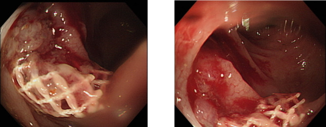
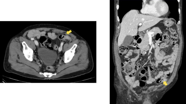
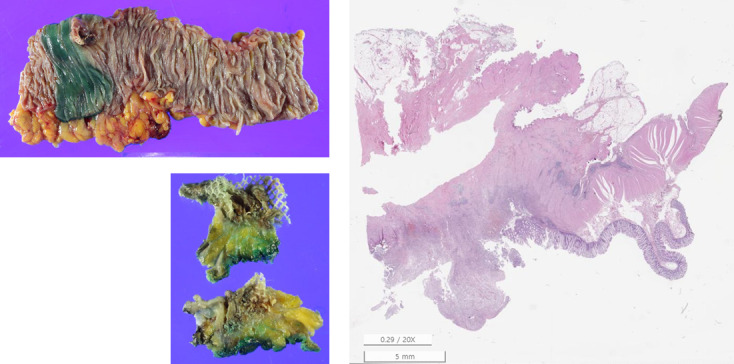
A 76-year-old man was referred to our clinic after a foreign body seen in his sigmoid colon during a colonoscopy. He had undergone three operations for a left inguinal hernia within the previous 8 years, and the first procedure was a laparoscopic totally extraperitoneal approach. Four years later, removal of migrated and infected mesh was conducted by open approach. He then had a positive stool occult blood test for routine check-up 4 years after the remnant mesh removal. An ill-defined lesion was identified on colonoscopy. CT revealed a 2.7 cm diameter enhancing lesion in the sigmoid colon. Laparoscopic sigmoidectomy was performed, and remnant mesh fragment was found in the sigmoid colon and removed. The migrated mesh could not be wholly removed by open abdominal approach and the remnant mesh fragment migrated to sigmoid colon. It suggests the importance of a laparoscopic approach to remove the entire mesh.
Keywords: gastrointestinal surgery; general surgery.
© BMJ Publishing Group Limited 2021. No commercial re-use. See rights and permissions. Published by BMJ.
Conflict of interest statement
Competing interests: None declared.
Figures
References
-
- Gossetti F, D’Amore L, Annesi E. Mesh-related visceral complications following inguinal herni repair: an imerging topic. Heria 2019;23:699–708. - PubMed
Publication types
MeSH terms
LinkOut - more resources
Full Text Sources
Other Literature Sources