

Proliferating trichilemmal tumour of the eyelid in a 7-year-old girl
- PMID: 33542009
- PMCID: PMC7868174
- DOI: 10.1136/bcr-2020-237476
Proliferating trichilemmal tumour of the eyelid in a 7-year-old girl
Abstract
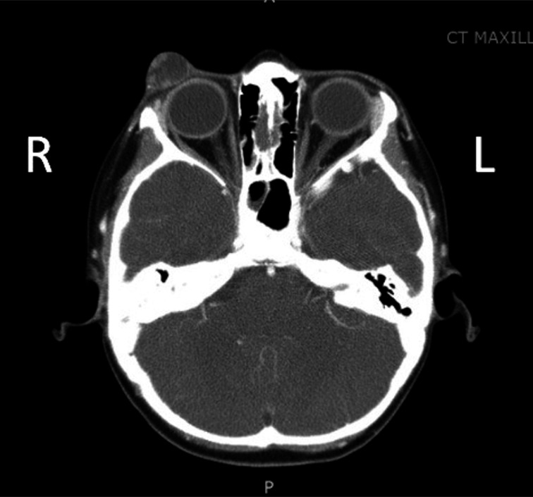
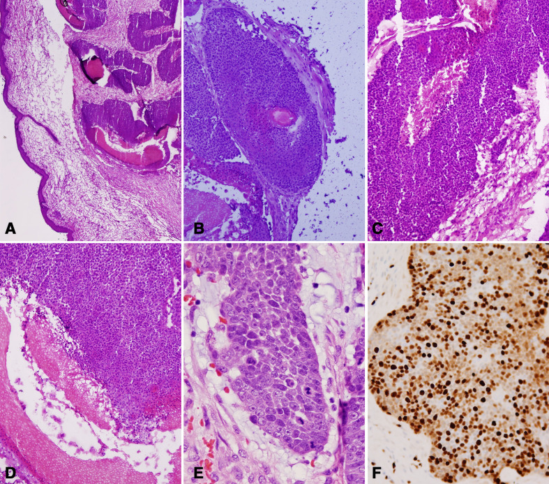
Proliferating trichilemmal tumours (PTTs) are rare cutaneous adnexal tumours derived from the hair shaft outer root sheath. We are reporting the first case of PTT in a young child. In this case, a 7-year-old girl presented with trichilemmal keratinisation consistent with PTT. The patient was monitored with no signs of recurrence. PTT is a rare tumour occurring primarily in adults and we present this case so that young patients with PTT can be diagnosed and treated appropriately with a painless, mobile, rapidly growing mass on the right upper eyelid. CT imaging showed well-circumscribed, heterogenous mass measuring 1.6 cm with fluid-filled appearance and no tissue invasion. Surgical excision was performed and pathology revealed an unencapsulated, well-demarcated tumour.
Keywords: dermatology; head and neck surgery; otolaryngology / ENT; pathology; plastic and reconstructive surgery.
© BMJ Publishing Group Limited 2021. No commercial re-use. See rights and permissions. Published by BMJ.
Conflict of interest statement
Competing interests: None declared.
Figures


References
-
- Fonseca TC, Bandeira CL, Sousa BA, et al. . Proliferating trichilemmal tumor: case report. J Bras Patol e Med Lab 2016;52:120–3. 10.5935/1676-2444.20160014 - DOI
Publication types
MeSH terms
LinkOut - more resources
Full Text Sources
Other Literature Sources
Research Materials
Miscellaneous