. 2021 May 1;27(9):2648-2662.
doi: 10.1158/1078-0432.CCR-20-2961. Epub 2021 Feb 4.

Therapeutic Targeting of Nemo-like Kinase in Primary and Acquired Endocrine-resistant Breast Cancer

Affiliations

Therapeutic Targeting of Nemo-like Kinase in Primary and Acquired Endocrine-resistant Breast Cancer

Xian Wang et al. Clin Cancer Res. .

Abstract

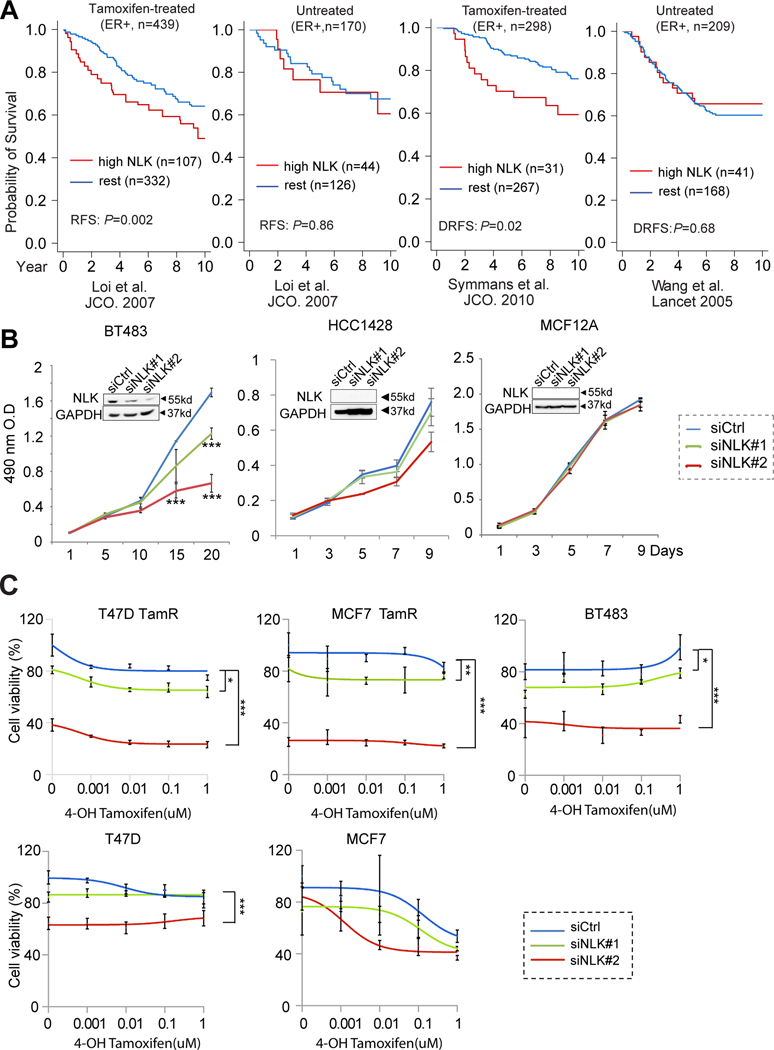
Purpose: Endocrine resistance remains a major clinical challenge in estrogen receptor (ER)-positive breast cancer. Despite the encouraging results from clinical trials for the drugs targeting known survival signaling, relapse is still inevitable. There is an unmet need to discover new drug targets in the unknown escape pathways. Here, we report Nemo-like kinase (NLK) as a new actionable kinase target that endows previously uncharacterized survival signaling in endocrine-resistant breast cancer.

Experimental design: The effects of NLK inhibition on the viability of endocrine-resistant breast cancer cell lines were examined by MTS assay. The effect of VX-702 on NLK activity was verified by kinase assay. The modulation of ER and its coactivator, SRC-3, by NLK was examined by immunoprecipitation, kinase assay, luciferase assay, and RNA sequencing. The therapeutic effects of VX-702 and everolimus were tested on cell line- and patient-derived xenograft (PDX) tumor models.

Results: NLK overexpression endows reduced endocrine responsiveness and is associated with worse outcome of patients treated with tamoxifen. Mechanistically, NLK may function, at least in part, via enhancing the phosphorylation of ERα and its key coactivator, SRC-3, to modulate ERα transcriptional activity. Through interrogation of a kinase profiling database, we uncovered and verified a highly selective dual p38/NLK inhibitor, VX-702. Coadministration of VX-702 with the mTOR inhibitor, everolimus, demonstrated a significant therapeutic effect in cell line-derived xenograft and PDX tumor models of acquired or de novo endocrine resistance.

Conclusions: Together, this study reveals the potential of therapeutic modulation of NLK for the management of the endocrine-resistant breast cancers with active NLK signaling.


Figures

Figure 1.
Figure 1.. Identification of NLK as a new kinase target in endocrine-resistant breast cancer.
(a) NLK overexpression predicts worse outcome in ER+ breast cancer patients treated with tamoxifen monotherapy but not in untreated patients. The association of NLK overexpression with recurrence free survival in tamoxifen-treated and untreated luminal breast cancer patients were assessed using public gene expression datasets by Loi et al., (14), Symmans et al., (15), and Wang et al., (16). DRFS, distant recurrence free survival. Here the cutoff for NLK overexpression is determined based on median plus one median absolute deviation (see Methods). (b) The effect of NLK inhibition by two independent siRNAs on the growth of the BT483 and HCC1428 luminal breast cancer cell lines, as well as the MCF12A benign breast epithelial cells. (c) The therapeutic effect of NLK silencing by two independent siRNAs on 4-OH tamoxifen treatment in the primary endocrine-resistant BT483 cells and the acquired tamoxifen resistant (TamR) clones of MCF7 and T47D cell lines as well as the parental MCF7 and T47D cells. *P<0.05; **P<0.01; ***P<0.001 based on t-test (time-point experiments) or ANOVA (tamoxifen dose curves) compared to siRNA controls. The error bars represent the standard deviations.
Figure 2.
Figure 2.. Identification of a novel dual p38 and NLK inhibitor VX-702, and its therapeutic effect to tamoxifen treatment in vitro.
(a) Heatmap of “hot-spot” drug-kinase assay identifying VX-702 as a potent dual p38 MAPK and NLK inhibitor with exclusive activity against p38 MAPKs and NLK. The percentage of kinase activity inhibition is shown in the red color scale. (b) Bar chart showing the inhibition efficacy of VX-702 over a panel of 300 kinases based on the same dataset as in A. (c) In vitro kinase assay using recombinant NLK protein and MBP as the substrate in the presence or absence of VX-702 treatment. (d) In vitro kinase assay for V5-NLK immuno-precipitated from VX-702 treated MCF7 cells using MBP as the substrate. In order to maintain the inhibition of NLK, the immunoprecipitation product was incubated in different doses of VX-702 throughout the in vitro kinase assay process. (e) The survival fraction of MCF7-TamR breast cancer cells and MCF10A non-cancerous breast epithelial cells following treatment with different doses of VX-702 for 7 days; 0.5 μM was determined as the effective concentration in vitro. (f) The therapeutic effect of VX-702 on 4-OH tamoxifen treatment in primary and acquired tamoxifen resistant breast cancer cell lines. The assays were carried out for 7 days under estrogen deprived condition. (g) Induction of ectopic NLK expression rescues the therapeutic effect of VX-702 to tamoxifen treatment in the MCF7-TamR and T47D-TamR cells. ***P<0.001 (based on two-way ANOVA). The error bars represent the standard deviations.
Figure 3.
Figure 3.. NLK phosphorylates ERα and its key coactivator SRC-3 and modulates ER transcriptional activity in endocrine-resistant breast cancer cells.
(a) ERE luciferase reporter assay showing the effect of NLK inhibition/overexpression or VX-702 treatment on ER transcriptional activity in BT483 cells under estrogen deprivation (ED) and 0.5uM tamoxifen treatment. (b) VX-702 significantly inhibits the ER transcriptional activity in MCF7-TamR and T47D-TamR cells in the presence E2, or ED plus different doses of 4-OH tamoxifen treatment. a-b, *P<0.05; **P<0.01, *** P<0.001. P-value was calculated by t-test. (c) Co-immunoprecipitation of ERα using anti-ERα antibody and WB using anti-NLK antibody or anti-ERα antibody in BT483 overexpressing endogenous NLK. (d) In vitro kinase assay of recombinant active NLK using ERα as substrate with or without VX-702 treatment. Bar chart presents the quantified band intensity. The error bars in 3a-c represent the standard deviations. (e) Western blot analysis of ER signaling in BT483 cells following NLK silencing and endocrine treatment. BT483 cells were seeded in phenol red-free medium with 5% charcoal-dextran-stripped FBS containing 0.5uM Tamoxifen or vehicle (ethanol) for 48 hours, and then reverse transfected with siCtrl, siNLK#1, siNLK#2 (10nM) for 72 hours. (f) NLK phosphorylates SRC-3. Upper panel, in vitro kinase assay using recombinant NLK and SRC-3 proteins after the indicated treatment. Middle panel, in vitro kinase assay using recombinant NLK and 1–6A mutant SRC-3 protein. Lower panel, in vitro kinase assay using recombinant NLK and SRC-3 protein with mutation at indicated site (1–6A mutation sites: T24A, S505A, S543A, S857A, S860A, or S867A). (g) ER target gene expression changes following NLK inhibition significantly correlates with their changes following tamoxifen treatment in BT483 and T47D TamR cells. Left, log2 ratio of ER target differential expression (DE) following Tamoxifen treatment correlated with siNLK #1/2 treatment in BT483 cells. Here we used the ER target genes (n=76) compiled from TRUST database (70) in the analysis. Middle, log2 ratio of ER target gene DE following Tamoxifen treatment compared to siNLK #1/2 treatment in T47D TamR cells. Here T47D specific ER target genes (n=83) provided by Lin et al are used in the analysis (71). Right, log2 ratios of ER target DE between Tamoxifen and VX-702 treatment in T47D TamR cells. The T47D specific ER target genes (n=83) are used in the analysis. The Pearson correlation coefficients are shown in the figure with all p-values less than 0.001.
Figure 4.
Figure 4.. The therapeutic effect of VX-702 treatment or in combination with mTOR inhibitor in an acquired tamoxifen-resistant tumor model derived from T47D cells.
(a) The effect of VX-702 treatment on the body weight measurements of mice from different treatment groups. NLK-high T47D tamoxifen-resistant tumors derived from #156L were transplanted into ovariectomized female nude mice and grown with tamoxifen. Upon tumor establishment, tamoxifen was withdrawn, and mice were randomized into six treatment arms: vehicle, VX-702 (50 mg/kg, o.g, BID), fulvestrant (Ful, 5 mg/mouse, s.c weekly), VX-702+Ful, everolimus (Eve, 5 mg/kg, o.g daily), or VX-702+everolimus. (b) The tumor growth curves for different treatment groups from the same in vivo experiment as in (a). Upper panel: the average tumor volumes of each treatment arm at different day points. The error bars represent the standard deviations. Lower panel: the tumor volumes of different treatment arms on Day 22. The Boxplot illustrates the distribution of the tumor volumes of each treatment group based on the following: minimum, first quartile, median, third quartile, the maximum, and the outliers. (c) VX-702 treated tumors collected at the endpoint were subjected to RPPA analysis. Tumors were sorted based on their endpoint volumes and were subdivided into two groups: large (volume >1000mm3) vs small (volume <1000mm3). Proteins that significantly differ between the two groups were plotted in a heat-map with the corresponding tumor volume for reference. The P-values are based on t-test. (d) Kaplan-Meier survival plot comparing the progression-free survival of different treatment arms. Progression-free survival was analyzed based on tumor-tripling time. *P<0.05; **P<0.01; *** P<0.001. “ns”, not significant. P-values were calculated based on two-way mixed ANOVA for comparing the tumor volumes and generalized Wilcoxon test for progression-free survival.
Figure 5.
Figure 5.. Reverse phase protein array analysis of T47D-TamR xenograft tumors harvested following 15 days of treatments.
(a) T47D-TamR xenograft tumors were harvested after 15 days of treatments and then subjected to RPPA analysis. The proteins that show a trend of altered expression or phosphorylation levels (P<0.1) following concomitant everolimus and VX-702 (Eve+VX) treatment compared to everolimus or VX-702 monotreatment was plotted in the heat-map. Each column represents a xenograft tumor sample, and each row represents an antibody against a specific protein or a phosphorylation site. (b) Heat-map of proteins that show a trend of altered expression or phosphorylation levels after VX-702 alone or VX-702+everolimus treatment compared to vehicle (P<0.1). (c) Heat-map of proteins that are changed (P<0.1) after everolimus alone or everolimus+VX-702 treatment compared to vehicle.
Figure 6.
Figure 6.. The therapeutic effect of VX-702 as a single agent or adjuvant to everolimus in the WHIM 11J PDX model with de novo tamoxifen resistance.
(a) WHIM 11J PDX tumors were transplanted into ovariectomized female nude mice in the absence of estrogen or tamoxifen supplementation. Upon tumor establishment, mice were randomized into four treatment arms: vehicle, VX-702 (50mg/kg, o.g BID), everolimus (Eve, 5 mg/kg, o.g daily), or VX-702+everolimus. Left: the average tumor volumes of each treatment group. Right: the tumor volumes of different treatment arms on Day 22. The Boxplot illustrates the distribution of the tumor volumes of each treatment group based on the following: minimum, first quartile, median, third quartile, the maximum, and the outliers. The error bars represent the standard deviations. (b) Kaplan-Meier survival plot comparing the progression-free survival of different treatment arms. Progression-free survival was analyzed based on tumor-tripling time. (c) The body weight measurements of mice in different treatment arms. *P<0.05; **P<0.01; *** P<0.001. P-values were calculated based on two-way mixed ANOVA for comparing the tumor volumes and generalized Wilcoxon test for progression-free survival.

Similar articles

Cited by

References

    1. Johnston SR. New strategies in estrogen receptor-positive breast cancer. Clin Cancer Res 2010;16(7):1979–87 doi 10.1158/1078-0432.CCR-09-1823. - DOI - PubMed
    1. Miller WR. Identification and mechanisms of endocrine resistance. Breast Cancer Res 2008;10 Suppl 4:S19 doi 10.1186/bcr2179. - DOI - PMC - PubMed
    1. Osborne CK, Schiff R. Mechanisms of endocrine resistance in breast cancer. Annual review of medicine 2011;62:233–47 doi 10.1146/annurev-med-070909-182917. - DOI - PMC - PubMed
    1. O’Sullivan CC. Overcoming Endocrine Resistance in Hormone-Receptor Positive Advanced Breast Cancer-The Emerging Role of CDK4/6 Inhibitors. Int J Cancer Clin Res 2015;2(4). - PMC - PubMed
    1. Osborne CK, Schiff R. Mechanisms of endocrine resistance in breast cancer. Annu Rev Med 2011;62:233–47 doi 10.1146/annurev-med-070909-182917. - DOI - PMC - PubMed

Publication types

MeSH terms