. 2021 Feb 2;12(2):136.
doi: 10.1038/s41419-021-03444-x.

A fine balance between Prpf19 and Exoc7 in achieving degradation of aggregated protein and suppression of cell death in spinocerebellar ataxia type 3

Affiliations

A fine balance between Prpf19 and Exoc7 in achieving degradation of aggregated protein and suppression of cell death in spinocerebellar ataxia type 3

Zhefan Stephen Chen et al. Cell Death Dis. .

Abstract

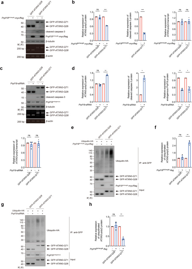
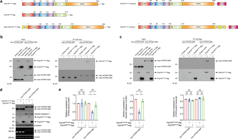
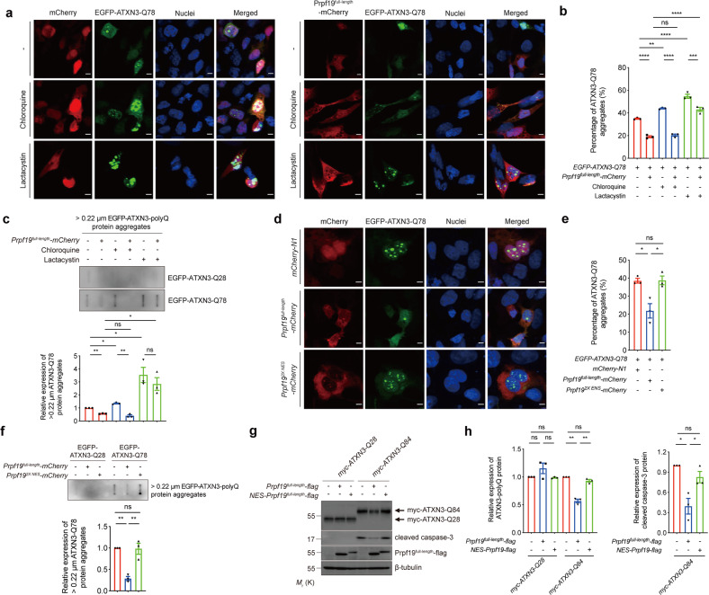
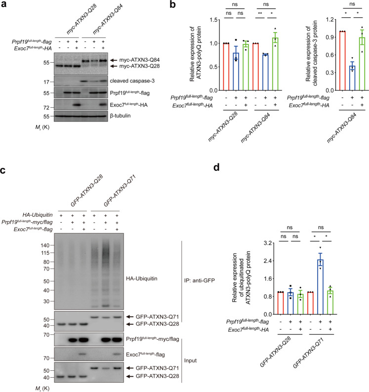
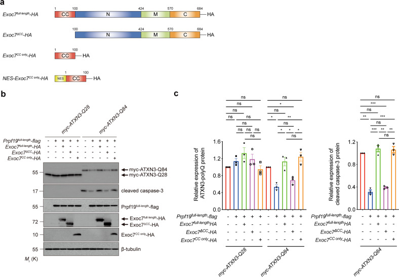
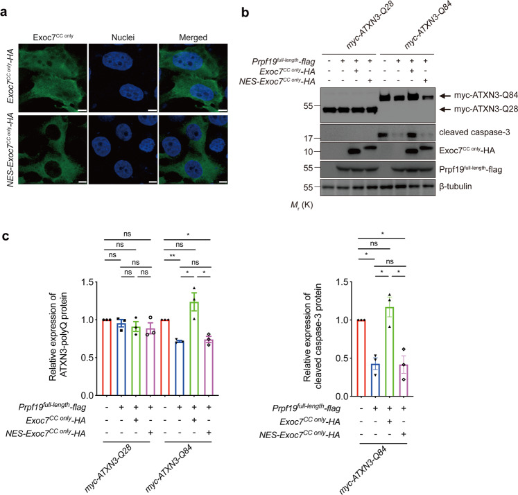
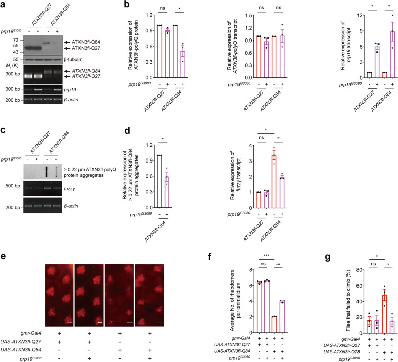
Polyglutamine (polyQ) diseases comprise Huntington's disease and several subtypes of spinocerebellar ataxia, including spinocerebellar ataxia type 3 (SCA3). The genomic expansion of coding CAG trinucleotide sequence in disease genes leads to the production and accumulation of misfolded polyQ domain-containing disease proteins, which cause cellular dysfunction and neuronal death. As one of the principal cellular protein clearance pathways, the activity of the ubiquitin-proteasome system (UPS) is tightly regulated to ensure efficient clearance of damaged and toxic proteins. Emerging evidence demonstrates that UPS plays a crucial role in the pathogenesis of polyQ diseases. Ubiquitin (Ub) E3 ligases catalyze the transfer of a Ub tag to label proteins destined for proteasomal clearance. In this study, we identified an E3 ligase, pre-mRNA processing factor 19 (Prpf19/prp19), that modulates expanded ataxin-3 (ATXN3-polyQ), disease protein of SCA3, induced neurodegeneration in both mammalian and Drosophila disease models. We further showed that Prpf19/prp19 promotes poly-ubiquitination and degradation of mutant ATXN3-polyQ protein. Our data further demonstrated the nuclear localization of Prpf19/prp19 is essential for eliciting its modulatory function towards toxic ATXN3-polyQ protein. Intriguingly, we found that exocyst complex component 7 (Exoc7/exo70), a Prpf19/prp19 interacting partner, modulates expanded ATXN3-polyQ protein levels and toxicity in an opposite manner to Prpf19/prp19. Our data suggest that Exoc7/exo70 exerts its ATXN3-polyQ-modifying effect through regulating the E3 ligase function of Prpf19/prp19. In summary, this study allows us to better define the mechanistic role of Exoc7/exo70-regulated Prpf19/prp19-associated protein ubiquitination pathway in SCA3 pathogenesis.


Conflict of interest statement

The authors declare that they have no conflict of interest.

Figures

Fig. 1
Fig. 1. Prpf19 modulates the poly-ubiquitination and degradation of expanded ATXN3-polyQ protein and also mediates ATXN3-polyQ-induced cytotoxicity.
a Overexpression of Prpf19 degraded the level of ATXN3-Q71 and suppressed the ATXN3-Q71-induced caspase-3 cleavage. The expression of ATXN3-Q28 was not affected, and the transcript level of ATXN3-polyQ was not altered when Prpf19 was overexpressed. n = 3 biological replicas. Each n represents an independent preparation of cell protein and RNA samples. b Quantification of ATXN3-polyQ protein, cleaved caspase-3 protein, and ATXN3-polyQ transcript levels in panel a. Error bars represent S.E.M. Statistical analysis was performed using two-tailed unpaired Student’s t test. ns denotes no significant difference and *** denotes P < 0.001. c Knockdown of Prpf19 increased the level of ATXN3-Q71 and enhanced the ATXN3-Q71-induced caspase-3 cleavage. The expression of ATXN3-Q28 was not affected, and the transcript level of ATXN3-polyQ was not altered when Prpf19 was knocked down. n = 3 biological replicas. Each n represents an independent preparation of cell protein and RNA samples. d Quantification of ATXN3-polyQ protein, cleaved caspase-3 protein, endogenous Prpf19 protein, and ATXN3-polyQ transcript levels in panel c. Error bars represent S.E.M. Statistical analysis was performed using two-tailed unpaired Student’s t test. ns denotes no significant difference, * denotes P < 0.05 and ** denotes P < 0.01. e Overexpression of Prpf19 promoted the poly-ubiquitination of ATXN3-Q71, and the level of ubiquitinated ATXN3-Q28 was not changed. n = 3 biological replicas. Each n represents an independent preparation of cell protein samples. f Quantification of the ubiquitination level of ATXN3-polyQ protein in panel e. Error bars represent S.E.M. Statistical analysis was performed using two-tailed unpaired Student’s t test. ns denotes no significant difference and ** denotes P < 0.01. g Knockdown of Prpf19 reduced the poly-ubiquitination of ATXN3-Q71 protein, and the level of ubiquitinated ATXN3-Q28 was not changed. n = 3 biological replicas. Each n represents an independent preparation of cell protein samples. h Quantification of the ubiquitination level of ATXN3-polyQ protein in panel g. Error bars represent S.E.M. Statistical analysis was performed using two-tailed unpaired Student’s t test. ns denotes no significant difference and ** denotes P < 0.01. Beta-actin or beta-tubulin was used as a loading control. Only representative gels and blots are shown.
Fig. 2
Fig. 2. The WD40 domain of Prpf19 protein is responsible for the degradation of expanded ATXN3-polyQ protein, and suppression of ATXN3-polyQ-induced cytotoxicity.
a Schematic representation of the domain composition of Prpf19 protein. U-box U-box domain, BD binding domain, LCR low complexity region, CC coiled-coil domain, GL globular domain, +++ charged region, WD40 WD40 domain. A nuclear export signal (NES) protein sequence was attached to the N-terminus of Prpf19full-length to generate NES-Prpf19full-length. A Flag tag was added to the C-terminus of Prpf19full-length, Prpf19ΔWD40, and NES-Prpf19full-length, respectively. Prpf19full-length protein sequence was inserted before the mCherry to generate Prpf19full-length-mCherry. Two NES protein sequences were inserted between Prpf19full-length and mCherry sequences to generate Prpf192X NES-mCherry. b Removal of the WD40 domain disrupted the interaction between Prpf19 and ATXN3-Q84 protein. “+” indicates that the anti-myc antibody was present in the immunoprecipitant, while “−” indicates that the anti-myc antibody was not included in the immunoprecipitant. n = 3 biological replicas. Each n represents an independent preparation of cell protein samples. c The interaction between Prpf19 and expanded ATXN3-Q84 protein was abolished when the WD40 domain was deleted. “+” indicates that the anti-flag antibody was present in the immunoprecipitant, while “−” indicates that the anti-flag antibody was not included in the immunoprecipitant. n = 3 biological replicas. Each n represents an independent preparation of cell protein samples. d Overexpression of Prpf19full-length, but not Prpf19ΔWD40, reduced the level of expanded ATXN3-Q84 protein and suppressed the ATXN3-Q84-induced caspase-3 cleavage. Overexpression of neither Prpf19full-length nor Prpf19ΔWD40 alters the level of ATXN3-Q28 or ATXN3-Q84 transcript. n = 3 biological replicas. Each n represents an independent preparation of cell protein and RNA samples. e Quantification of ATXN3-polyQ protein, cleaved caspase-3 protein, and ATXN3-polyQ transcript levels in panel d. Error bars represent S.E.M. Statistical analysis was performed using one-way ANOVA followed by post hoc Tukey’s test. ns denotes no significant difference, * denotes P < 0.05, and ** denotes P < 0.01. Beta-actin or beta-tubulin was used as a loading control. Only representative gels and blots are shown.
Fig. 3
Fig. 3. Inhibition of proteasomal function partially disrupts the effect of Prpf19 on degrading ATXN3-polyQ protein aggregates, and the nuclear Prpf19 is essential for mediating the degradation of expanded ATXN3-polyQ protein, and suppression of ATXN3-polyQ-induced cytotoxicity.
a Treatment of chloroquine or lactacystin increased the percentage of cells with ATXN3-Q78 (green) protein aggregates. Overexpression of Prpf19 (red) reduced the percentage of cells with ATXN3-Q78 (green) protein aggregates either in the absence or presence of chloroquine. However, treatment of lactacystin abolished the effect of Prpf19 on modulating ATXN3-Q78 protein aggregation. Cell nuclei (blue) were stained with Hoechst 33342. Scale bars: 5 μm. n = 3 biological replicas. Each n represents an independent preparation of immunocytochemistry samples. b Quantification of the percentage of cells with ATXN3-Q78 aggregates in panel a. At least 100 cells were counted in each control or experimental group in an independent experiment. Error bars represent S.E.M. Statistical analysis was performed using one-way ANOVA followed by post hoc Tukey’s test. ns denotes no significant difference, ** denotes P < 0.01, *** denotes P < 0.001, and **** denotes P < 0.0001. c Overexpression of Prpf19full-length reduced the level of > 0.22 µm ATXN3-Q78 protein aggregates, while such modulatory effect was abolished when cells were treated with lactacystin, but not chloroquine. n = 3 biological replicas. Each n represents an independent preparation of cell protein samples. Error bars represent S.E.M. Statistical analysis was performed using one-way ANOVA followed by post hoc Tukey’s test. ns denotes no significant difference, * denotes P < 0.05, and ** denotes P < 0.01. d The Prpf19full-length (red) protein localized in both nuclear and cytoplasmic compartments, while the Prpf192X NES (red) protein predominantly localized in the cytoplasm. Overexpression of Prpf19full-length, but not Prpf192X NES reduced the percentage of cells with ATXN3-Q78 (green) protein aggregates. Cell nuclei (blue) were stained with Hoechst 33342. Scale bars: 5 μm. n = 3 biological replicas. Each n represents an independent preparation of immunocytochemistry samples. e Quantification of the percentage of cells with ATXN3-Q78 aggregates in panel c. At least 100 cells were counted in each control or experimental group in an independent experiment. Error bars represent S.E.M. Statistical analysis was performed using one-way ANOVA followed by post hoc Tukey’s test. ns denotes no significant difference and * denotes P < 0.05. f Overexpression of Prpf19full-length, but not Prpf192X NES reduced the level of > 0.22 µm ATXN3-Q78 protein aggregates. n = 3 biological replicas. Each n represents an independent preparation of cell protein samples. Error bars represent S.E.M. Statistical analysis was performed using one-way ANOVA followed by post hoc Tukey’s test. ns denotes no significant difference and ** denotes P < 0.01. g Overexpression of Prpf19, but not NES-Prpf19, led to the degradation of ATXN3-Q84 protein, and suppression of ATXN3-Q84-induced caspase-3 cleavage. Overexpression of neither Prpf19 nor NES-Prpf19 caused the change in the level of ATXN3-Q28 protein. n = 3 biological replicas. Each n represents an independent preparation of cell protein samples. h Quantification of the ATXN3-polyQ protein and cleaved caspase-3 protein levels in panel g. Error bars represent S.E.M. Statistical analysis was performed using one-way ANOVA followed by post hoc Tukey’s test. ns denotes no significant difference, * denotes P < 0.05 and ** denotes P < 0.01. Beta-tubulin was used as a loading control. Only representative images and blots are shown.
Fig. 4
Fig. 4. Overexpression of Exoc7 perturbs the Prpf19-mediated expanded ATXN3-polyQ protein ubiquitination, degradation, and suppression of ATXN3-polyQ-induced cytotoxicity.
a Reduction of ATXN3-Q84 protein level and cytotoxicity mediated by Prpf19 overexpression was resumed when Exoc7 was co-overexpressed. n = 3 biological replicas. Each n represents an independent preparation of cell protein samples. b Quantification of ATXN3-polyQ protein and cleaved caspase-3 protein levels in panel a. Error bars represent S.E.M. Statistical analysis was performed using one-way ANOVA followed by post hoc Tukey’s test. ns denotes no significant difference, * denotes P < 0.05, and ** denotes P < 0.01. c Overexpression of Prpf19 promoted the poly-ubiquitination of ATXN3-Q71 protein, whereas the ubiquitination level of ATXN3-Q28 protein was not altered. Co-overexpression of Exoc7 suppressed the Prpf19-mediated increased poly-ubiquitination of ATXN3-Q71 protein. Meanwhile, the ubiquitination level of ATXN3-Q28 protein was not affected. n = 3 biological replicas. Each n represents an independent preparation of cell protein samples. d Quantification of the ubiquitinated ATXN3-polyQ protein levels in panel c. Error bars represent S.E.M. Statistical analysis was performed using one-way ANOVA followed by post hoc Tukey’s test. ns denotes no significant difference and * denotes P < 0.05. Beta-tubulin was used as a loading control. Only representative blots are shown.
Fig. 5
Fig. 5. Overexpression of Exoc7 coiled-coil domain perturbs the Prpf19-mediated expanded ATXN3-polyQ protein degradation and suppression of ATXN3-polyQ-induced cytotoxicity.
a Schematic representation of the domain composition of Exoc7 protein. CC coiled-coil domain, N N-terminal domain, M middle domain, C C-terminal domain. A nuclear export signal (NES) protein sequence was attached to the N-terminus of Exoc7CC only to generate NES-Exoc7CC only. A HA tag was added to the C-terminus of Exoc7full-length, Exoc7ΔCC, Exoc7CC only, and NES-Exoc7CC only, respectively. b Overexpression of Prpf19 reduced the expression of ATXN3-Q84 protein and suppressed ATXN3-Q84-induced caspase-3 cleavage. Co-overexpression of either Exoc7full-length or Exoc7CC only, but not Exoc7ΔCC, abolished the Prpf19-mediated ATXN3-Q84 protein degradation and suppression of ATXN3-Q84-induced cytotoxicity. n = 3 biological replicas. Each n represents an independent preparation of cell protein samples. c Quantification of ATXN3-polyQ protein and cleaved caspase-3 protein levels in panel b. Error bars represent S.E.M. Statistical analysis was performed using one-way ANOVA followed by post hoc Tukey’s test. ns denotes no significant difference, * denotes P < 0.05, ** denotes P < 0.01, and *** denotes P < 0.001. Beta-tubulin was used as a loading control. Only representative blots are shown.
Fig. 6
Fig. 6. The nuclear Exoc7CC only is responsible for counteracting the Prpf19-mediated expanded ATXN3-polyQ degradation and suppression of ATXN3-polyQ-induced cytotoxicity.
a The Exoc7CC only (green) localized evenly in both the nuclear and cytoplasmic compartments, while the addition of the NES protein sequence caused the cytoplasmic retention of Exoc7CC only. Cell nuclei (blue) were stained with Hoechst 33342. Scale bars: 5 μm. n = 3 biological replicas. Each n represents an independent preparation of immunocytochemistry samples. b Overexpression of Prpf19 led to the reduction of ATXN3-Q84 protein level and suppression of ATXN3-Q84-induced caspase-3 cleavage. Co-overexpression of Exoc7CC only, but not NES-Exoc7CC only, suppressed the Prpf19’s modulatory effects. n = 3 biological replicas. Each n represents an independent preparation of cell protein samples. c Quantification of ATXN3-polyQ protein and cleaved caspase-3 protein levels in panel b. Error bars represent S.E.M. Statistical analysis was performed using one-way ANOVA followed by post hoc Tukey’s test. ns denotes no significant difference, * denotes P < 0.05 and ** denotes P < 0.01. Beta-tubulin was used as a loading control. Only representative images and blots are shown.
Fig. 7
Fig. 7. Knockdown of prp19 enhances the expanded ATXN3-polyQ protein level and neurodegeneration in in vivo Drosophila model.
a Knockdown of prp19 enhanced the ATXN3fl-Q84 protein level, but it did not alter the levels of ATXN3fl-Q27 protein and ATXN3-polyQ transcripts. Two independent prp19 double-strand RNA (dsRNA) fly lines (GD22147 and GD41438) were used. The knockdown effect was confirmed by RT-PCR. n = 3 biological replicas. Each n represents an independent preparation of fly protein or RNA samples, in which ten fly heads were homogenized to extract proteins or RNAs in each control or experimental group. All flies were raised at 21.5 °C and assayed at 1-day post eclosion (dpe). b Quantification of the ATXN3-polyQ protein, ATXN3-polyQ transcript, and prp19 transcript levels in panel a. Error bars represent S.E.M. Statistical analysis was performed using one-way ANOVA followed by post hoc Tukey’s test. ns denotes no significant difference, * denotes P < 0.05 and ** denotes P < 0.01. c Knockdown of prp19 resulted in the increased levels of > 0.22 µm ATXN3fl-Q84 protein aggregates and fuzzy transcript. n = 3 biological replicas. Each n represents an independent preparation of fly protein or RNA samples, in which ten fly heads were homogenized to extract proteins or RNAs in each control or experimental group. All flies were raised at 21.5 °C and assayed at 1 dpe. d Quantification of the > 0.22 µm ATXN3fl-Q84 protein aggregates and fuzzy transcript levels in panel c. Error bars represent S.E.M. Statistical analysis was performed using one-way ANOVA followed by post hoc Tukey’s test. ns denotes no significant difference, * denotes P < 0.05. e Knockdown of prp19 led to an enhancement of ATXN3fl-Q84 neurodegeneration in Drosophila. Two independent prp19 dsRNA fly lines (GD22147 and GD41438) were used. Scale bars: 5 μm. n = 3 biological replicas. Each n represents an independent trial of pseudo pupil assay, in which a total number of 20 fly eyes were examined, and 10 ommatidia per fly-eye were counted in each control or experimental group. All flies were raised at 21.5 °C and assayed at 1 dpe. f Quantification of the average number of rhabdomeres per ommatidium in panel e. Error bars represent S.E.M. Statistical analysis was performed using one-way ANOVA followed by post hoc Tukey’s test. ns denotes no significant difference, ** denotes p < 0.01 and *** denotes p < 0.001. The flies were of genotypes w; gmr-Gal4 UAS-ATXN3fl-Q27/ + ; +/+, w; gmr-Gal4 UAS-ATXN3fl-Q27/UAS-prp19-dsRNAGD22147; +/+, w; gmr-Gal4 UAS-ATXN3fl-Q27/+; UAS-prp19-dsRNAGD41438/+, w; gmr-Gal4/+; UAS-ATXN3fl-Q84/ + , w; gmr-Gal4/UAS-prp19-dsRNAGD22147; UAS-ATXN3fl-Q84/ + , w; gmr-Gal4/+; UAS-ATXN3fl-Q84/UAS-prp19-dsRNAGD41438. g Knockdown of prp19 enhanced the climbing defects in ATXN3tr-Q78 flies. n = 3 biological replicas. Each n represents an independent trial of the climbing assay, in which the climbing activity of at least 24 flies were measured in each biological replicate. All flies were raised at 25 °C and assayed at 5 dpe. Error bars represent S.E.M. Statistical analysis was performed using one-way ANOVA followed by post hoc Tukey’s test. ns denotes no significant difference, * denotes P < 0.05, ** denotes P < 0.01, and **** denotes P < 0.0001. The flies used for climbing assay were of genotypes elav-Gal4/+; +/+; UAS-ATXN3tr-Q27/ + , elav-Gal4/+; UAS-prp19-dsRNAGD22147/+; UAS-ATXN3tr-Q27/ + , elav-Gal4/+; +/+; UAS-ATXN3tr-Q27/UAS-prp19-dsRNAGD41438, elav-Gal4/+; UAS-ATXN3tr-Q78/ + ; + / + , elav-Gal4/+; UAS-ATXN3tr-Q78/UAS-prp19-dsRNAGD22147; +/+, elav-Gal4/+; UAS-ATXN3tr-Q78/ + ; UAS-prp19-dsRNAGD41438/+. Beta-actin or beta-tubulin was used as a loading control. Only representative images, gels, and blots are shown.
Fig. 8
Fig. 8. Overexpression of prp19 suppresses the expanded ATXN3-polyQ protein level and neurodegeneration in in vivo Drosophila model.
a Overexpression of prp19 (prp19G3080) reduced the ATXN3fl-Q84 protein level, but it did not alter the levels of ATXN3fl-Q27 protein and ATXN3-polyQ transcripts. Overexpression of prp19 was confirmed by RT-PCR. n = 3 biological replicas. Each n represents an independent preparation of fly protein or RNA samples, in which ten fly heads were homogenized to extract proteins or RNAs in each control or experimental group. All flies were raised at 21.5 °C and assayed at 12 dpe. b Quantification of the ATXN3-polyQ protein, ATXN3-polyQ transcript, and prp19 transcript levels in panel a. Error bars represent S.E.M. Statistical analysis was performed using two-tailed unpaired Student’s t test. ns denotes no significant difference and * denotes P < 0.05. c Overexpression of prp19 resulted in the decreased levels of > 0.22 µm ATXN3fl-Q84 protein aggregates and fuzzy transcript. n = 3 biological replicas. Each n represents an independent preparation of fly protein or RNA samples, in which ten fly heads were homogenized to extract proteins or RNAs in each control or experimental group. All flies were raised at 21.5 °C and assayed at 12 dpe. d Quantification of the > 0.22 µm ATXN3fl-Q84 protein aggregates and fuzzy transcript levels in panel c. Error bars represent S.E.M. Statistical analysis was performed using two-tailed unpaired Student’s t test. * denotes P < 0.05. e The ATXN3fl-Q84-induced neurodegeneration was suppressed by prp19 overexpression (prp19G3080) in Drosophila. Scale bars: 5 μm. n = 3 biological replicas. Each n represents an independent trial of pseudo pupil assay, in which a total number of 20 fly eyes were examined, and 10 ommatidia per fly-eye were counted in each control or experimental group. All flies were raised at 21.5 °C and assayed at 12 dpe. f Quantification of the average number of rhabdomeres per ommatidium in panel e. Error bars represent S.E.M. Statistical analysis was performed using one-way ANOVA followed by post hoc Tukey’s test. ns denotes no significant difference, ** denotes P < 0.01 and *** denotes P < 0.001. The flies were of genotypes w; gmr-Gal4 UAS-ATXN3fl-Q27/ + ; + / + , w; gmr-Gal4 UAS-ATXN3fl-Q27/prp19G3080; +/+, w; gmr-Gal4/+; UAS-ATXN3fl-Q84/ + , w; gmr-Gal4/prp19G3080; UAS-ATXN3fl-Q84/ + . g Overexpression of prp19 alleviated the climbing defects in ATXN3tr-Q78 flies. n = 3 biological replicas. Each n represents an independent trial of the climbing assay, in which the climbing activity of at least 20 flies was measured in each biological replicate. All flies were raised at 25 °C and assayed at 10 dpe. Error bars represent S.E.M. Statistical analysis was performed using one-way ANOVA followed by post hoc Tukey’s test. ns denotes no significant difference and * denotes P < 0.05. The flies used for climbing assay were of genotypes elav-Gal4/+; +/+; UAS-ATXN3tr-Q27/ + , elav-Gal4/+; prp19G3080/+; UAS-ATXN3tr-Q27/ + , elav-Gal4/+; UAS-ATXN3tr-Q78/ + ; + / + , elav-Gal4/+; UAS-ATXN3tr-Q78/prp19G3080; +/+. Beta-actin or beta-tubulin was used as loading control. Only representative images, gels, and blots are shown.

Similar articles

Cited by

References

    1. Lieberman AP, Shakkottai VG, Albin RL. Polyglutamine repeats in neurodegenerative diseases. Annu Rev. Pathol. 2019;14:1–27. doi: 10.1146/annurev-pathmechdis-012418-012857. - DOI - PMC - PubMed
    1. Stoyas CA, La Spada AR. The CAG-polyglutamine repeat diseases: a clinical, molecular, genetic, and pathophysiologic nosology. Handb. Clin. Neurol. 2018;147:143–170. doi: 10.1016/B978-0-444-63233-3.00011-7. - DOI - PubMed
    1. McLoughlin HS, Moore LR, Paulson HL. Pathogenesis of SCA3 and implications for other polyglutamine diseases. Neurobiol. Dis. 2020;134:104635. doi: 10.1016/j.nbd.2019.104635. - DOI - PMC - PubMed
    1. Seidel K, et al. On the distribution of intranuclear and cytoplasmic aggregates in the brainstem of patients with spinocerebellar ataxia type 2 and 3. Brain Pathol. 2017;27:345–355. doi: 10.1111/bpa.12412. - DOI - PMC - PubMed
    1. Matos CA, de Almeida LP, Nobrega C. Machado-Joseph disease/spinocerebellar ataxia type 3: lessons from disease pathogenesis and clues into therapy. J. Neurochem. 2019;148:8–28. doi: 10.1111/jnc.14541. - DOI - PubMed

Publication types

MeSH terms