. 2021 Feb 1;12(2):148.
doi: 10.1038/s41419-021-03403-6.

The interplay of UBE2T and Mule in regulating Wnt/β-catenin activation to promote hepatocellular carcinoma progression

Affiliations

The interplay of UBE2T and Mule in regulating Wnt/β-catenin activation to promote hepatocellular carcinoma progression

Nicole Pui Yu Ho et al. Cell Death Dis. .

Abstract

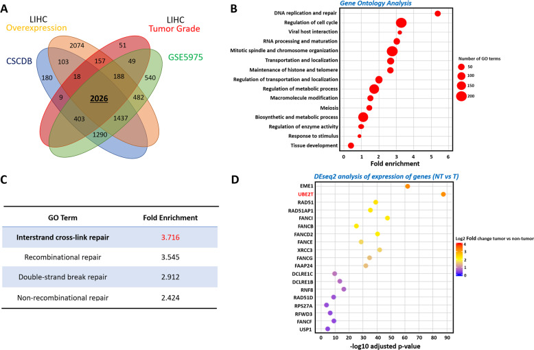
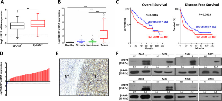
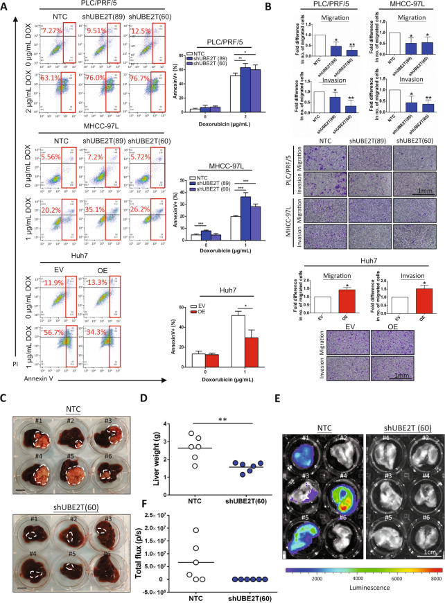
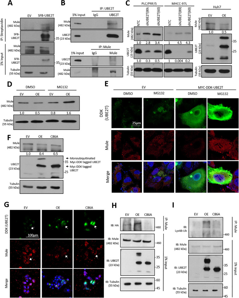
Emerging evidence indicates the role of cancer stem cells (CSCs) in tumor relapse and therapeutic resistance in patients with hepatocellular carcinoma (HCC). To identify novel targets against liver CSCs, an integrative analysis of publicly available datasets involving HCC clinical and stemness-related data was employed to select genes that play crucial roles in HCC via regulation of liver CSCs. We revealed an enrichment of an interstrand cross-link repair pathway, in which ubiquitin-conjugating enzyme E2 T (UBE2T) was the most significantly upregulated. Consistently, our data showed that UBE2T was upregulated in enriched liver CSC populations. Clinically, UBE2T overexpression in HCC was further confirmed at mRNA and protein levels and was correlated with advanced tumor stage and poor patient survival. UBE2T was found to be critically involved in the regulation of liver CSCs, as evidenced by increases in self-renewal, drug resistance, tumorigenicity, and metastasis abilities. Mule, an E3 ubiquitin ligase, was identified to be the direct protein binding partner of UBE2T. Rather than the canonical role of acting as a mediator to transfer ubiquitin to E3 ligases, UBE2T is surprisingly able to physically bind and regulate the protein expression of Mule via ubiquitination. Mule was found to directly degrade β-catenin protein, and UBE2T was found to mediate liver CSC functions through direct regulation of Mule-mediated β-catenin degradation; this effect was abolished when the E2 activity of UBE2T was impaired. In conclusion, we revealed a novel UBE2T/Mule/β-catenin signaling cascade that is involved in the regulation of liver CSCs, which provides an attractive potential therapeutic target for HCC.


Conflict of interest statement

The authors declare that they have no conflict of interest.

Figures

Fig. 1
Fig. 1. UBE2T was upregulated in enriched liver CSC populations.
a An integrative analysis of three publicly available datasets, CSCdB, TCGA (LIHC), and GSE5975, was performed to search for genes that were commonly upregulated between liver cancer cells and liver CSCs compared to normal liver cells. A Venn diagram analysis showed that a total of 2026 genes were commonly upregulated. b GO analysis of these 2026 genes with BiNGO followed by clustering with AutoAnnotate revealed enrichment of DNA replication and repair pathways. c Within the replication and repair pathway, the interstrand cross-link repair pathway had the highest and most significant fold enrichment. d Upon DEseq2 analysis, UBE2T was the most significantly upregulated gene among 19 genes in the interstrand cross-link repair pathway.
Fig. 2
Fig. 2. UBE2T is upregulated in the enriched CSC population and contributes to poor patient survival.
a UBE2T expression was significantly upregulated in CSC-enriched EpCAM+ populations in the GSE5975 dataset (**p < 0.01, t test). b In the analysis of dataset GSE25097, we found upregulation of UBE2T mRNA in a cohort of 243 paired HCC and nontumor samples (***p < 0.001, t test). In addition, UBE2T was upregulated in cirrhotic samples compared with healthy donor samples (*p < 0.05, t test). c In the analysis of the Cancer Genome Atlas (TCGA), the overall and disease-free survival rates of HCC patients with high UBE2T overexpression were significantly lower than those of patients with low UBE2T expression (p = 0.0016 and p = 0.0013, respectively; log-rank test). d qPCR analysis showed that 55% of HCC patient specimens (45 out of 82 cases) showed an eightfold increase in UBE2T mRNA expression compared to that in adjacent nontumor liver tissues. e A representative photo (case #12457) showing the boundary between the nontumor (NT) and tumor (T) regions (M indicates the margin, scale bar: 200 µm). f UBE2T protein overexpression was found in 87.5% (7/8) of HCC specimens (n = 8). Immunoblots are representative of two individual experiments.
Fig. 3
Fig. 3. UBE2T regulates the liver CSC characteristics of HCC cells.
a Two different shUBE2T sequences (89 and 60) and one sequence for nontarget control (NTC) were used. Western blotting showed the successful knockdown of UBE2T in PLC/PRF/5 and MHCC-97L cells. b Successful overexpression of UBE2T was also evidenced in Huh7 cells. c Knockdown of UBE2T also reduced the size and number of hepatospheres formed by PLC/PRF/5 and MHCC-97L cells (*p < 0.05 and ***p < 0.001, t test), while overexpression of UBE2T increased the size and number of hepatospheres formed by Huh7 cells (*p < 0.05, t test; scale bar: 100 µm). d Knockdown of UBE2T in PLC/PRF/5 and MHCC-97L cell lines suppressed tumorigenicity compared with that in NTC cells. Representative photos showing the injection of 50000 and 10000 cells derived from PLC/PRF/5 and MHCC-97L cells. Overexpression of UBE2T led to increased tumorigenicity of Huh7 cells. Representative photographs showing the injection of 10000 cells (scale bar: 1 cm). e Knockdown of UBE2T decreased the expression of CD47, CD90, and CD133 in both PLC/PRF/5 and MHCC-97L cells compared with that in NTC cells (*p < 0.05 and **p < 0.01, t test). Overexpression of UBE2T increased the expression of CD47, CD90, and CD133 in Huh7 cells (*p < 0.05 and **p < 0.01, t test). Immunoblots, FACS and sphere formation assay represent means ± SD in at least three independent experiments (n = 3–4).
Fig. 4
Fig. 4. UBE2T regulates the chemoresistance, migration, invasion, and metastatic potential of HCC cells.
a Compared to NTC cells, shUBE2T cells derived from PLC/PRF/5 and MHCC-97L cells showed a higher percentage of annexin V-positive cells in response to doxorubicin treatment at 2 and 1 µg/mL for 24 h. Compared to EV control cells, UBE2T OE cells showed a lower percentage of annexin V-positive cells in response to doxorubicin treatment at 1 µg/mL for 24 h (*p < 0.05, **p < 0.01, and ***p < 0.001, t test). b Knockdown of UBE2T reduced the number of migratory and invasive HCC cells in uncoated and Matrigel-coated Transwell assays, respectively (*p < 0.05 and **p < 0.01, t test). UBE2T OE cells showed a higher number of migratory and invasive HCC cells than EV control cells (*p < 0.05, t test). c In an orthotopic HCC metastatic model, livers with corresponding tumors derived from NTC cells and shUBE2T (60) cells were excised (scale bar: 1 cm). d Liver weight was shown as a dot plot (**p < 0.05, t test). e Suppression of UBE2T reduced the incidence of lung metastasis (0/6 vs. 4/6) in MHCC-97L cells in vivo. f Signal intensity of the lungs was shown as a dot plot. Apoptotic assay, migration and invasion assays represent means ± SD in at least three independent experiments (n = 3–6).
Fig. 5
Fig. 5. Functional significance of the interaction of UBE2T with Mule.
a The interaction between SFB-UBE2T and Mule was demonstrated by coimmunoprecipitation in Huh7 cells. Mule was detected after the pull-down of SFB-UBE2T. b Reciprocal coimmunoprecipitation demonstrated the interaction between endogenous UBE2T and Mule in MHCC-97L cells. c Mule protein levels were upregulated upon UBE2T knockdown in PLC/PRF/5 and MHCC-97L cells. UBE2T overexpression led to the downregulation of Mule in Huh7 cells. d UBE2T-overexpressing Huh7 cells were subjected to MG132 treatment. Western blot analysis showed that the proteasome inhibitor MG132 rescued the downregulation of Mule in UBE2T-overexpressing cells. e IF analysis showed a reduction in Mule expression in UBE2T OE cells compared to control cells. MG132 treatment increased the level of Mule in both the cytoplasm and nucleus, indicating that UBE2T regulates Mule by proteasomal degradation (scale bar: 25 μm). f Impaired E2 activity of UBE2T reduced the degradation of Mule. The arrow indicates the absence of monoubiquitination in the C86A mutant. g By IF staining, the C86A mutation rescued the suppression of Mule expression in the UBE2T OE clones of Huh7 cells (scale bar: 100 μm). h HA-ubiquitin together with wild-type Myc-DDK-UBE2T, C86A mutant or control vector were co-transfected into Huh7 cells. After treatment with 20 μM MG132 for 6 h, Mule was pulled down to investigate the effect of UBE2T on the abundance of ubiquitinated Mule. Overexpression of wild-type UBE2T promoted the ubiquitination of Mule, while C86A mutation significantly suppressed ubiquitination (IP: Mule, IB: HA). i Immunoprecipitation assay showing an increase in K48-linked ubiquitination of Mule protein (IP: Mule; IB: Lys48-Ub). Immunoblots and IF stainings representative of two or more independent experiments.
Fig. 6
Fig. 6. β-catenin as a downstream effect of the UBE2T/Mule complex.
a Knockdown of Mule led to upregulation of β-catenin expression in Huh7 cells. b Knockdown of UBE2T in PLC/PRF/5 and MHCC-97L cells resulted in upregulation of Mule and suppression of β-catenin. c IF staining of β-catenin showed a reduction in the total expression and nuclear expression of β-catenin in UBE2T knockdown HCC cells (scale bar: 25 μm). Arrows indicate nuclear expression of β-catenin. d Overexpression of UBE2T enhanced β-catenin expression, possibly via degradation of Mule. C86A-transfected HCC cells showed attenuated effects on Mule-mediated β-catenin degradation. e, f IF staining showed marked upregulation of β-catenin in the nucleus and cytoplasm in OE Huh7 transfectant cells but not C86A mutant cells. Arrows indicate nuclear expression of β-catenin. β-Catenin expression in the nucleus and cytoplasm of Huh7 cells derived from EV, OE, and C86A was quantified. g By TOP/FOP assay, transactivating activity of β-catenin was examined in UBE2T-overexpressing and knockdown HCC cells (NTC vs. shUBE2T; EV vs. OE/C86A; OE vs. C86A) (*p < 0.05 and **p < 0.01, t test). h By qPCR analysis, the expression of β-catenin downstream genes, including Cyclin D1 and c-Myc, was examined in UBE2T-overexpressing and knockdown HCC cells (NTC vs. shUBE2T; EV vs. OE/C86A; OE vs. C86A) (*p < 0.05, **p < 0.01, and ***p < 0.001, t test). Immunoblots and IF stainings representative of two or more independent experiments. Immunoblots, IF staining, qPCR, and TOP/FOP assay represent means ± SD in three or more independent experiments (n = 3–4).
Fig. 7
Fig. 7. UBE2T regulated liver CSCs through the Wnt/β-catenin signaling cascade.
a A tissue microarray consisting of 51 tumor tissues and corresponding nontumor liver tissues was subjected to IHC analysis. Cases #23 and #45 showed high expression of both UBE2T and β-catenin, while cases #15 and #20 showed low expression of these proteins. b UBE2T expression was significantly correlated with β-catenin expression in a cohort of 51 HCC clinical samples (***p < 0.001, Fisher’s exact test). c The addition of CHIR99021 at a dose of 1 μM abolished the inhibitory effect of UBE2T knockdown on the size and number of spheres formed from PLC/PRF/5 cells (*p < 0.05, t test). d The addition of CHIR99021 at a dose of 1 μM increased the expression of CD47, CD90, and CD133 upon UBE2T knockdown in PLC/PRF/5 cells (*p < 0.05 and **p < 0.01, t test). FACS and sphere formation assay represent means ± SD in at least three independent experiments (n = 3–4).

Similar articles

Cited by

References

    1. Bray F, et al. Global cancer statistics 2018: GLOBOCAN estimates of incidence and mortality worldwide for 36 cancers in 185 countries. Ca. Cancer J. Clin. 2018;68:394–424. doi: 10.3322/caac.21492. - DOI - PubMed
    1. Kumari R, Sahu MK, Tripathy A, Uthansingh K, Behera M. Hepatocellular carcinoma treatment: hurdles, advances and prospects. Hepat. Oncol. 2018;5:HP98. - PMC - PubMed
    1. Lapidot T, et al. A cell initiating human acute myeloid leukaemia after transplantation into SCID mice. Nature. 1994;367:645–648. doi: 10.1038/367645a0. - DOI - PubMed
    1. O’Brien CA, Pollett A, Gallinger S, Dick JE. A human colon cancer cell capable of initiating tumor growth in immunodeficient mice. Nature. 2007;445:106–110. doi: 10.1038/nature05372. - DOI - PubMed
    1. Singh SK, et al. Identification of a cancer stem cell in human brain tumors. Cancer Res. 2003;63:5821–5828. - PubMed

Publication types