. 2021 Feb 1;12(2):142.
doi: 10.1038/s41419-021-03419-y.

CircRILPL1 promotes muscle proliferation and differentiation via binding miR-145 to activate IGF1R/PI3K/AKT pathway

Affiliations

CircRILPL1 promotes muscle proliferation and differentiation via binding miR-145 to activate IGF1R/PI3K/AKT pathway

Xuemei Shen et al. Cell Death Dis. .

Abstract

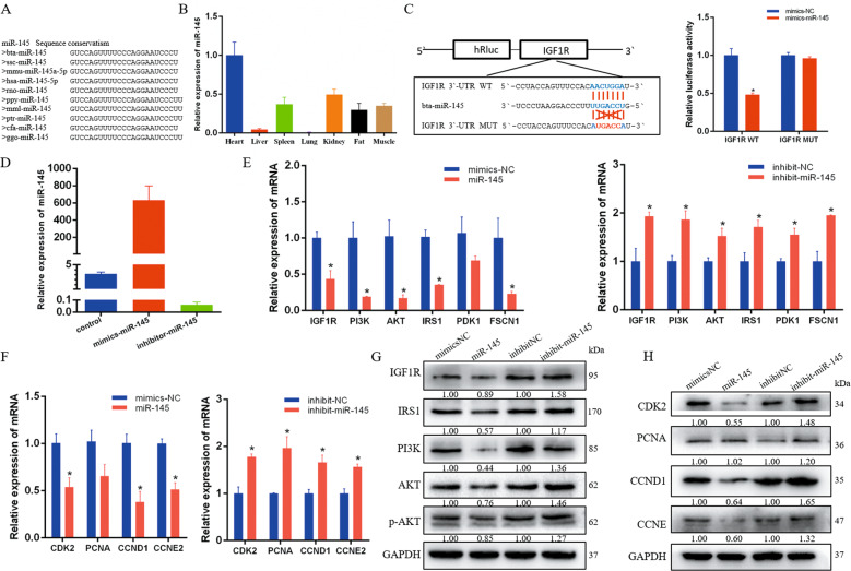
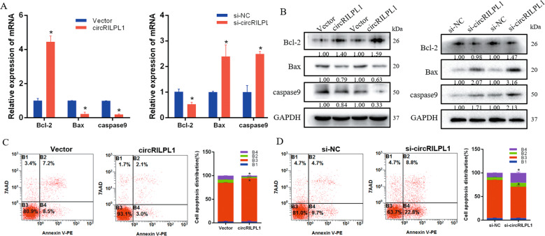
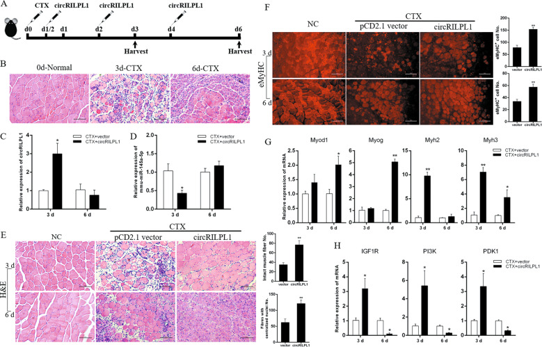
Many novel non-coding RNAs, such as microRNAs (miRNAs) and circular RNAs (circRNAs), are involved in various physiological and pathological processes. The PI3K/AKT signaling pathway is important for its role in regulating skeletal muscle development. In this study, molecular and biochemical assays were used to confirm the role of miRNA-145 (miR-145) in myoblast proliferation and apoptosis. Based on sequencing data and bioinformatics analysis, we identified a new circRILPL1, which acts as a sponge for miR-145. The interactions between circRILPL1 and miR-145 were examined by bioinformatics, a luciferase assay, and RNA immunoprecipitation. Mechanistically, knockdown or exogenous expression of circRILPL1 in the primary myoblasts was performed to prove the functional significance of circRILPL1. We investigated the inhibitory effect of miR-145 on myoblast proliferation by targeting IGF1R to regulate the PI3K/AKT signaling pathway. A novel circRILPL1 was identified that could sponge miR-145 and is related to AKT activation. In addition, circRILPL1 was positively correlated with muscle proliferation and differentiation in vitro and could inhibit cell apoptosis. The newly identified circRILPL1 functions as a miR-145 sponge to regulate the IGF1R gene and rescue the inhibitory effect of miR-145 on the PI3K/AKT signaling pathway, thereby promoting myoblast growth.


Conflict of interest statement

The authors declare that they have no conflict of interest.

Figures

Fig. 1
Fig. 1. miR-145 targets IGF1R to inhibit the PI3K/AKT signaling pathway in bovine myocytes.
A Sequence conservation analysis of miR-145 among different species. B miR-145 expression pattern in different cattle tissues. C The dual fluorescence reporter system verified the targeting relationship between miR-145 and the IGF1R gene. D Real-time qPCR detected miR-145 overexpression and interference efficiency in bovine primary muscle cells. E, G The mRNA and protein levels of PI3K/AKT pathway-related genes in bovine myocytes with miR-145 overexpression and inhibition. F, H The mRNA and protein levels of cell proliferation-related genes in bovine myocytes with miR-145 overexpression and inhibition. n = 3. *P < 0.05.
Fig. 2
Fig. 2. miR-145 promotes the apoptosis of bovine primary myocytes.
A, B The expression of Bcl-2, Bax, caspase9, and p53 was detected by real-time qPCR and western blots. C, D Cell apoptosis was determined by Annexin V/7-AAD dual staining followed by flow cytometry. E, F Cells were stained with JC-1 and images were acquired using a fluorescence microscope. Scale bars, 200 µm. Data are shown as means ± SEM for three individuals. n = 3. *P < 0.05.
Fig. 3
Fig. 3. circRILPL1 serves as a miR-145 sponge.
A CircRNAs with miR-145 absorption sites were screened from high-throughput sequencing data. B Luciferase activity of psiCheck2-miR-145 sensor in HEK293T cells co-transfected with miR-145 mimics and circRNAs, which are putatively binding to the miR-145 sequence. Luciferase activity was normalized by firefly luciferase activity. C Effect of circRILPL1 on the abundance of miRNAs. D Ago2-RIP assay for the amount of circRILPL1 and miR-145 in bovine myoblasts. E CircRNA pull-down assays were performed using a specific biotin-labeled circRILPL1 probe in myoblasts. Real-time qPCR was used to detect the expression levels of circRILPL1 and miR-145 in immunoprecipitates. n = 3. **P < 0.01. Compared with negative control (NC) probe. F Predicted circRILPL1 secondary structure and binding site to miR-145. G Luciferase reporter activity of circRILPL1-WT and circRILPL1-mut in HEK293T cells co-transfected with miR-145 mimics or NC mimics. H The miR-145 biosensor (psiCHECK-2-miR-145 2×) was transfected into HEK293T cells, together with mimics-NC, miR-145 mimics, pCD2.1-non, or pCD2.1-circRILPL1, and luciferase activities were measured after transfection. I Luciferase reporter activity of IGF1R-3’UTR in HEK293T cells with circRILPL1 knockdown or overexpression. J IGF1R expression in myocytes transfected with miR-145 mimics alone or co-transfected with circRILPL1. *P < 0.05. **P < 0.01.
Fig. 4
Fig. 4. circRILPL1 identification and expression pattern in bovine skeletal muscle.
A The genomic locus of circRILPL1 (top). The back-splice junction (arrow) of circRILPL1 was identified by Sanger sequencing (bottom). B PCR analysis of divergent and convergent primers in cDNA and genomic DNA (gDNA). C, D Agarose gel electrophoresis and real-time qPCR analysis of circRILPL1 and RILPL1 levels in myoblasts with and without RNase R treatment. E Real-time qPCR for the abundance of circRILPL1 and RILPL1 in myocytes treated with actinomycin D. F FISH detection of circRILPL1 in myoblasts. Scale bars, 50 µm. G Levels of circRILPL1 in the nuclear and cytoplasmic fractions of myoblasts. H The expression of circRILPL1 in different tissues of cattle at three developmental stages. I Real-time qPCR was used to detect the overexpression and interference efficiency of circRILPL1. Data are presented as means ± SEM. n = 3. **P < 0.01. ***P < 0.001.
Fig. 5
Fig. 5. circRILPL1 promotes cell proliferation by affecting the PI3K/AKT signaling pathway.
A, B The expression levels of cell proliferation marker genes were detected by real-time qPCR and western blots. C, D The expression levels of PI3K/AKT pathway-related genes were detected by real-time qPCR and western blots. E, F CCK-8 analysis after overexpression and interference with circRILPL1. G EdU analysis after overexpression and interference with circRILPL1. Scale bars, 200 µm. H, I Cell cycle analysis of myocytes with overexpressing or silencing circRILPL1. *P < 0.05.
Fig. 6
Fig. 6. circRILPL1 inhibits the apoptosis of bovine primary myocytes.
A, B The expression of Bcl-2, Bax, and caspase9 was detected by real-time qPCR and western blots. C, D Cell apoptosis was determined by Annexin V/7-AAD dual staining followed by flow cytometry.
Fig. 7
Fig. 7. circRILPL1 regulates IGF1R through miR-145.
A CCK-8 assay for myocytes transfected with miR-145 mimics alone or co-transfected with circRILPL1. n = 6. B CCK-8 assay for myocytes with circRILPL1 overexpression and knockdown of IGF1R. n = 6. C, D EdU assay for myocytes transfected with miR-145 mimics alone or co-transfected with circRILPL1. Scale bars, 200 µm. E Cell proliferation and PI3K/AKT pathway-related protein analysis for myocytes transfected with circRILPL1 alone or co-transfected with si-IGF1R and miR-145 mimics. F The expression of MyoD, MyoG, and MyHC was detected by real-time qPCR. G Myocytes were transfected with circRILPL1 alone or co-transfected with si-IGF1R, immunofluorescence (MyHC) was used to analyze the level of muscle cell differentiation. Scale bars, 500 µm. H After 4 days of differentiation, the percentage of nuclei present in MyHC-positive myotubes with the indicated number of nuclei were quantified in cells. Data are presented as mean ± SEM of three independent experiments. I The protein expression of MyoD, MyoG, and MyHC was detected by western blots.
Fig. 8
Fig. 8. CircRILPL1 improves mouse muscle regeneration in vivo.
A Injection scheme for circRILPL1 into CTX-injured muscles. N = 3 mice for each group. B H&E staining of TA muscle at 0, 3, and 6 days after injection of CTX. Scale bars, 100 µm. C The expression of circRILPL1 in the above-injected muscles after injection of overexpression plasmid. The data were normalized to GAPDH mRNA. D The expression of mmu-miR-145a-5p in the above-injected muscles after injection of circRILPL1 plasmid. The data were normalized to U6. E After circRILPL1 expression plasmid injection, muscle H&E staining was performed on the 3rd and 6th day of muscle injury. The empty pCD2.1 vector was injected as a control. Mice in which CTX-muscle injury was not induced served as negative controls (NC). Scale bars, 100 µm. Fibers with intact structure (3 d) or centrally localized nuclei (CLN, 6 d) were quantified. F The above fibers were stained for eMyHC. Scale bars, 100 µm. The positively stained fibers were quantified. G, H The expression of IGF1R, PI3K, PDK1, and myogenic markers in the above-injected muscles. The data were normalized to GAPDH mRNA and represent the average of three independent experiments. Data are presented as means ± SEM. n = 3. *P < 0.05. **P < 0.01.

References

    1. Jansen K, Pavlath G. Molecular control of mammalian myoblast fusion. Methods Mol. Biol. 2008;475:115. doi: 10.1007/978-1-59745-250-2_7. - DOI - PubMed
    1. Bentzinger CF, Wang YX, Rudnicki MA. Building muscle: molecular regulation of myogenesis. Cold Spring Harb. Perspect. Biol. 2012;4:a008342. doi: 10.1101/cshperspect.a008342. - DOI - PMC - PubMed
    1. Braun T, Gautel M. Transcriptional mechanisms regulating skeletal muscle differentiation, growth and homeostasis. Nat. Rev. Mol. Cell Biol. 2011;12:349. doi: 10.1038/nrm3118. - DOI - PubMed
    1. Mok GF, Sweetman D. Many routes to the same destination: lessons from skeletal muscle development. Reproduction. 2011;141:301. doi: 10.1530/REP-10-0394. - DOI - PubMed
    1. Engel AG, Shen X-M, Selcen D, Sine SM. Congenital myasthenic syndromes: pathogenesis, diagnosis, and treatment. Lancet Neurol. 2015;14:420–434. doi: 10.1016/S1474-4422(14)70201-7. - DOI - PMC - PubMed

Publication types

LinkOut - more resources