. 2021 Feb 4;12(1):792.
doi: 10.1038/s41467-021-21078-7.

Engineering advanced logic and distributed computing in human CAR immune cells

Affiliations

Engineering advanced logic and distributed computing in human CAR immune cells

Jang Hwan Cho et al. Nat Commun. .

Abstract

The immune system is a sophisticated network of different cell types performing complex biocomputation at single-cell and consortium levels. The ability to reprogram such an interconnected multicellular system holds enormous promise in treating various diseases, as exemplified by the use of chimeric antigen receptor (CAR) T cells as cancer therapy. However, most CAR designs lack computation features and cannot reprogram multiple immune cell types in a coordinated manner. Here, leveraging our split, universal, and programmable (SUPRA) CAR system, we develop an inhibitory feature, achieving a three-input logic, and demonstrate that this programmable system is functional in diverse adaptive and innate immune cells. We also create an inducible multi-cellular NIMPLY circuit, kill switch, and a synthetic intercellular communication channel. Our work highlights that a simple split CAR design can generate diverse and complex phenotypes and provide a foundation for engineering an immune cell consortium with user-defined functionalities.


Conflict of interest statement

The authors declare the following competing interests: Boston University has filed a patent application, WO2017091546A1, (Methods and compositions relating to chimeric antigen receptors) with J.H.C, and W.W.W. as the named inventor based on this work. W.W.W. is a co-founder and shareholder of Senti Biosciences, and received research support from Senti Biosciences. J.J.C. is a co-founder and shareholder of Senti Biosciences. A.O. is a current employee of Hitachi, Ltd. J.H.C. is a current employee of Spark Therapeutics. K.S. is current employee of KSQ Therapeutics. All other authors declare no competing interests.

Figures
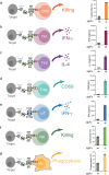
Fig. 1
Fig. 1. The panel of cell types redirected by SUPRA CAR.
a Cytotoxicity by RR zipCAR-expressing CD8+ T cells. (Right) Nalm6 cells expressing Her2 were co-cultured in vitro with RR zipCAR-expressing CD8+ human primary T cells with and without α-Her2-EE zipFv (n = 3, data are represented as mean ± SD). b IFN-γ cytokine level from RR zipCAR expressing in vitro differentiated Th1 cells. (Right) Nalm6 cells expressing Her2 were co-cultured with RR zipCAR-expressing Th1 cells with and without α-Her2-EE zipFv (n = 3, data are represented as mean ± SD). c IL-4 cytokine level from RR zipCAR expressing in vitro differentiated Th2 cells. (Right) Nalm6 cells expressing Her2 were co-cultured with RR zipCAR-expressing Th2 cells with and without α-Her2-EE zipFv (n = 3, data are represented as mean ± SD). d CD69 expression level from RR zipCAR-FoxP3 expressing isolated Treg cells (CD4 + CD25hiCD127low). (Right) Nalm6 cells expressing Her2 were co-cultured with RR zipCAR-expressing Treg cells with and without α-Her2-EE zipFv (n = 3, data are represented as mean ± SD). e IFN-γ cytokine level from FOS zipCAR-expressing isolated γδ T cells. (Right) Nalm6 cells expressing Her2 were co-cultured with FOS zipCAR-expressing γδ T cells with and without α-Her2-SYN9 zipFv (n = 3, data are represented as mean ± SD). f Cytotoxicity by RR zipCAR-expressing NK-92MI cells. (Right) Nalm6 cells expressing Her2 were co-cultured in vitro with RR zipCAR-expressing NK cells with and without α-Her2-EE zipFv (n = 3, data are represented as mean ± SD). g Phagocytosis by RR zipCAR-expressing THP-1 macrophages. (Right) Nalm6 cells expressing Her2 were co-cultured in vitro with RR zipCAR-expressing THP-1 macrophages with and without α-Her2-EE zipFv (n = 3, data are represented as mean ± SD).
Fig. 2
Fig. 2. Engineering endogenous immune system with SUPRA CAR-expressing different T-cell subtypes.
a Schematic of controlling macrophage polarization by zipCAR-expressing Th1 and Th2 cells. The RR zipCAR and FOS zipCAR control activity of Th1 and Th2 cells, respectively. α-Axl-EE zipFv binds to RR zipCAR and activates Th1 cells. α-Her2-SYN9 zipFv binds to the FOS zipCAR and activates Th2 cells. Activation of Th1 and Th2 CD4+ T cells leads to secretion of IFN-γ and IL-4, respectively. Macrophage polarizes to M1 (proinflammatory) when exposed to IFN-γ secreted by Th1 cell and it polarized to M2 (anti-inflammatory) when exposed to IL-4 secreted by Th2 cells. b IFN-γ (Top) and IL-4 (Bottom) production from RR zipCAR-expressing Th1 cells and FOS zipCAR-expressing Th2 cells with or without 5 nM α-Her2-SYN9 zipFv and 5 nM α-Axl-EE zipFv (n = 3, data are represented as the mean ± SD). c HLA-DR (Top left) and CCR7 (Top right) expression levels in THP-1 macrophages were measured by flow cytometer 24 h after staring co-culture. (Bottom) CD206 in THP-1 macrophages was also analyzed at the same time as detecting M2 marker (n = 3, data are represented as the mean ± SD).
Fig. 3
Fig. 3. The intracellular AND logic with different signaling domains.
a Diagram of intracellular AND logic. b Primary human CD8+ T cells were transduced with FOS zipCAR-containing CD3ζ domain and RR zipCAR-containing CD28 domain. Cytotoxicity against Her2- and Axl-expressing Nalm6 was measured 24 h after adding α-Her2-SYN9 and/or α-Axl-EE zipFvs. The heatmap indicates cytotoxicity at varying zipFv concentrations (n = 3, data are represented as mean). c Cytotoxicity of CD8+ T cells transduced with FOS zipCAR-containing CD3ζ domain and RR zipCAR-containing 4-1BB domain. The heatmap indicates cytotoxicity at varying zipFv concentrations (n = 3, data are represented as mean). d (Left) Isolated Treg cells were transduced with two zipCAR constructs: SYN6-CD3ζ-P2A-FOXP3 and SYN1-CD28-P2A-puro. After puromycin selection (2 μg/mL), Treg cells were co-cultured with Her2- and Axl-expressing Nalm6 target cells (Right) The heatmap shows surface CTLA-4 expression detected after 48 h by flow cytometry at varying zipFv concentrations (α-Axl-SYN5 and α-Her2-SYN2) (n = 3, data are represented as mean).
Fig. 4
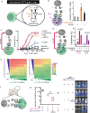
Fig. 4. The intracellular NOT logic with BTLA in different cell types.
a Diagram of intracellular NOT logic with BTLA co-inhibitory signaling domain. b IFN-γ production from CD4+ T cells transduced with FOS-CD28-CD3ζ and RR zipCAR with BTLA co-inhibitory domain. (Right) Supernatants were collected 24 h after adding 1.2 nM α-Her2-SYN9 zipFv and/or 12 nM α-Axl-EE zipFv (n = 2; data are represented as the mean + SD). c Effect of concentration of α-Her2-SYN9 zipFv on cytotoxicity performed by FOS zipCAR-expressing NK-92MI cells with various activation domains (magenta, CD3ζ; light purple, CD28-CD3ζ; dark purple, 2B4; yellow, DAP12; blue, CD28; black with square, ICOS; black with circle, NKG2D; n = 3; data are represented as the mean ± SD). d Suppression of cytotoxicities by BTLA. NK-92MI cells expressing FOS zipCAR with activating domains (CD3ζ or 2B4) and RR zipCAR with BTLA co-inhibitory domain were co-cultured with Her2 and Axl-expressing Nalm6 target cells in the presence of different combinations of zipFvs (α-Axl-SYN9, α-Her2-EE). (Right) Live target cells were measured by flow cytometry 24 h after co-culture (magenta, CD3ζ; dark purple, 2B4; n = 3; data are represented as the mean + SD; the statistical significance was determined by two-tailed student’s t-test). e Effect of concentration of α-Her2-SYN9 and/or α-Axl-EE zipFvs on cytotoxicity performed by NK-92MI cells. SUPRA CAR-NK cells express FOS zipCAR with BTLA co-inhibitory domain and RR zipCAR with CD3ζ domain (Left) and with 2B4 activation domain (Right) (n = 3, data are represented as the mean). f Schematic of the xenograft mouse model for verification of NOT gate NK cells. NK-92MI cells were expressing both FOS zipCAR with CD3ζ and RR zipCAR with BTLA. Target SKOV3-luc cells have high-level Her2 expression. We also administrated α-Her2-EE zipFv, α-Her2-SYN9 zipFv, and α-Her2 scFv (Mock) to specified groups in Fig. 4g. g (Left) Box and whiskers graph shows tumor burden in each group at 22 days after tumor injection; tumor only without NK cells (black), tumor and NK cells without zipFv (gray), tumor and NK cells with α-Her2-EE zipFv and α-Her2 scFv (magenta), and tumor and NK cells with α-Her2-EE zipFv and α-Her2-SYN9 zipFv (blue). Each zipFvs were injected intraperitoneally every day for 21 days. (Right) Representative bioluminescence images at day 22. Box plots indicate median (middle line), 25th, 75th percentile (box), and 5th and 95th percentile (whiskers). n = 4 (4th group is n = 2 because 2 mice died before imaging); mean ± SD; the statistical significance was determined by Multiple t-test; **p = 0.01.
Fig. 5
Fig. 5. Tunable 3-input multilogic in the single cell.
a Design of orthogonal SUPRA CARs that control CD3ζ, CD28, and BTLA signaling domains inducibly and independently. Primary CD8+ T cells were engineered to express FOS zipCAR, SYN6 zipCAR and SYN1 zipCAR that contain CD3ζ domain, CD28, and BTLA signaling domain, respectively. In addition, α-Meso-SYN9 zipFv, α-Axl-SYN5 zipFv, and α-Her2-SYN2 zipFv lead to activation of CD3ζ, CD28, and BTLA, respectively. (Right) IFN-γ secretion was measured after co-culturing with Her2, Axl, and Meso expressing Nalm6 target cells with different zipFv combinations (n = 3, data are represented as the mean + SD, the statistical significance was determined by Student’s t-test). b Primary CD4+ T cells expressing FOS-CD3ζ, SYN6-CD28, and SYN1-BTLA were co-cultured with Her2, Axl, and CD19 expressing Nalm6 target cells. The 3D heatmap shows IFN-γ production from 3-input CD4+ T cells at varying concentrations of three different corresponding zipFvs (n = 2, data are represented as the mean).
Fig. 6
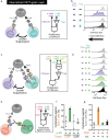
Fig. 6. The intercellular NOT gate with regulatory T (Treg) cells.
a Diagram of intercellular NOT gate with Treg cells. The RR zipCAR and FOS zipCAR control the activity of Treg and conventional CD4+ T cells (Tconv), respectively. α-Axl-EE zipFv binds to the RR zipCAR and activates Treg cells. α-Her2-SYN9 zipFv binds to the FOS zipCAR and activates CD4+ Tconv cells. Activation of Treg cells led to the suppression of CD4+ Tconv cells. b Suppression of growth of CD4+ Tconv cell by SUPRA CAR equipped Treg cells. CD4+ Tconv cells expressing FOS zipCAR were prelabeled with CellTrace Violet dye. When activated by CAR signaling, the fluorescence intensity of labeled cells decrease with cellular growth. Cells were analyzed by flow cytometer after 4 days of the co-culture period. The left shift of peaks indicates T-cell proliferation. Each plot shows dye fluorescence of CD4+ Tconv cells with different zipFv combinations (representative of three biological replicates). c Diagram of A AND NOT (B AND C) logic gate with AND gate Treg cells. Target Nalm6 cells express Her2, Axl, and CD19. CD4+ Tconv cells expressing FOS zipCAR were prelabeled with CellTrace Violet dye. Treg cells expressed SYN1-CD28 and SYN6-CD3ζ (see also Supplementary Fig. 3b). d Suppression of CD4+ conventional T-cell growth by AND gate Treg cells. α-Her2-SYN9 zipFv activated CD4+ conv T cells. α-CD19 SYN2 zipFv and α-Her2-SYN5 zipFv activated CD28 and CD3ζ in Treg cells, respectively. Histograms indicate the divided cells in CD4+ conv cells measured by fluorescent dye dilution for 4 days. e Diagram of the kill switch with CAR-targeting NK cells. Target NALM6 cells expressed Her2. CD8+ T cells and NK cells expressed SYN6-V5 tag zipCAR and α-FITC-CAR, respectively. f Cytotoxicity against CD8+ cells. α-V5-FITC was added to co-culture of CD8+ T cells and α-FITC-CAR-NK cells. The graph compares the cytotoxicity against SYN6-V5 zipCAR+ CD8+ T cells to CAR negative CD8+ T cells (n = 3; Mean + SD). g Cytotoxicity against target cells. α-Her2-SYN5 and/or α-V5-FITC were added to co-culture of SYN6-V5 zipCAR+ CD8+ T cells, α-FITC-CAR-NK cells, and target Nalm6 cells simultaneously. Live target cells are counted by flow cytometer 24 h after co-culture (n = 3; Mean + SD).
Fig. 7
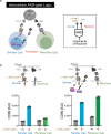
Fig. 7. The intercellular AND gate using zipFv secretion system.
a Diagram of intercellular AND gate logic. CD4+ Sender cells express SYN6 CAR and secrete α-Axl-SYN2 zipFv when activated by α-Her2 zipFv. CD4+ receiver cells get activated by α-Axl-SYN2 zipFv secreted by sender cells. b. (Left) Addition of α-Her2 zipFv will activate sender cells that contain zipFv secretion module and secrete α-Axl-SYN2 zipFv, which will activate receiver cells as measured by CD69 expression level (Right) Activation of sender cells without zipFv secretion module does not lead to activation of receiver cells (n = 3, data are represented as the mean + SD).

References

    1. Ellebrecht CT, et al. Reengineering chimeric antigen receptor T cells for targeted therapy of autoimmune disease. Science. 2016;353:179–184. doi: 10.1126/science.aaf6756. - DOI - PMC - PubMed
    1. Ribas A, Wolchok JD. Cancer immunotherapy using checkpoint blockade. Science. 2018;359:1350–1355. doi: 10.1126/science.aar4060. - DOI - PMC - PubMed
    1. Wei SC, Duffy CR, Allison JP. Fundamental mechanisms of immune checkpoint blockade therapy. Cancer Discov. 2018;8:1069–1086. doi: 10.1158/2159-8290.CD-18-0367. - DOI - PubMed
    1. Feucht J, et al. Calibration of CAR activation potential directs alternative T cell fates and therapeutic potency. Nat. Med. 2019;25:82–88. doi: 10.1038/s41591-018-0290-5. - DOI - PMC - PubMed
    1. Grupp SA, et al. Chimeric antigen receptor-modified T cells for acute lymphoid leukemia. N. Engl. J. Med. 2013;368:1509–1518. doi: 10.1056/NEJMoa1215134. - DOI - PMC - PubMed

Publication types

MeSH terms

Substances