. 2021 Feb 4;11(1):3063.
doi: 10.1038/s41598-021-82341-x.

Chondrogenesis of human amniotic fluid stem cells in Chitosan-Xanthan scaffold for cartilage tissue engineering

Affiliations

Chondrogenesis of human amniotic fluid stem cells in Chitosan-Xanthan scaffold for cartilage tissue engineering

Carolina C Zuliani et al. Sci Rep. .

Abstract

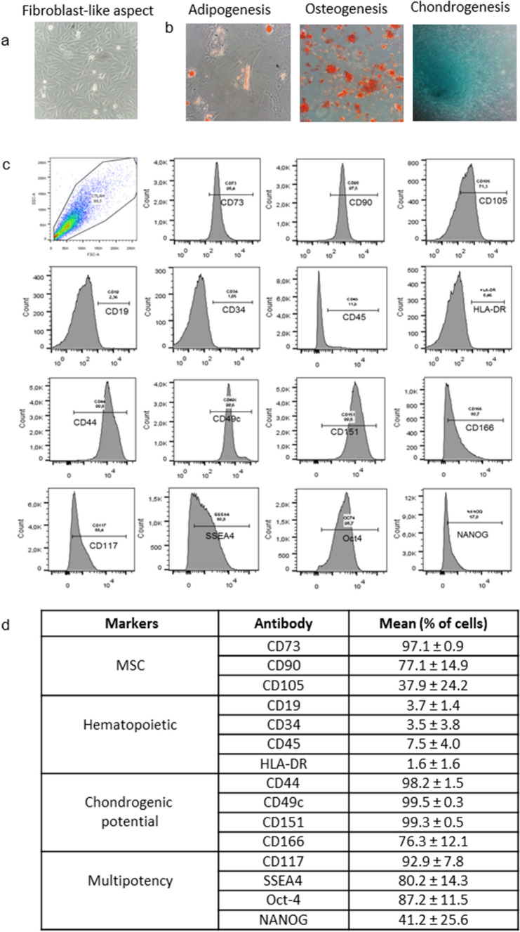
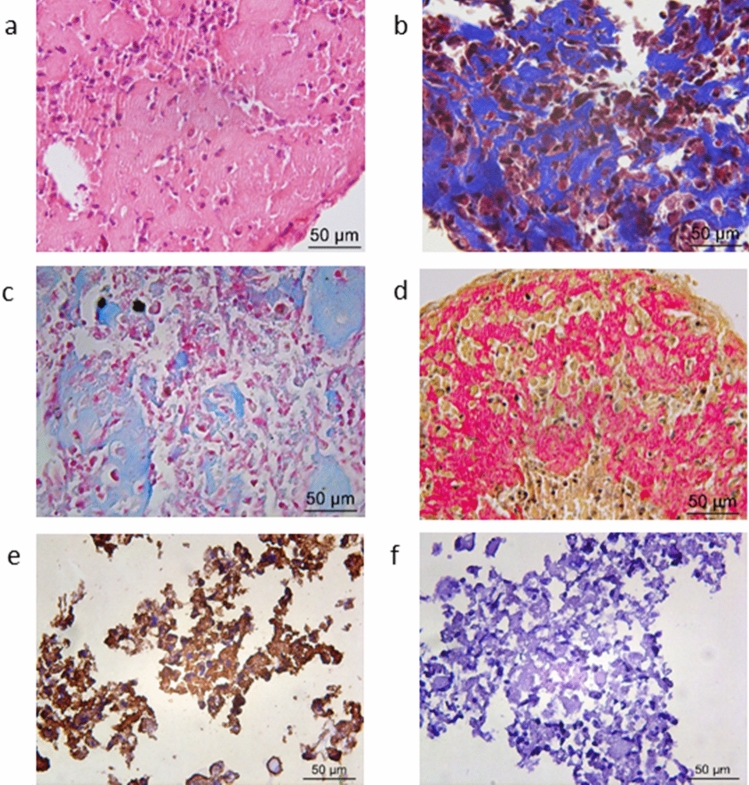
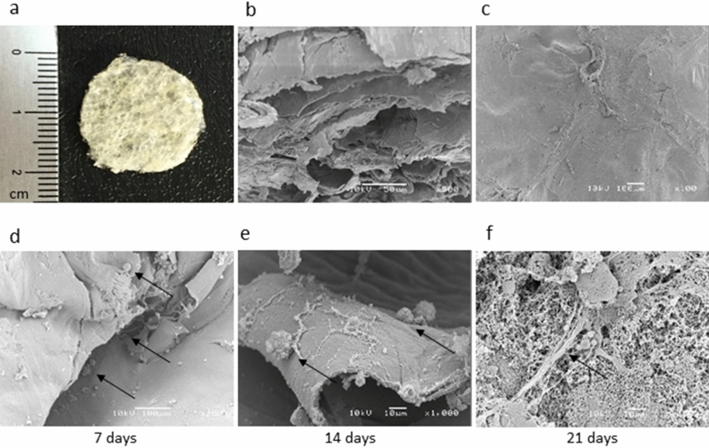
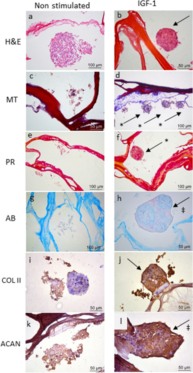
Articular chondral lesions, caused either by trauma or chronic cartilage diseases such as osteoarthritis, present very low ability to self-regenerate. Thus, their current management is basically symptomatic, progressing very often to invasive procedures or even arthroplasties. The use of amniotic fluid stem cells (AFSCs), due to their multipotentiality and plasticity, associated with scaffolds, is a promising alternative for the reconstruction of articular cartilage. Therefore, this study aimed to investigate the chondrogenic potential of AFSCs in a micromass system (high-density cell culture) under insulin-like growth factor 1 (IGF-1) stimuli, as well as to look at their potential to differentiate directly when cultured in a porous chitosan-xanthan (CX) scaffold. The experiments were performed with a CD117 positive cell population, with expression of markers (CD117, SSEA-4, Oct-4 and NANOG), selected from AFSCs, after immunomagnetic separation. The cells were cultured in both a micromass system and directly in the scaffold, in the presence of IGF-1. Differentiation to chondrocytes was confirmed by histology and by using immunohistochemistry. The construct cell-scaffold was also analyzed by scanning electron microscopy (SEM). The results demonstrated the chondrogenic potential of AFSCs cultivated directly in CX scaffolds and also in the micromass system. Such findings support and stimulate future studies using these constructs in osteoarthritic animal models.


Conflict of interest statement

The authors declare no competing interests.

Figures

Figure 1
Figure 1
Characterization of human AFSCs: (a) cells showing adherent growth, exhibiting fibroblast-like aspect (400 ×); (b) differentiation potential in mesenchymal lineage after culture in specific medium (400 ×): adipogenic, oil Red O staining indicating lipidic vesicles; osteogenic, alizarin red staining indicating calcium matrix formation and chondrogenic, Alcian blue staining evidencing glycosaminoglycan presence; (c) immunophenotype analysis of AFSCs by flow cytometry; (d) percentage of cells showing specific markers of MSC, hematopoietic, multipotency, and chondrogenic potential in different samples.
Figure 2
Figure 2
Micromass results stimulated with IGF-1 for 21 days by chondrogenic differentiation: Histological analysis (400 ×): H&E stain (neoformed matrix appears in pink) (a), Masson’s trichrome (collagens appear in blue) (b), Alcian Blue stain (glycosaminoglycans appear in light blue) (c), and Picrosirius Red stain (collagens fibers appears in red) (d); positive immunohistochemical staining for specific Type II collagen (e) and negative control (f).
Figure 3
Figure 3
Macroscopic aspect of the CX scaffold (a) SEM micrographs of CX: section porous structure (b); surface and morphology (c). Aspect of CD117+ AFSCs cultured in CX analyzed in different periods. The black arrows indicate the cells adhered to the scaffold with 7 days (d), 14 days (e), and 21 days of culture, exhibiting many adhered cells and evident matrix network production (f).
Figure 4
Figure 4
Chondrogenic differentiation of CD117+ AFSCs seeded into CX scaffold for 21 days with and without IGF-1 stimulation. In the group stimulated by IGF-1 (b,d,f,h,j,l) more compact agglomeration of the cells is observed,with a higher amount of extracellular material compared to that noticed in the non-stimulated control group (a,c,e,g,i,k). H&E staining (a,b); collagen (indicated with *) stained in blue by Masson´s trichrome (MT) (c,d); collagen fibers shown in red by Picrosirius red (PR) (e,f); glycosaminoglycans (‡) stained light blue using Alcian blue (AB) (g,h). Labeling with anti-collagen type II antibody (COL II) (i,j) and anti-aggrecan antibody (ACAN) (k,l) in the stimulated group compared to the control group.

References

    1. Sgaglione NA, Miniaci A, Gillogly SD, Carter TR. Update on advanced surgical techniques in the treatment of traumatic focal articular cartilage lesions in the knee. Arthroscopy. 2002;18:9–32. doi: 10.1053/jars.2002.31783. - DOI - PubMed
    1. Chiang H, Jiang CC. Repair of articular cartilage defects: Review and perspectives. J. Formos. Med. Assoc. 2009;108:87–101. doi: 10.1016/S0929-6646(09)60039-5. - DOI - PubMed
    1. Mobasheri A, Bay-Jensen AC, van Spil WE, Larkin J, Levesque MC. Osteoarthritis year in review 2016: Biomarkers (biochemical markers) Osteoarth. Cartil. 2017;25:199–208. doi: 10.1016/j.joca.2016.12.016. - DOI - PubMed
    1. Fibel KH, Hillstrom HJ, Halpern BC. State-of-the-Art management of knee osteoarthritis. World J. Clin. Cases. 2015;3:89–101. doi: 10.12998/wjcc.v3.i2.89. - DOI - PMC - PubMed
    1. Cheng NC, Estes BT, Awad HA, Guilak F. Chondrogenic differentiation of adipose-derived adult stem cells by a porous scaffold derived from native articular cartilage extracellular matrix. Tissue Eng. Part A. 2009;15:231–241. doi: 10.1089/ten.tea.2008.0253. - DOI - PMC - PubMed

Publication types

MeSH terms