. 2021 Feb 4;11(1):3022.
doi: 10.1038/s41598-021-82622-5.

Photons detected in the active nerve by photographic technique

Affiliations

Photons detected in the active nerve by photographic technique

Andrea Zangari et al. Sci Rep. .

Abstract

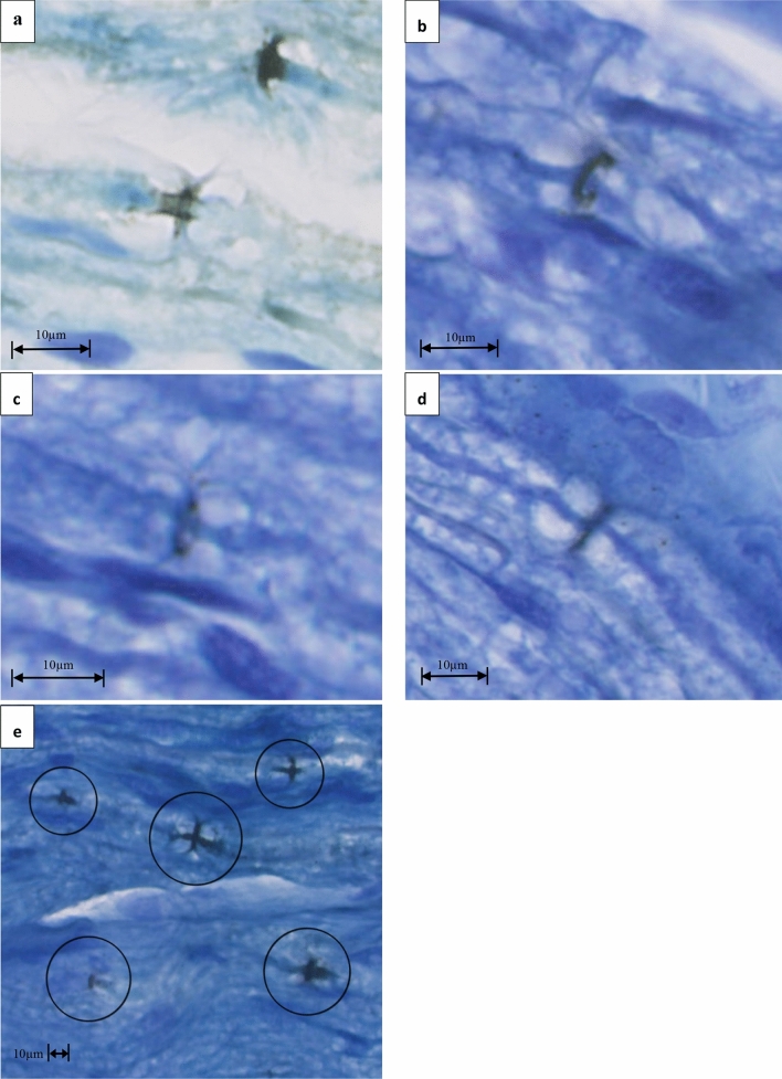
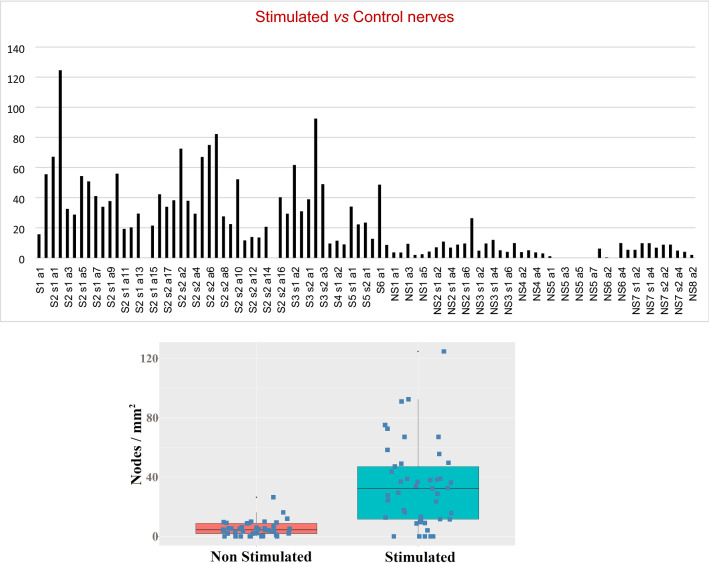
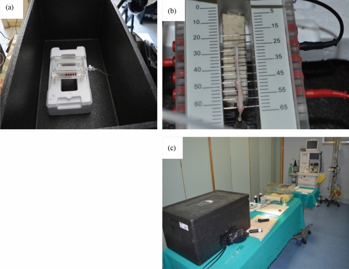
The nervous system is one of the most complex expressions of biological evolution. Its high performance mostly relies on the basic principle of the action potential, a sequential activation of local ionic currents along the neural fiber. The implications of this essentially electrical phenomenon subsequently emerged in a more comprehensive electromagnetic perspective of neurotransmission. Several studies focused on the possible role of photons in neural communication and provided evidence of the transfer of photons through myelinated axons. A hypothesis is that myelin sheath would behave as an optical waveguide, although the source of photons is controversial. In a previous work, we proposed a model describing how photons would arise at the node of Ranvier. In this study we experimentally detected photons in the node of Ranvier by Ag+ photoreduction measurement technique, during electrically induced nerve activity. Our results suggest that in association to the action potential a photonic radiation takes place in the node.


Conflict of interest statement

The authors declare no competing interests.

Figures

Figure 1
Figure 1
Examples of nodes where Ag deposited after electrical stimulation of rat sciatic nerves. In (ad) a zoom of some annular Ag depositions in the nodes are displayed. Circles in (e) put in evidence Ag deposition in the nodes and along paranodal segments.
Figure 2
Figure 2
Examples of rat sciatic nerve sections (a,b). Node count, by small circle markings, in delimited fascicular areas in sections from non-stimulated nerve (c) and stimulated nerve (d).
Figure 3
Figure 3
Up: Nodes’ density in sample areas in control nerves and nodes’ density in sample areas in stimulated nerves. NS non-stimulated (control nerve), S stimulated nerve, s section, a sample area (n integer, number of nerve, section and sample respectively); Down: Node density of “non stimulated” and “stimulated” sample groups represented as a boxplot including average values and standard deviation.
Figure 4
Figure 4
Two different examples (left and right) of Ag deposition (black) in the paranode after electrical stimulation of the rat sciatic nerves.
Figure 5
Figure 5
Experimental setup for nerve stimulation and chemical processing. (a) The Biopac nerve chamber in acrylic (Biopac ®); it features 15 stainless steel pins; each pin is spaced 5 mm apart to provide a variety of recording and stimulation configurations; the sockets accept 2 mm plugs and interface with stimulation and recording cables; the nerve chamber is secured within the polystyrene dark box to prevent environmental light from hitting the nerve. (b) Stimulation and measurement electrodes connected to the steel pins and a rat nerve placed on the pins. (c) The final lab setup: by means of a tube the different solutions for the chemical treatment of nerves can be introduced or extracted without opening the box; wires from the electrodes and the tube are passed through holes in the box wall and isolated by envelopes; the dark box is closed and ready for the experiment.

References

    1. Hodgkin AL, Huxley AF. A quantitative description of membrane current and its application to conduction and excitation in nerve. J. Physiol. 1952;117:500–544. doi: 10.1113/jphysiol.1952.sp004764. - DOI - PMC - PubMed
    1. Anastassiou C, Perin R, Markram H, et al. Ephaptic coupling of cortical neurons. Nat. Neurosci. 2011;14:217–223. doi: 10.1038/nn.2727. - DOI - PubMed
    1. Wu F, Ma J, Zhang G. A new neuron model under electromagnetic field. Appl. Math. Comput. 2019;347:590–599.
    1. Cifra M, Fields JZ, Farhadi A. Electromagnetic cellular interactions. Prog. Biophys. Mol. Biol. 2011;105:223–246. doi: 10.1016/j.pbiomolbio.2010.07.003. - DOI - PubMed
    1. Buzsáki G, Anastassiou CA, Koch C. The origin of extracellular fields and currents—EEG, ECoG, LFP and spikes. Nat. Rev. Neurosci. 2012;13:407–420. doi: 10.1038/nrn3241. - DOI - PMC - PubMed

LinkOut - more resources