

The intestinal neuro-immune axis: crosstalk between neurons, immune cells, and microbes
- PMID: 33542493
- PMCID: PMC8075967
- DOI: 10.1038/s41385-020-00368-1
The intestinal neuro-immune axis: crosstalk between neurons, immune cells, and microbes
Abstract
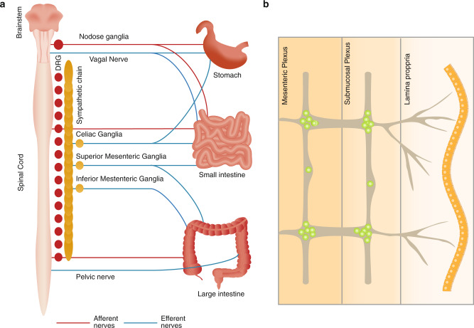
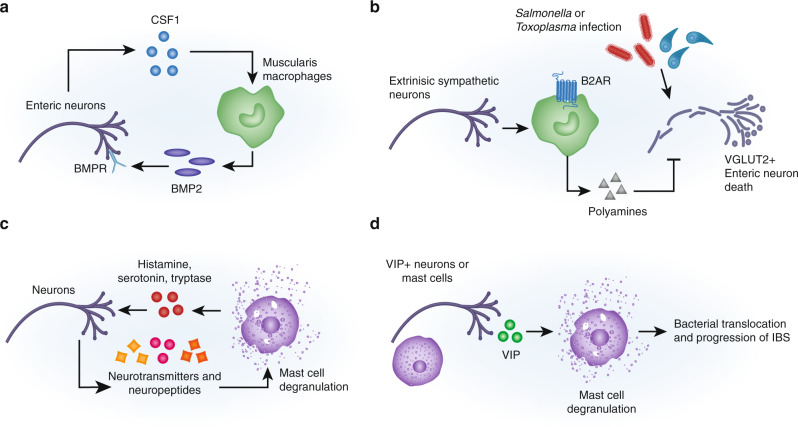
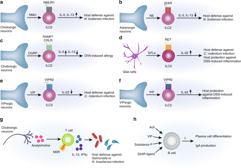
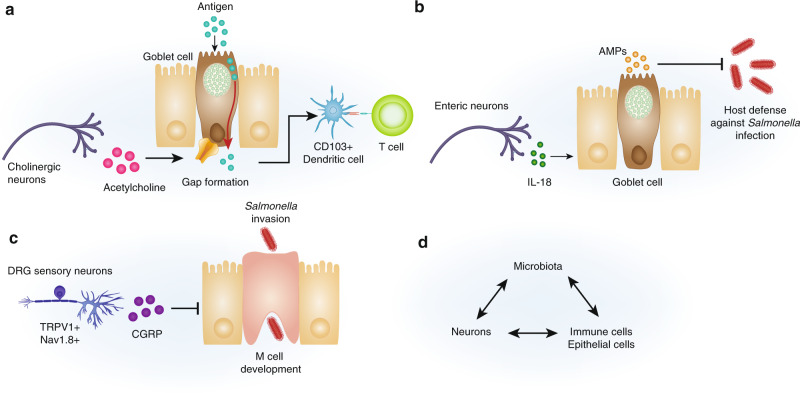
The gastrointestinal tract is densely innervated by a complex network of neurons that coordinate critical physiological functions. Here, we summarize recent studies investigating the crosstalk between gut-innervating neurons, resident immune cells, and epithelial cells at homeostasis and during infection, food allergy, and inflammatory bowel disease. We introduce the neuroanatomy of the gastrointestinal tract, detailing gut-extrinsic neuron populations from the spinal cord and brain stem, and neurons of the intrinsic enteric nervous system. We highlight the roles these neurons play in regulating the functions of innate immune cells, adaptive immune cells, and intestinal epithelial cells. We discuss the consequences of such signaling for mucosal immunity. Finally, we discuss how the intestinal microbiota is integrated into the neuro-immune axis by tuning neuronal and immune interactions. Understanding the molecular events governing the intestinal neuro-immune signaling axes will enhance our knowledge of physiology and may provide novel therapeutic targets to treat inflammatory diseases.
Conflict of interest statement
I.M.C consults for GSK, Kallyope, and Senda Pharmaceuticals, and his lab receives sponsored research support from GSK and Allergan Pharmaceuticals. The other authors declare no conflicts of interest.
Figures
Similar articles
-
Neuroimmune Interactions in the Intestine.Annu Rev Immunol. 2024 Jun;42(1):489-519. doi: 10.1146/annurev-immunol-101921-042929. Annu Rev Immunol. 2024. PMID: 38941607 Review.
-
Neuro-immune cell interactions in the regulation of intestinal immune homeostasis.Curr Opin Gastroenterol. 2025 Jan 1;41(1):38-45. doi: 10.1097/MOG.0000000000001065. Epub 2024 Oct 17. Curr Opin Gastroenterol. 2025. PMID: 39417780 Review.
-
Enteric neuro-immune interactions in intestinal health and disease.Semin Immunol. 2023 Nov;70:101819. doi: 10.1016/j.smim.2023.101819. Epub 2023 Aug 25. Semin Immunol. 2023. PMID: 37632991 Review.
-
Neuro-Immune Circuits Regulate Immune Responses in Tissues and Organ Homeostasis.Front Immunol. 2020 Mar 20;11:308. doi: 10.3389/fimmu.2020.00308. eCollection 2020. Front Immunol. 2020. PMID: 32265899 Free PMC article. Review.
-
Gut Microbiota Modulation on Intestinal Mucosal Adaptive Immunity.J Immunol Res. 2019 Oct 3;2019:4735040. doi: 10.1155/2019/4735040. eCollection 2019. J Immunol Res. 2019. PMID: 31687412 Free PMC article. Review.
Cited by
-
Necrotizing Enterocolitis and the Preterm Infant Microbiome.Adv Exp Med Biol. 2024;1449:29-41. doi: 10.1007/978-3-031-58572-2_2. Adv Exp Med Biol. 2024. PMID: 39060729 Review.
-
Probiotics in Postoperative Pain Management.J Pers Med. 2023 Nov 25;13(12):1645. doi: 10.3390/jpm13121645. J Pers Med. 2023. PMID: 38138872 Free PMC article. Review.
-
Immunotoxicity of nanomaterials in health and disease: Current challenges and emerging approaches for identifying immune modifiers in susceptible populations.Wiley Interdiscip Rev Nanomed Nanobiotechnol. 2022 Nov;14(6):e1804. doi: 10.1002/wnan.1804. Wiley Interdiscip Rev Nanomed Nanobiotechnol. 2022. PMID: 36416020 Free PMC article. Review.
-
APE1/Ref-1 as a Therapeutic Target for Inflammatory Bowel Disease.Biomolecules. 2023 Oct 24;13(11):1569. doi: 10.3390/biom13111569. Biomolecules. 2023. PMID: 38002251 Free PMC article. Review.
-
The enteric nervous system.Physiol Rev. 2023 Apr 1;103(2):1487-1564. doi: 10.1152/physrev.00018.2022. Epub 2022 Dec 15. Physiol Rev. 2023. PMID: 36521049 Free PMC article. Review.
References
-
- Goltz F, Freusberg A. Ueber gefässerweiternde Nerven. Pflüger. Arch. 1874;9:174–197. doi: 10.1007/BF01612335. - DOI
-
- Wein, S. S. S. B. K. A. W. 1876 Untersuchungen uber die Gefasswurzeln dis Ischiadicus.
-
- IX. Experiments in examination of the peripheral distribution of the fibres of the posterior roots of some spinal nerves. Philos. Trans. R. Soc. Lond. B. 1893;184:641–763. doi: 10.1098/rstb.1893.0009. - DOI
Publication types
MeSH terms
Grants and funding
LinkOut - more resources
Full Text Sources
Other Literature Sources
