

Nocardia endophthalmitis in a child: Distinct clinical and imaging features on orbital CT scan
- PMID: 33542988
- PMCID: PMC7849851
- DOI: 10.4103/1319-4534.301164
Nocardia endophthalmitis in a child: Distinct clinical and imaging features on orbital CT scan
Abstract
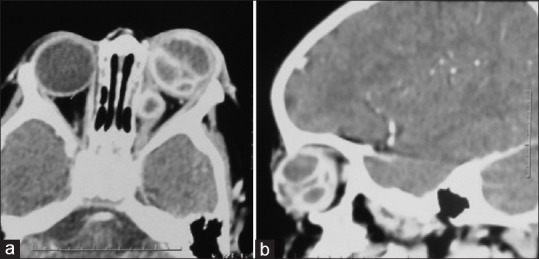
Nocardia is a rare cause of endophthalmitis in immunocompetent individuals with poor visual outcomes. We, herein report a 15 month otherwise healthy child, who presented with hyphema, vitreous hemorrhage and secondary glaucoma following a vague history of trauma in the left eye 2 months before presentation. He presented a week later with features of panophthalmitis which were confirmed on B-scan and orbital CT scan. CT scan with contrast revealed the presence of multiple ring enhancing abscesses in the vitreous cavity and also in the intraconal space. Evisceration was done and smear and cultures revealed Nocardia. Rare presentation in a healthy pediatric patient and typical CT scan findings are discussed.
Keywords: Nocardia endophthalmitis; orbital abscess; orbital imaging; pediatric.
Copyright: © 2020 Saudi Journal of Ophthalmology.
Conflict of interest statement
There are no conflicts of interest.
Figures


Similar articles
-
A rare presentation of Klebsiella pneumoniae endogenous panophthalmitis with optic neuritis and orbital cellulitis from a urinary tract infection.IDCases. 2021 Sep 23;26:e01289. doi: 10.1016/j.idcr.2021.e01289. eCollection 2021. IDCases. 2021. PMID: 34646733 Free PMC article.
-
Post-traumatic endophthalmitis caused by Nocardia nova.JMM Case Rep. 2019 Feb 20;6(2):e005175. doi: 10.1099/jmmcr.0.005175. eCollection 2019 Feb. JMM Case Rep. 2019. PMID: 30886723 Free PMC article.
-
Evisceration with Primary Orbital Implant in Fulminant Endophthalmitis/Panophthalmitis.Orbit. 2015;34(5):279-83. doi: 10.3109/01676830.2015.1078366. Epub 2015 Aug 26. Orbit. 2015. PMID: 26308681
-
Isolated Nocardia exalbida endogenous endophthalmitis.Ocul Immunol Inflamm. 2011 Aug;19(4):237-9. doi: 10.3109/09273948.2011.563898. Ocul Immunol Inflamm. 2011. PMID: 21770800 Review.
-
Salmonella Typhi Associated Endogenous Endophthalmitis: A Case Report and a Review of Literature.Ocul Immunol Inflamm. 2018;26(4):527-532. doi: 10.1080/09273948.2017.1306085. Epub 2017 Apr 28. Ocul Immunol Inflamm. 2018. PMID: 28453408 Review.
Cited by
-
Nocardia keratitis presenting as an anterior chamber ball of exudates and its management.BMJ Case Rep. 2023 Feb 21;16(2):e251647. doi: 10.1136/bcr-2022-251647. BMJ Case Rep. 2023. PMID: 36810335 Free PMC article.
References
-
- Khan S, Athwal L, Zarbin M, Bhagat N. Pediatric infectious endophthalmitis: A review. J Pediatr Ophthalmol Strabismus. 2014;51:140–53. - PubMed
-
- Bansal Pooja, Venkatesh Pradeep, Sharma Yograj. Posttraumatic endophthalmitis in children: Epidemiology, diagnosis, management, and prognosis Semin. Ophthalmol. 2016. pp. 1–9. https://doi.org/10.1080/08820538.2016.1238095 . Epub ahead of print. - PubMed
-
- Al-Rashaed SA, Abu El-Asrar AM. Exogenous endophthalmitis in pediatric age group. Ocul Immunol Inflamm. 2006;14:285–92. - PubMed
-
- Good WV, Hing S, Irvine AR, et al. Postoperative endophthalmitis in children following cataract surgery. J Pediatr Ophthalmol Strabismus. 1990;27:283–5. - PubMed
-
- Decroos FC, Garg P, Reddy AK, et al. Hyderabad endophthalmitis research group optimizing diagnosis and management of Nocardia keratitis, scleritis, and endophthalmitis: 11-year microbial and clinical overview. Ophthalmology. 2011;118:1193–200. - PubMed
Publication types
LinkOut - more resources
Full Text Sources