Review
. 2021 Apr;22(4):568-576.
doi: 10.3348/kjr.2020.0981. Epub 2020 Dec 21.

How to Treat Peripheral Arteriovenous Malformations

Affiliations
Review

How to Treat Peripheral Arteriovenous Malformations

Ran Kim et al. Korean J Radiol. 2021 Apr.

Abstract

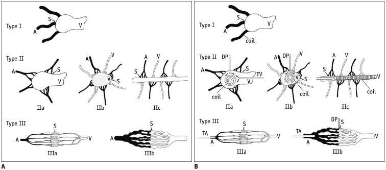
Arteriovenous malformations (AVMs) are direct communications between primitive reticular networks of dysplastic vessels that have failed to mature into capillary vessels. Based on angiographic findings, peripheral AVMs can be classified into six types: type I, type IIa, type IIb, type IIc, type IIIa, and type IIIb. Treatment strategies vary with the types. Type I is treated by embolizing the fistula between the artery and the vein with coils. Type II (IIa, IIb, and IIc) AVM is treated as follows: first, reduce the blood flow velocity in the venous segment of the AVM with coils; second, perform ethanol embolotherapy of the residual shunts. Type IIIa is treated by transarterial catheterization of the feeding arteries and injection of diluted ethanol. Type IIIb is treated by transarterial or direct puncture approaches. A high concentration of ethanol is injected through the transarterial catheter or direct puncture needle. When the fistula is large, coil insertion is required to reduce the amount of ethanol. Type I and type II AVMs showed the best clinical results; type IIIb showed a satisfactory response rate. However, type IIIa showed the poorest response rate, either alone or in combination with other types. Clinical success can be achieved by using different treatment strategies for different angiographic AVM types.

Keywords: Angiography; Arteriovenous malformation; Embolotherapy.


Conflict of interest statement

The authors have no potential conflicts of interest to disclose.

Figures

Fig. 1
Fig. 1. Angiographic classification and treatment strategy of peripheral AVMs.
A. Angiographic classification of AVMs. Type I: arteriovenous fistulae with three or less feeding arteries and a single draining vein. Type IIa: multiple arterioles shunt to the focal segment of the single draining vein. Type IIb: multiple arterioles shunt to the venous sac with multiple draining veins. Type IIc: multiple arterioles shunt along the long segment of the draining vein. Type IIIa: multiple arterioles shunt to multiple draining veins through multiple fine fistulae. Type IIIb: multiple arterioles shunt to multiple draining veins through multiple enlarged fistulae. B. Strategies for embolotherapy based on the types of AVMs. Type I: arteriovenous fistula is embolized with coils. Type IIa: the focal venous sac is embolized with coils or core-removed guidewires through a direct puncture or a transvenous catheter approach. Type IIb: the venous sac is embolized with coils or core-removed guidewires through the direct puncture approach. Type IIc: the long segment of the draining vein is occluded with coils or core-removed guidewires using the transvenous approach or a direct puncture. Type IIIa: diluted ethanol (50–60%) is injected through the transarterial catheter. Type IIIb: a high concentration of ethanol (80–100%) is injected through the transarterial catheter or the direct puncture needle. When the fistula is large, coil insertion is required to reduce the amount of ethanol. A = artery, AVM = arteriovenous malformation, DP = direct puncture, S = shunt, TA = transarterial, TV = transvenous, V = vein
Fig. 2
Fig. 2. Images of a 42-year-old woman with a type I renal AVM.
A. Venous phase of the pretreatment angiogram shows the direct arteriovenous fistula formation at two intrarenal branches. B. A selective angiogram of the proximal portion of arteriovenous fistula using a microcatheter shows a type I AVM more clearly. Arteriovenous fistulae were embolized with coils using the intra-arterial approach. A total of 12 coils were inserted into the feeding arteries of individual arteriovenous fistulae. C. Completion angiogram shows complete obliteration of the AVM. Other normal intrarenal branches were completely spared without flow disturbance. The treatment was completed in a single session. The AVM did not recur during the 6-year follow-up.
Fig. 3
Fig. 3. Images of a 30-year-old man with a type IIa pelvic AVM.
A. The venous phase of the pretreatment angiogram shows multiple feeding arterioles from the inferior mesenteric artery and both internal iliac arteries shunting to the focal segment of the single draining vein in the pelvis. B. Selective angiogram of the small feeding arteriole from the inferior mesenteric artery using microcatheter shows the shunt to the single draining vein more clearly (asterisk). C. A direct puncture of the draining vein was performed using a transabdominal approach, and a 6-Fr guiding sheath was inserted. D. Embolotherapy was performed with four regular guidewires: 115 coils using the transvenous approach and a total of 63 cc of pure ethanol using the intra-arterial approach during three sessions of treatment. E. Completion angiogram shows complete obliteration of the AVM. The AVM did not recur during the 6-year follow-up.
Fig. 4
Fig. 4. Images of a 27-year-old man with a type IIb foot AVM.
A. The venous phase of a pretreatment angiogram shows multiple arterioles from the anterior and posterior tibial arteries shunting to the venous sac with multiple small draining veins. There are two small aneurysmal changes (asterisks) in the anterior tibial artery. B. A selective angiogram after the puncture of the venous sac shows the venous sac with multiple small draining veins more clearly. C. A total of 15 cc of pure ethanol was injected slowly, and a blood pressure cuff was inflated to 150 mm Hg. D. A completion angiogram shows complete obliteration of the AVM. The treatment was completed in a single session. The AVM did not recur during the 6-year follow-up.
Fig. 5
Fig. 5. Images from a 6-year-old boy with type IIc AVM in the thigh.
Arterial (A) and venous (B) phases of a pretreatment superficial femoral artery angiogram show multiple arterioles shunting along the long segment of the draining vein (asterisk). The long segment of the draining vein was occluded with coils using the transvenous and direct puncture approach. After slowing the flow within the AVM, pure ethanol was injected using the transvenous and intra-arterial approaches to eradicate the residual AVM lesion (C). A completion angiogram of the common femoral artery shows complete obliteration of the AVM. The AVM did not recur during the 6-year follow-up (D).
Fig. 6
Fig. 6. Images of a 16-year-old girl with a type IIIa AVM in the shoulder.
A. Venous phase of a pretreatment angiogram shows the AVM of the nidus as a diffuse blush and early draining veins. B. Superselective angiogram shows microfistulae between the arterioles and venules as a blush. C. Completion angiogram (after five sessions of intra-arterial injection of 50–70% diluted ethanol through the fine feeding arteries) shows a markedly improved AVM.
Fig. 7
Fig. 7. Images of a 43-year-old man with a type IIIb AVM in the foot.
A. Venous phase of a pretreatment angiogram shows multiple dilated fistulae and dilated draining veins. B. Direct puncture of one of the dilated fistulae shows typical hypertrophy of the fistula, which was compatible with a type IIIb AVM. Pure ethanol was injected directly through the needle, and the blood pressure cuff was inflated to 150 mm Hg. C. Completion angiogram (after three sessions of an injection of 80–100% ethanol through the needle) shows near-complete embolization of the foot AVM with an intact dorsalis pedis branch (arrow).

References

    1. Lee BB, Baumgartner I, Berlien HP, Bianchini G, Burrows P, Do YS, et al. Consensus document of the international union of angiology (IUA)-2013. Current concept on the management of arterio-venous malformations. Int Angiol. 2013;32:9–36. - PubMed
    1. Park HS, Do YS, Park KB, Kim DI, Kim YW, Kim MJ, et al. Ethanol embolotherapy of hand arteriovenous malformations. J Vasc Surg. 2011;53:725–731. - PubMed
    1. Do YS, Kim YW, Park KB, Kim DI, Park HS, Cho SK, et al. Endovascular treatment combined with embolosclerotherapy for pelvic arteriovenous malformations. J Vasc Surg. 2012;55:465–471. - PubMed
    1. Cho SK, Do YS, Shin SW, Kim DI, Kim YW, Park KB, et al. Arteriovenous malformations of the body and extremities: analysis of therapeutic outcomes and approaches according to a modified angiographic classification. J Endovasc Ther. 2006;13:527–538. - PubMed
    1. Flye MW, Jordan BP, Schwartz MZ. Management of congenital arteriovenous malformations. Surgery. 1983;94:740–747. - PubMed

MeSH terms