Review
. 2021 May;64(5):985-993.
doi: 10.1007/s00125-021-05395-6. Epub 2021 Feb 5.

Hypothalamic glucose-sensing mechanisms

Affiliations
Review

Hypothalamic glucose-sensing mechanisms

Nal Ae Yoon et al. Diabetologia. 2021 May.

Abstract

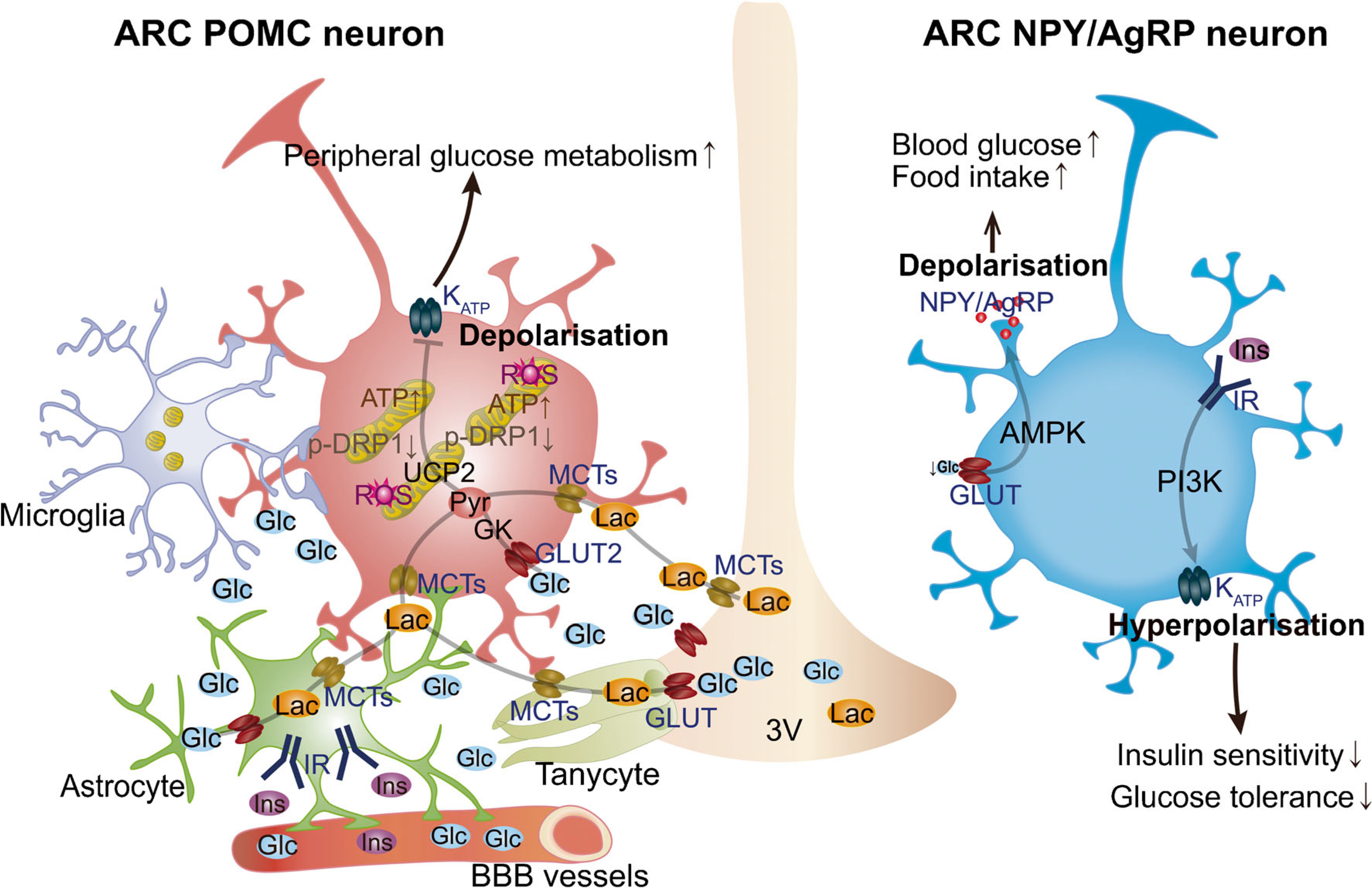
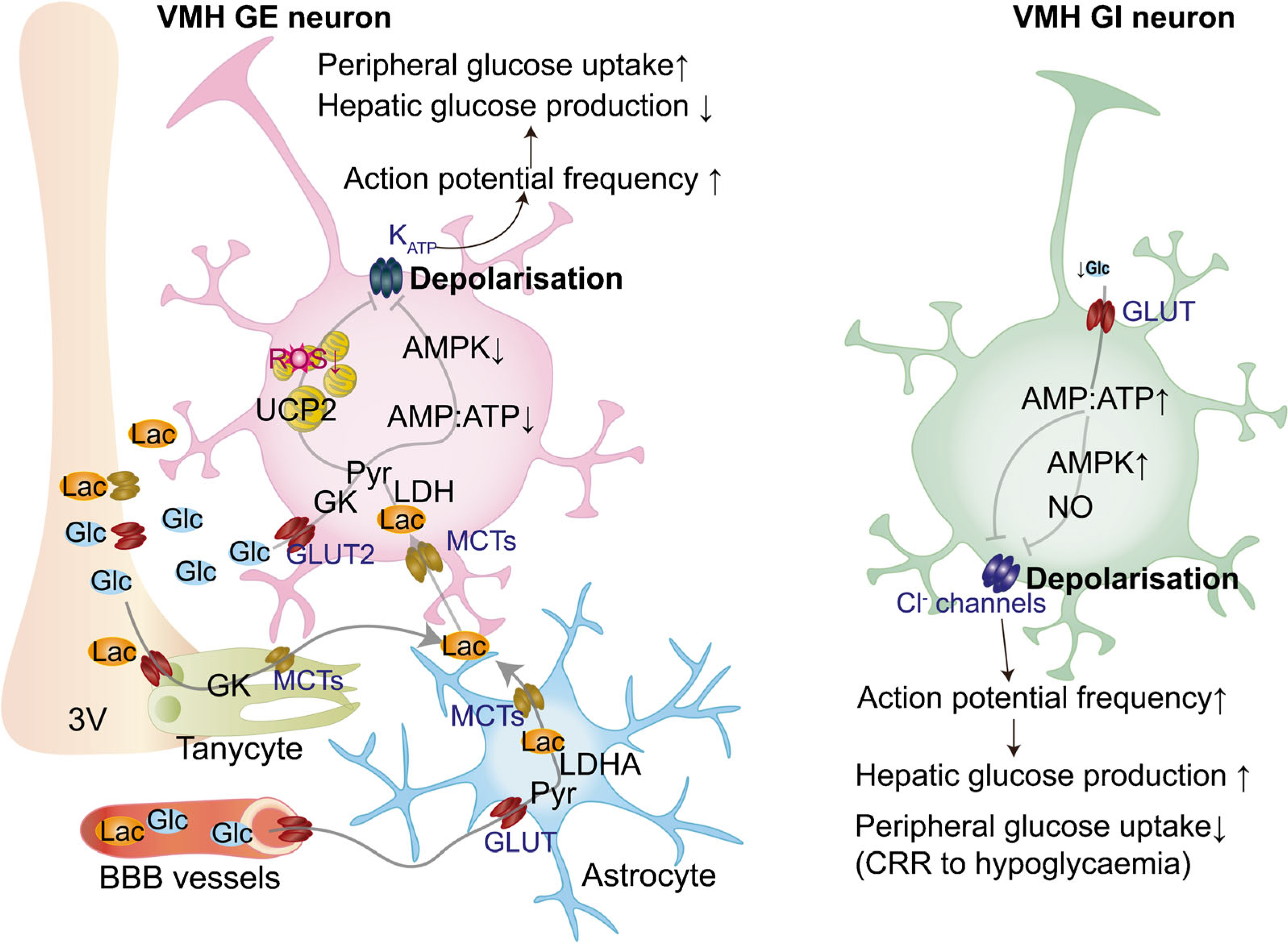
Chronic metabolic diseases, including diabetes and obesity, have become a major global health threat of the twenty-first century. Maintaining glucose homeostasis is essential for survival in mammals. Complex and highly coordinated interactions between glucose-sensing mechanisms and multiple effector systems are essential for controlling glucose levels in the blood. The central nervous system (CNS) plays a crucial role in regulating glucose homeostasis. Growing evidence indicates that disruption of glucose sensing in selective CNS areas, such as the hypothalamus, is closely interlinked with the pathogenesis of obesity and type 2 diabetes mellitus. However, the underlying intracellular mechanisms of glucose sensing in the hypothalamus remain elusive. Here, we review the current literature on hypothalamic glucose-sensing mechanisms and discuss the impact of alterations of these mechanisms on the pathogenesis of diabetes.

Keywords: Astrocytes; Brain; Counterregulatory responses; Diabetes; Glucose-sensing; Hypothalamus; Neurons; Obesity; Review; Tanycytes.


Figures

Fig. 1
Fig. 1
Glucose-sensing mechanisms in the hypothalamic ARC. The ability of POMC and NPY/AgRP neuronal populations in ARC to alter energy metabolism is due to their sensitivity to several circulating signals, including hormones and nutrients (e.g. glucose and lactate). Hypothalamic astrocytes provide neurons with structural support and nutrients. Astrocytic insulin signalling regulates glucose sensing via control of glucose uptake across the blood–brain barrier (BBB). Glucose is delivered to the extracellular space and transported to glial cells, such as astrocytes and tanycytes, through GLUTs and then metabolised to lactate. When released, lactate is taken up by neurons via monocarboxylate transporters (MCTs) to serve as a glycolytic substrate and metabolised into pyruvate. Hypothalamic microglia have been shown to affect POMC neuronal activation and synaptic input organisation in diet-induced obesity. Changes in glucose levels have been shown to affect POMC activity via KATP channels. Mitochondrial dynamics in hypothalamic POMC neurons has been also shown to play a pivotal role in regulating neuronal activity in response to glucose fluctuations. The activity of the ARC glucose-inhibited NPY/AgRP neurons is regulated by AMPK activity in response to decreased glucose levels, while insulin has been shown to induce hyperpolarisation of these neurons. Glc, glucose; Ins, insulin; IR, insulin receptor; Lac, lactate; PI3K, phosphoinositide 3-kinase; Pyr, pyruvate; ROS, reactive oxygen species; 3V, third ventricle.
Fig. 2
Fig. 2
Glucose-sensing mechanisms in the hypothalamic VMH. In vivo, glucose is released in the extracellular space and transported to glial cells, such as astrocytes and tanycytes, through GLUTs and then metabolised to lactate. Once released, lactate enters neurons via monocarboxylate transporters (MCTs) to serve as a glycolytic substrate and is metabolised into pyruvate via the enzyme LDH. Mitochondrial pyruvate uptake leads to increased ATP levels that, in turn, affect ATP-dependent K+ (KATP) channels, resulting in plasma membrane depolarisation. Glucose-excited (GE) neurons can also uptake glucose through GLUT2, leading to the phosphorylation of glucose by GK, a decrease in the AMP:ATP ratio, decreased AMPK activity and closing of KATP channel. Closure of KATP channels leads to membrane depolarisation and increased action potential frequency. The activation of VMH GE neurons leads to decreased hepatic glucose production and increased peripheral glucose uptake. VMH glucose-inhibited (GI) neurons are activated in response to hypoglycaemia. Decreased glucose entry into neurons leads to the rise in the cellular AMP:ATP ratio and activation of AMPK, which induces formation of NO. Increased AMP:ATP also inhibits Cl channels, leading to membrane depolarisation and increased action potential frequency, which leads to activation of CRRs to hypoglycaemia to increase hepatic glucose production and decrease peripheral glucose uptake. BBB, blood–brain barrier; Glc, glucose; Lac, lactate; LDHA, LDH isoenzyme A; Pyr, pyruvate; ROS, reactive oxygen species; 3V, third ventricle.

Similar articles

Cited by

References

    1. Czech MP (2017) Insulin action and resistance in obesity and type2 diabetes. Nat Med 23(7):804–814. 10.1038/nm.4350 - DOI - PMC - PubMed
    1. Roder PV, Wu B, Liu Y, Han W (2016) Pancreatic regulation of glucose homeostasis. Exp Mol Med 48:e219. 10.1038/emm.2016.6 - DOI - PMC - PubMed
    1. Ritter S (2017) Monitoring and maintenance of brain glucose supply importance of hindbrain catecholamine neurons in this multifaceted task. In: Harris RBS (ed) Appetite and food intake: Central control, 2nd edn. CRC Press/Taylor& Francis, Boca Raton, pp 177–204 - PubMed
    1. Timper K, Bruning JC (2017) Hypothalamic circuits regulating appetite and energy homeostasis: Pathways to obesity. Dis Model Mech 10(6):679–689. 10.1242/dmm.026609 - DOI - PMC - PubMed
    1. Routh VH (2010) Glucose sensing neurons in the ventromedial hypothalamus. Sensors 10(10):9002–9025. 10.3390/s101009002 - DOI - PMC - PubMed

Publication types

LinkOut - more resources