

4-hydroxytamoxifen does not deteriorate cardiac function in cardiomyocyte-specific MerCreMer transgenic mice
- PMID: 33544211
- PMCID: PMC7864833
- DOI: 10.1007/s00395-020-00841-9
4-hydroxytamoxifen does not deteriorate cardiac function in cardiomyocyte-specific MerCreMer transgenic mice
Abstract
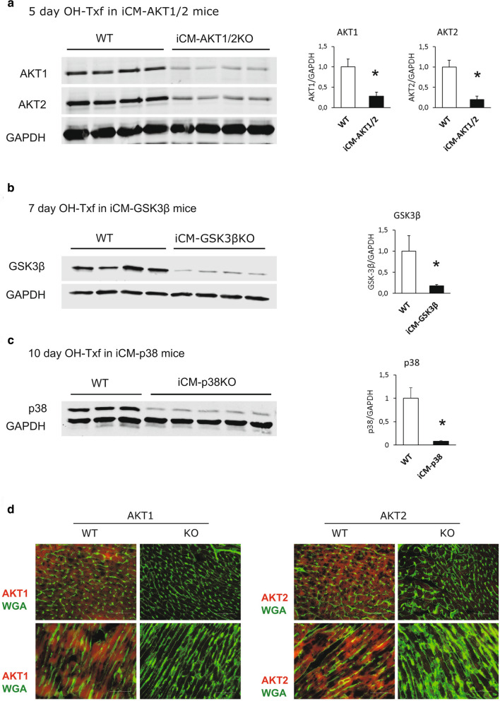
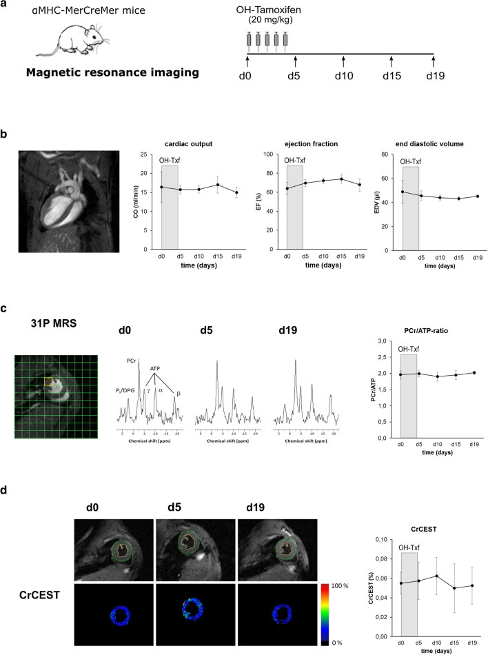
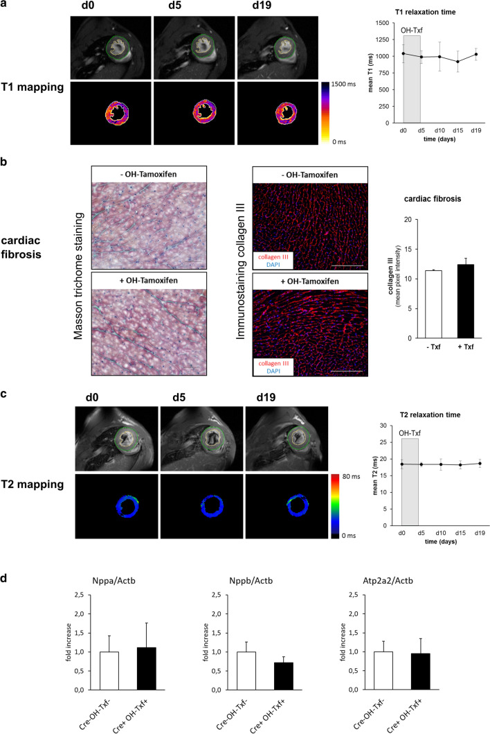
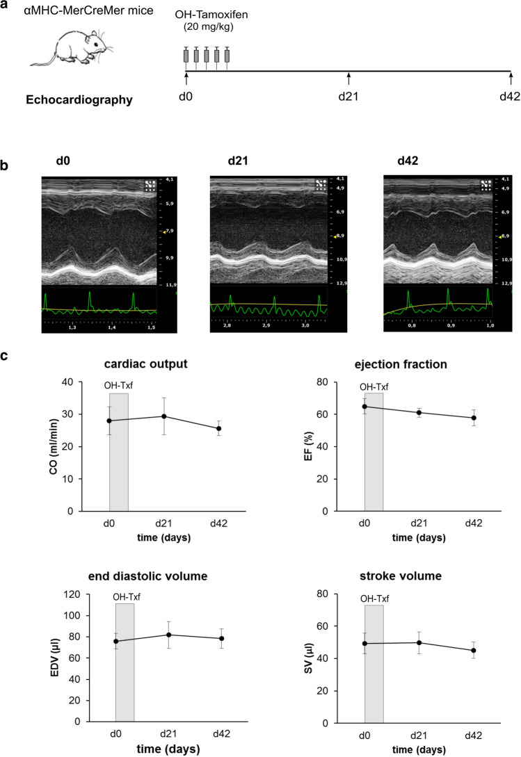
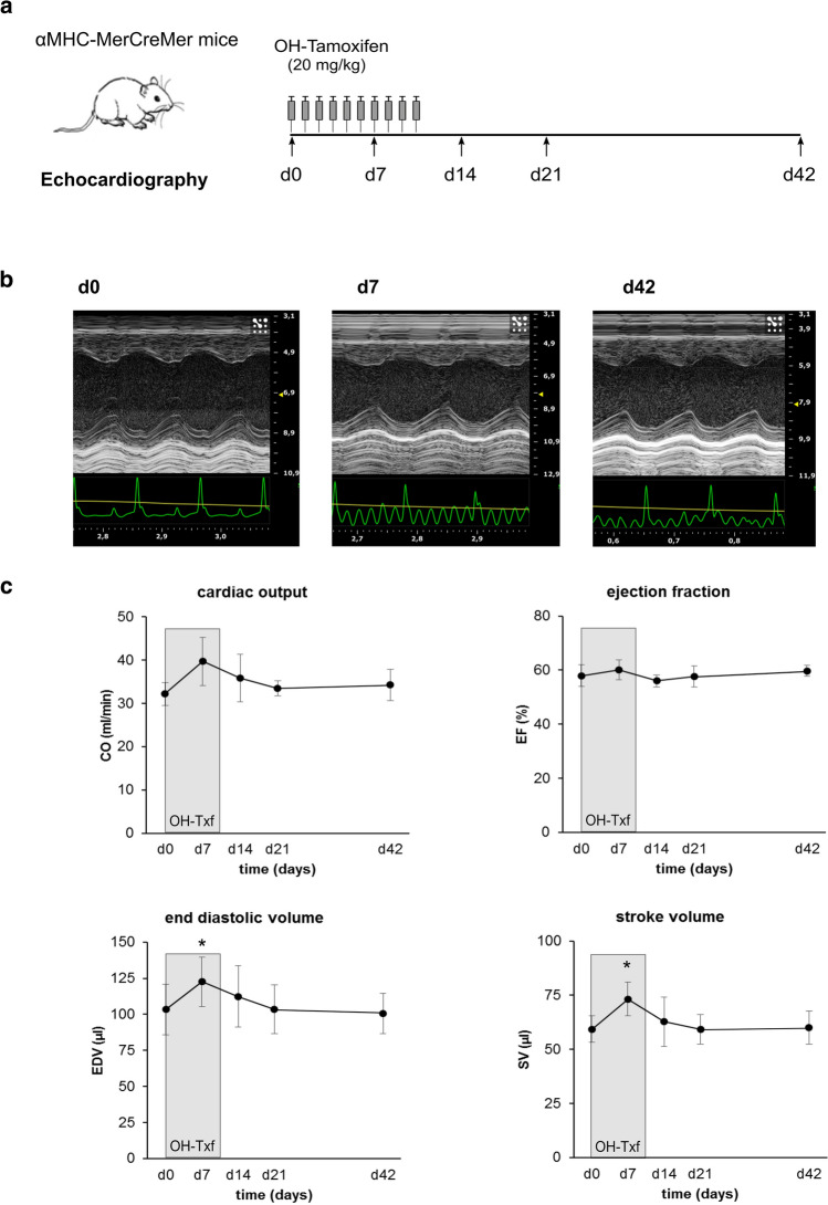
Conditional, cell-type-specific transgenic mouse lines are of high value in cardiovascular research. A standard tool for cardiomyocyte-restricted DNA editing is the αMHC-MerCreMer/loxP system. However, there is an ongoing debate on the occurrence of cardiac side effects caused by unspecific Cre activity or related to tamoxifen/oil overload. Here, we investigated potential adverse effects of DNA editing by the αMHC-MerCreMer/loxP system in combination with a low-dose treatment protocol with the tamoxifen metabolite 4-hydroxytamoxifen (OH-Txf). αMHC-MerCreMer mice received intraperitoneally OH-Txf (20 mg/kg) for 5 or 10 days. These treatment protocols were highly efficient to induce DNA editing in adult mouse hearts. Multi-parametric magnetic resonance imaging revealed neither transient nor permanent effects on cardiac function during or up to 19 days after 5 day OH-Txf treatment. Furthermore, OH-Txf did not affect cardiac phosphocreatine/ATP ratios assessed by in vivo 31P MR spectroscopy, indicating no Cre-mediated side effects on cardiac energy status. No MRI-based indication for the development of cardiac fibrosis was found as mean T1 relaxation time was unchanged. Histological analysis of myocardial collagen III content after OH-Txf confirmed this result. Last, mean T2 relaxation time was not altered after Txf treatment suggesting no pronounced cardiac lipid accumulation or tissue oedema. In additional experiments, cardiac function was assessed for up to 42 days to investigate potential delayed side effects of OH-Txf treatment. Neither 5- nor 10-day treatment resulted in a depression of cardiac function. Efficient cardiomyocyte-restricted DNA editing that is free of unwanted side effects on cardiac function, energetics or fibrosis can be achieved in adult mice when the αMHC-MerCreMer/loxP system is activated by the tamoxifen metabolite OH-Txf.
Keywords: 4-hydroxytamoxifen; Cardiac energetics; Cardiac function; Cardiomyopathy; aMHC-MerCreMer/loxP system.
Conflict of interest statement
The authors declare that they have no conflict of interest.
Figures
Similar articles
-
Myh6-driven Cre recombinase activates the DNA damage response and the cell cycle in the myocardium in the absence of loxP sites.Dis Model Mech. 2020 Dec 18;13(12):dmm046375. doi: 10.1242/dmm.046375. Dis Model Mech. 2020. PMID: 33106234 Free PMC article.
-
Moderate and high amounts of tamoxifen in αMHC-MerCreMer mice induce a DNA damage response, leading to heart failure and death.Dis Model Mech. 2013 Nov;6(6):1459-69. doi: 10.1242/dmm.010447. Epub 2013 Aug 7. Dis Model Mech. 2013. PMID: 23929941 Free PMC article.
-
Systolic dysfunction in cardiac-specific ligand-inducible MerCreMer transgenic mice.Am J Physiol Heart Circ Physiol. 2011 Jul;301(1):H253-60. doi: 10.1152/ajpheart.00786.2010. Epub 2011 May 2. Am J Physiol Heart Circ Physiol. 2011. PMID: 21536850 Free PMC article.
-
Cre-loxP DNA recombination is possible with only minimal unspecific transcriptional changes and without cardiomyopathy in Tg(alphaMHC-MerCreMer) mice.Am J Physiol Heart Circ Physiol. 2010 Nov;299(5):H1671-8. doi: 10.1152/ajpheart.01155.2009. Epub 2010 Aug 27. Am J Physiol Heart Circ Physiol. 2010. PMID: 20802136
-
Avoidance of transient cardiomyopathy in cardiomyocyte-targeted tamoxifen-induced MerCreMer gene deletion models.Circ Res. 2009 Jul 2;105(1):12-5. doi: 10.1161/CIRCRESAHA.109.198416. Epub 2009 Jun 11. Circ Res. 2009. PMID: 19520971 Free PMC article.
Cited by
-
PANoptosis is a prominent feature of desmoplakin cardiomyopathy.J Cardiovasc Aging. 2023 Feb;3(1):3. doi: 10.20517/jca.2022.34. Epub 2023 Jan 1. J Cardiovasc Aging. 2023. PMID: 36818425 Free PMC article.
-
The Pitfalls of in vivo Cardiac Physiology in Genetically Modified Mice - Lessons Learnt the Hard Way in the Creatine Kinase System.Front Physiol. 2021 May 14;12:685064. doi: 10.3389/fphys.2021.685064. eCollection 2021. Front Physiol. 2021. PMID: 34054587 Free PMC article. Review.
-
Effects of tamoxifen inducible MerCreMer on gene expression in cardiac myocytes in mice.J Cardiovasc Aging. 2022;2:8. doi: 10.20517/jca.2021.30. Epub 2022 Jan 5. J Cardiovasc Aging. 2022. PMID: 35079750 Free PMC article.
-
Prolonged tamoxifen-enriched diet is associated with cardiomyopathy and nutritional frailty in mice.Exp Physiol. 2024 Apr;109(4):513-523. doi: 10.1113/EP091668. Epub 2024 Jan 30. Exp Physiol. 2024. PMID: 38291801 Free PMC article.
-
Fibroblast growth factor 18 alleviates stress-induced pathological cardiac hypertrophy in male mice.Nat Commun. 2023 Mar 4;14(1):1235. doi: 10.1038/s41467-023-36895-1. Nat Commun. 2023. PMID: 36871047 Free PMC article.
References
-
- Andersson KB, Birkeland JA, Finsen AV, Louch WE, Sjaastad I, Wang Y, Chen J, Molkentin JD, Chien KR, Sejersted OM, Christensen G. Moderate heart dysfunction in mice with inducible cardiomyocyte-specific excision of the Serca2 gene. J Mol Cell Cardiol. 2009;47:180–187. doi: 10.1016/j.yjmcc.2009.03.013. - DOI - PubMed
-
- Bersell K, Choudhury S, Mollova M, Polizzotti BD, Ganapathy B, Walsh S, Wadugu B, Arab S, Kuhn B. Moderate and high amounts of tamoxifen in alphaMHC-MerCreMer mice induce a DNA damage response, leading to heart failure and death. Dis Model Mech. 2013;6:1459–1469. doi: 10.1242/dmm.010447. - DOI - PMC - PubMed
Publication types
MeSH terms
Substances
Grants and funding
LinkOut - more resources
Full Text Sources
Other Literature Sources
