Review
. 2021 Apr 1;94(1120):20201013.
doi: 10.1259/bjr.20201013. Epub 2021 Feb 5.

Magnetic resonance imaging in the evaluation of pathologic nipple discharge: indications and imaging findings

Affiliations
Review

Magnetic resonance imaging in the evaluation of pathologic nipple discharge: indications and imaging findings

Naziya Samreen et al. Br J Radiol. .

Abstract

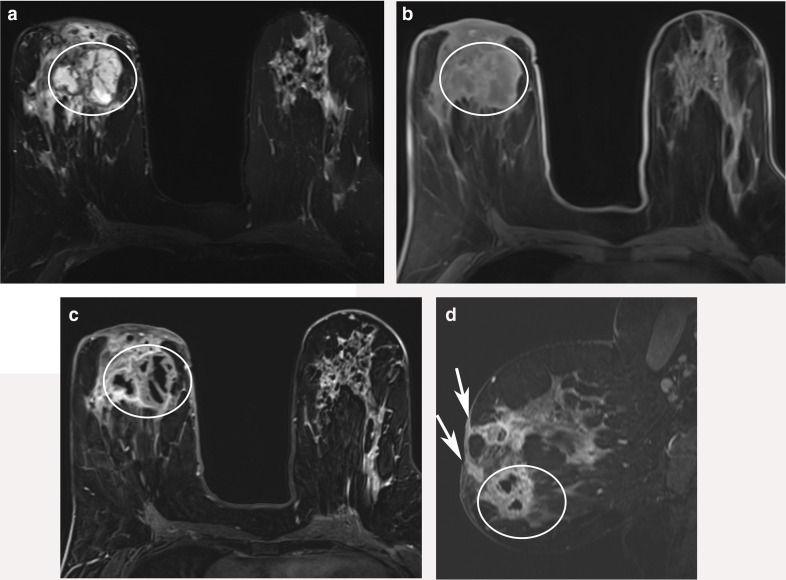
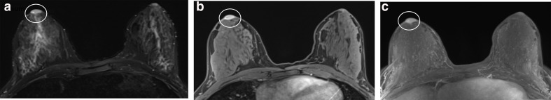
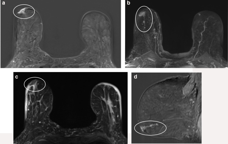
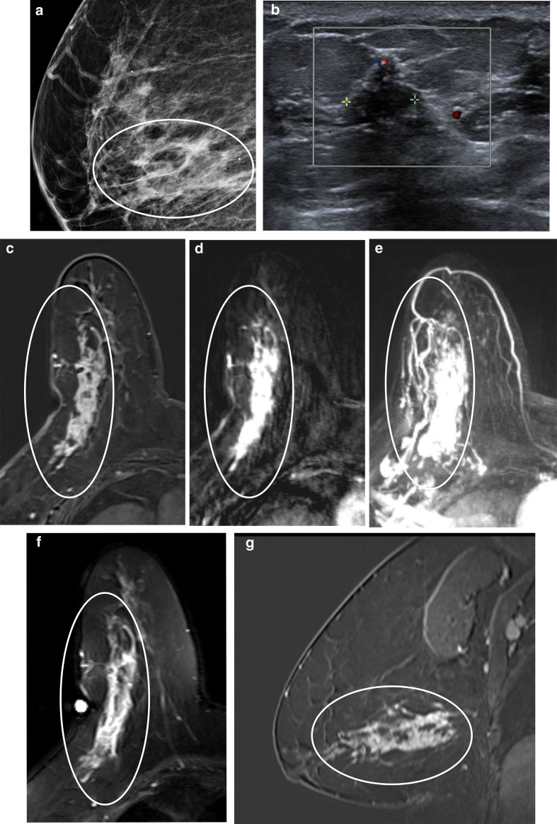
Pathologic nipple discharge (PND) is typically unilateral, spontaneous, involves a single duct, and is serous or bloody in appearance. In patients with PND, breast MRI can be helpful as an additional diagnostic tool when conventional imaging with mammogram and ultrasound are negative. MRI is able to detect the etiology of nipple discharge in 56-61% of cases when initial imaging with mammogram and ultrasound are negative. Advantages to using MRI in evaluation of PND include good visualization of the retroareolar breast and better evaluation of posterior lesions which may not be well evaluated on mammograms and galactograms. It is also less invasive compared to central duct excision. Papillomas and nipple adenomas are benign breast masses that can cause PND and are well visualized on MRI. Ductal ectasia, and infectious etiologies such as mastitis, abscess, and fistulas are additional benign causes of PND that are well evaluated with MRI. MRI is also excellent for evaluation of malignant causes of PND including Paget's disease, ductal carcinoma in-situ and invasive carcinoma. MRI's high negative predictive value of 87-98.2% is helpful in excluding malignant etiologies of PND.


Figures

Figure 1.
Figure 1.
Normal nipple enhancement: The most superficial layer, known as SLE, consists of blood vessels and typically enhances greater than adjacent skin. The next layer is the NEZ, which is comprised predominantly of connective tissue. Deep to that is the INE which consists of lactiferous duct bundles and smooth muscles, and represents the majority of nipple enhancement on MRI. INE, internal nipple enhancement; NEZ, non-enhancing zone; SLE, superficial linear enhancement.
Figure 2.
Figure 2.
Ductal ectasia: 47-year-old female with left bloody nipple discharge. No abnormality was noted on mammogram, and ultrasound demonstrated dilated ducts with fluid. T1 weighted fat suppressed (a), T1 weighted non-fat suppressed (b) and T2 weighted (c) sequences demonstrate proteinaceous debris/blood (arrow) in the branching, mildly dilated ductal system upper inner periareolar region. No abnormal enhancement in the ducts or periductal region (circle) is noted on the ultrafast fat suppressed contrast-enhanced sequence (d) or the axial T1 weighted fat suppressed contrast-enhanced fat suppressed sequence (e). Surgical consultation was recommended due to atypia noted on cytology from expressed fluid. Ductal excision was performed which demonstrated benign findings including mammary duct ectasia, cystic apocrine hyperplasia, and a 1 mm small intraductal papilloma.
Figure 3.
Figure 3.
Papilloma: 39-year-old female with clear left nipple discharge and outside ultrasound demonstrating no sonographic abnormality in the left retroareolar region. MRI was performed for further evaluation. In the left breast at 8:00, 4 cm posterior to the nipple, a 0.8 cm oval mass enhancing mass (circle) seen on the axial fat suppressed subtraction contrast-enhanced sequence (a). On ultrafast imaging axial post-contrast subtraction sequence (b), the mass (circle) demonstrated rapid wash-in. The mass (circle) was seen on the maximum intensity projection of the ultrafast sequences (c). The mass (circle) is hyperintense on the T2 weighted sequence (d). (e) An MRI directed ultrasound was performed which demonstrated a 0.8 × 0.7 x 0.6 cm oval, hypoechoic mass (circle) with indistinct margins, correlating to the finding on recent breast MRI. Ultrasound guided biopsy was performed which demonstrated a papilloma. Patient was referred for surgical excision.
Figure 4.
Figure 4.
Mastitis/Chronic inflammation: 47-year-old female with history of right nipple discharge. (A) Axial T2 weighted sequence demonstrates dilated retroareolar ducts (arrow) containing hyperintense material. The proteinaceous material within the ducts (arrow) is also hyperintense on the noncontrast fat suppressed T1 weighted sequence (B). There is segmental clumped non-mass enhancement (arrow) surrounding the ducts on the contrast-enhanced T1 weighted sequence (C). Patient underwent MRI-guided biopsy which demonstrated ductal ectasia and periductal chronic inflammation. Follow-up MRI demonstrated decreased enhancement also supporting benign etiology.
Figure 5.
Figure 5.
Granulomatous inflammation/abscess: 58-year-old female with history of right breast palpable lump and nipple discharge, status post-ultrasound-guided core biopsy yielding acute and chronic non-necrotizing granulomatous inflammation. MRI was performed for worsening symptoms. (A) MRI demonstrates a T2-hyperintense fluid collection with septations (circle). (B) The fluid collection is hypointense on the fat suppressed T1 weighted sequence (circle). (C) Axial fat suppressed contrast-enhanced sequence demonstrates enhancement within the rim of the fluid collection as well as within the septations (circle). (D) On the sagittal contrast-enhanced sequence, the abscess is again seen (circle), and multiple fistulas are noted extending to the skin (arrow). Findings are compatible with granulomatous abscess and fistula formation.
Figure 6.
Figure 6.
Nipple adenoma: 23-year-old female with family history of breast cancer. MRI was obtained due to right nipple pain, discharge and swelling. (A) MRI demonstrated an enlarged nipple on the right that was hyperintense on the T2 weighted sequence (circle). (B) On the fat suppressed contrast-enhanced sequence, there is asymmetric enhancement and enlargement of the right nipple (circle) with no additional abnormal enhancement within the right breast. (C) The asymmetric nipple enhancement and enlargement is well seen on the maximum intensity projection (circle). A right nipple incisional biopsy was performed which demonstrated nipple adenoma.
Figure 7.
Figure 7.
Paget’s disease: 83-year-old female presenting with right nipple erythema, discharge, and skin changes. (A) Axial fat suppressed contrast-enhanced MRI sequence demonstrates marked enhancement of the right nipple (circle) suspicious for Paget’s disease. There is linear non-mass enhancement extending posteriorly, well seen on the maximum intensity projection image (B) and the sagittal sequence (D), highly concerning for additional disease. (C) The enlarged right nipple with Paget’s disease is hypointense on the T2 weighted sequence. Right nipple punch biopsy demonstrated Paget’s disease. Lumpectomy demonstrated DCIS with involvement of the nipple epidermis, consistent with Paget’s disease. DCIS, ductal carcinoma in situ.
Figure 8.
Figure 8.
DCIS: 64-year-old postmenopausal female with right breast clear nipple discharge. (a) Mammogram demonstrated loosely grouped coarse heterogenous calcifications in the lower central right breast with associated focal asymmetry (circle). (b) Ultrasound demonstrated a 1.8 × 0.7 x 0.9 cm hypoechoic mass with occasional angular margins (square), corresponding to the focal asymmetry and calcifications on mammography. Ultrasound-guided biopsy was performed which demonstrated ductal carcinoma in situ, papillary and cribriform type, intermediate grade, with central necrosis. MRI performed for surgical planning. (c) The axial contrast-enhanced subtraction VIBE sequence demonstrated right breast extensive segmental non-mass enhancement on (circle) measuring 10.3 cm in the AP dimension, consistent with biopsy-proven DCIS. (d) Similar pattern of contrast enhancement is noted on the ultrafast sequence (circle), which demonstrates a slightly lower resolution. (e) The maximum intensity projection from the ultrafast sequence well demonstrates the non-mass enhancement (circle). (f) The biopsy proven DCIS is hyperintense on the T2 weighted sequence (circle). (g) The sagittal demonstrates also demonstrates the non-mass enhancement (circle). AP, Anteroposterior; DCIS, Ductal carcinoma in situ; VIBE, Volumetric interpolated breath-hold,
Figure 9.
Figure 9.
Inflammatory breast carcinoma: 30-year-old post-partum female with pain and swelling in the left breast, treated with antibiotics without improvement. Skin thickening was noted on physical exam and punch biopsy demonstrated inflammatory carcinoma. MRI was obtained to evaluate extent of disease. (a) T2 weighted sequence demonstrates diffuse left breast skin thickening and edema (circle) which appears hyperintense and asymmetric to right. (b) Axial and sagittal (d) fat suppressed contrast-enhanced sequence demonstrates enhancement in the skin as well as extensive non-mass enhancement within the breast which extends to the nipple anteriorly (circle). (c) The skin thickening and non-mass enhancement in the breast is well seen on the maximum intensity projection image. Enlarged left axillary lymph nodes are also noted (arrow).

Similar articles

Cited by

References

    1. Milon A, Wahab CA, Kermarrec E, Bekhouche A, Taourel P, Thomassin-Naggara I. Breast MRI: is faster better? AJR Am J Roentgenol 2020; 214: 282–95. doi: 10.2214/AJR.19.21924 - DOI - PubMed
    1. Peters NHGM, Borel Rinkes IHM, Zuithoff NPA, Mali WPTM, Moons KGM, Peeters PHM. Meta-analysis of Mr imaging in the diagnosis of breast lesions. Radiology 2008; 246: 116–24. doi: 10.1148/radiol.2461061298 - DOI - PubMed
    1. Medeiros LR, Duarte CS, Rosa DD, Edelweiss MI, Edelweiss M, Silva FR, et al. . Accuracy of magnetic resonance in suspicious breast lesions: a systematic quantitative review and meta-analysis. Breast Cancer Res Treat 2011; 126: 273–85. doi: 10.1007/s10549-010-1326-9 - DOI - PubMed
    1. Bennani-Baiti B, Baltzer PA. Mr imaging for diagnosis of malignancy in mammographic microcalcifications: a systematic review and meta-analysis. Radiology 2017; 283: 692–701. doi: 10.1148/radiol.2016161106 - DOI - PubMed
    1. ACoR. Acrpractice parameterfor the performance of contrast-enhanced magnetic resonance imaging (MRI) of the breast. 2018. Available from: https://www.acr.org/-/media/ACR/Files/Practice-Parameters/MR-Contrast-Br....