. 2021 Mar-Apr;41(2):321-337.
doi: 10.1148/rg.2021200101. Epub 2021 Feb 5.

Digital Breast Tomosynthesis: Update on Technology, Evidence, and Clinical Practice

Affiliations

Digital Breast Tomosynthesis: Update on Technology, Evidence, and Clinical Practice

Yiming Gao et al. Radiographics. 2021 Mar-Apr.

Abstract

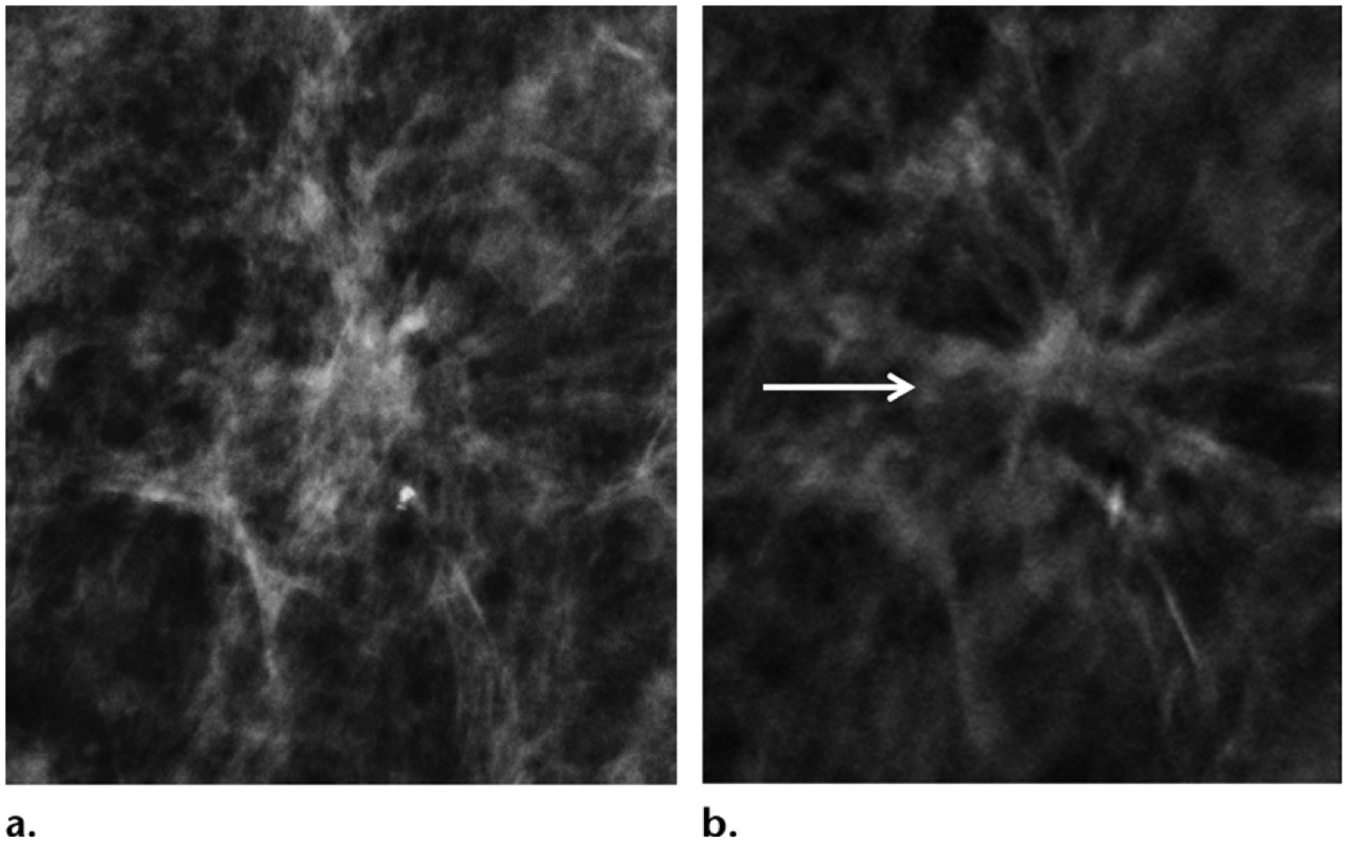
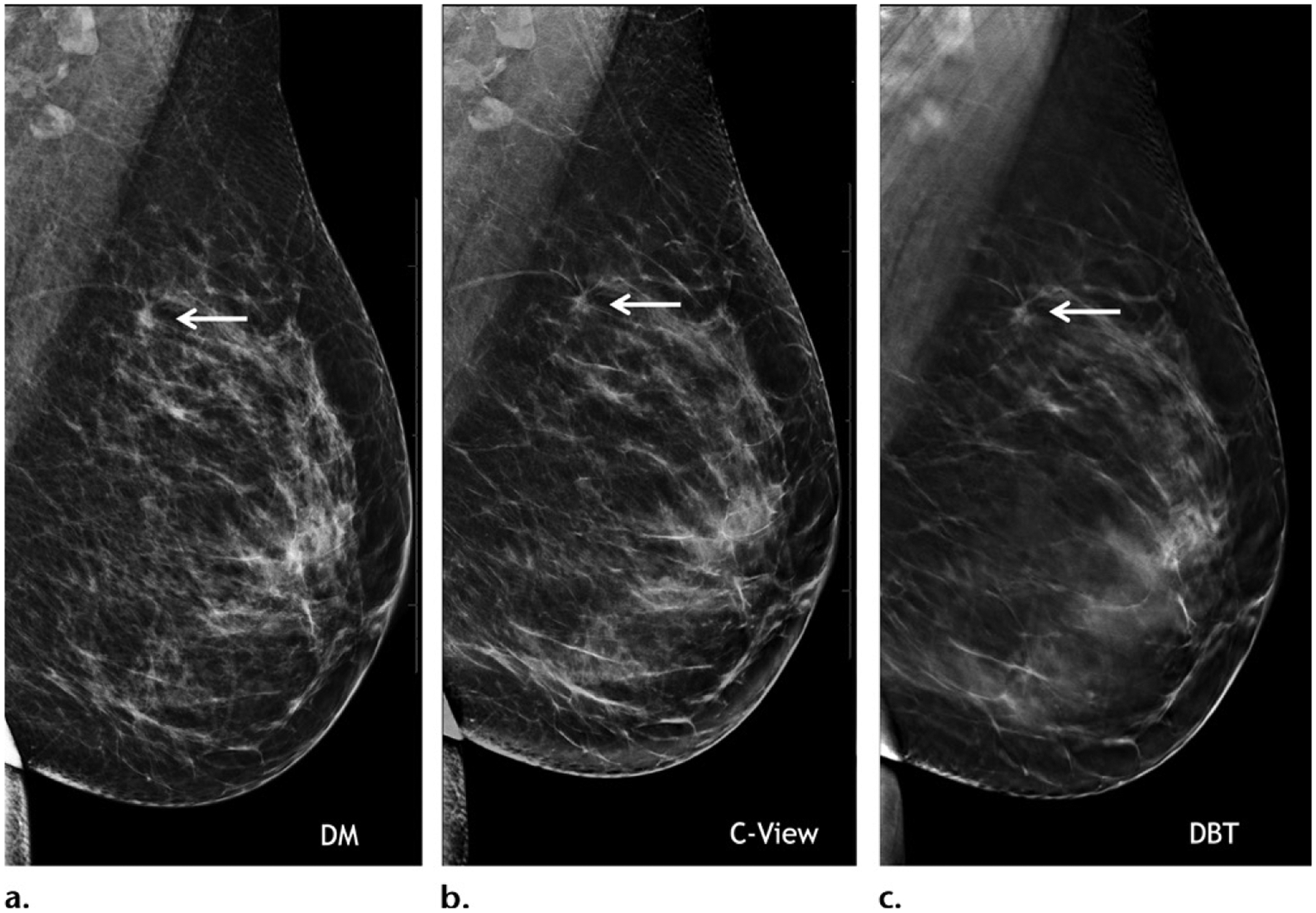
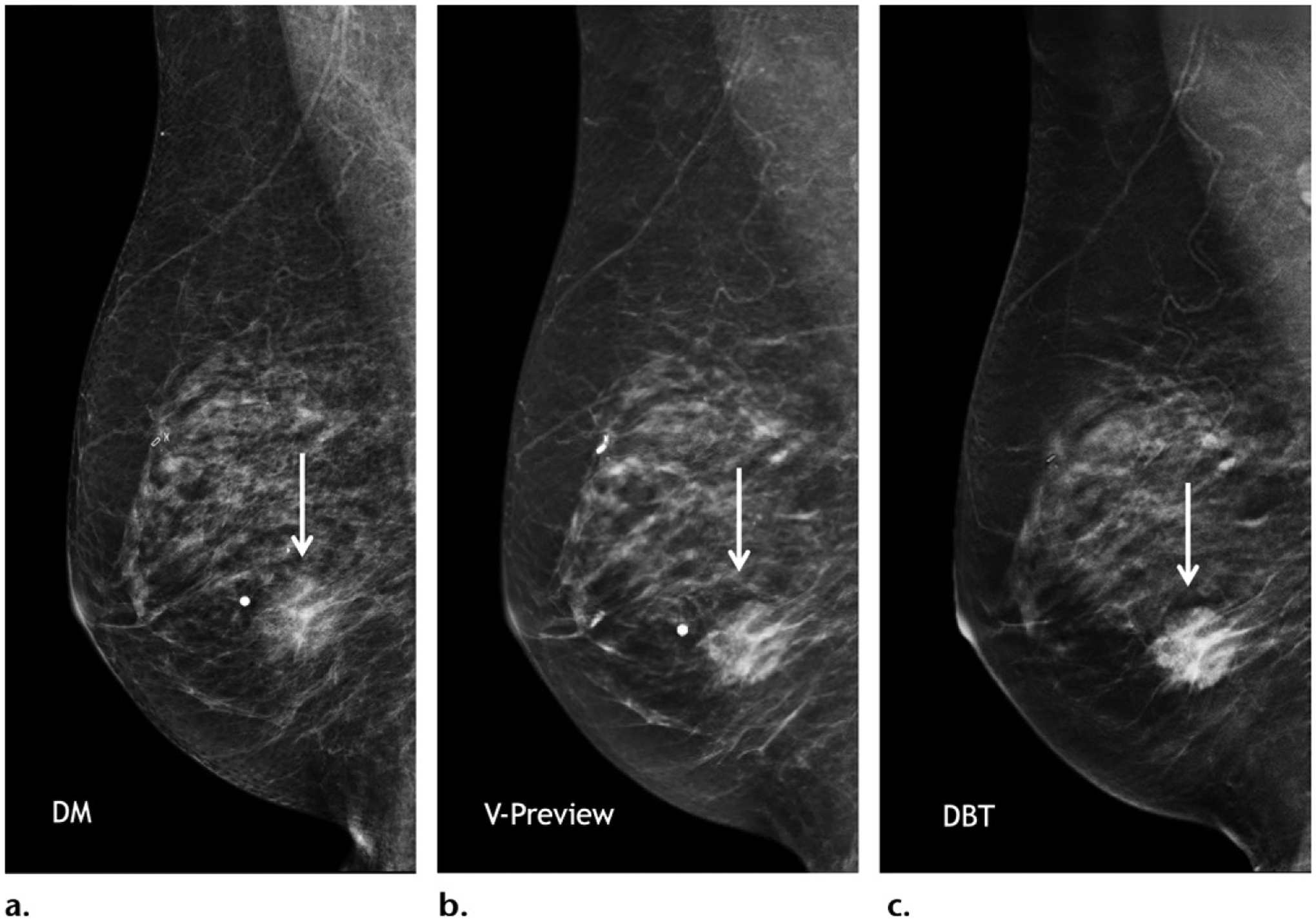
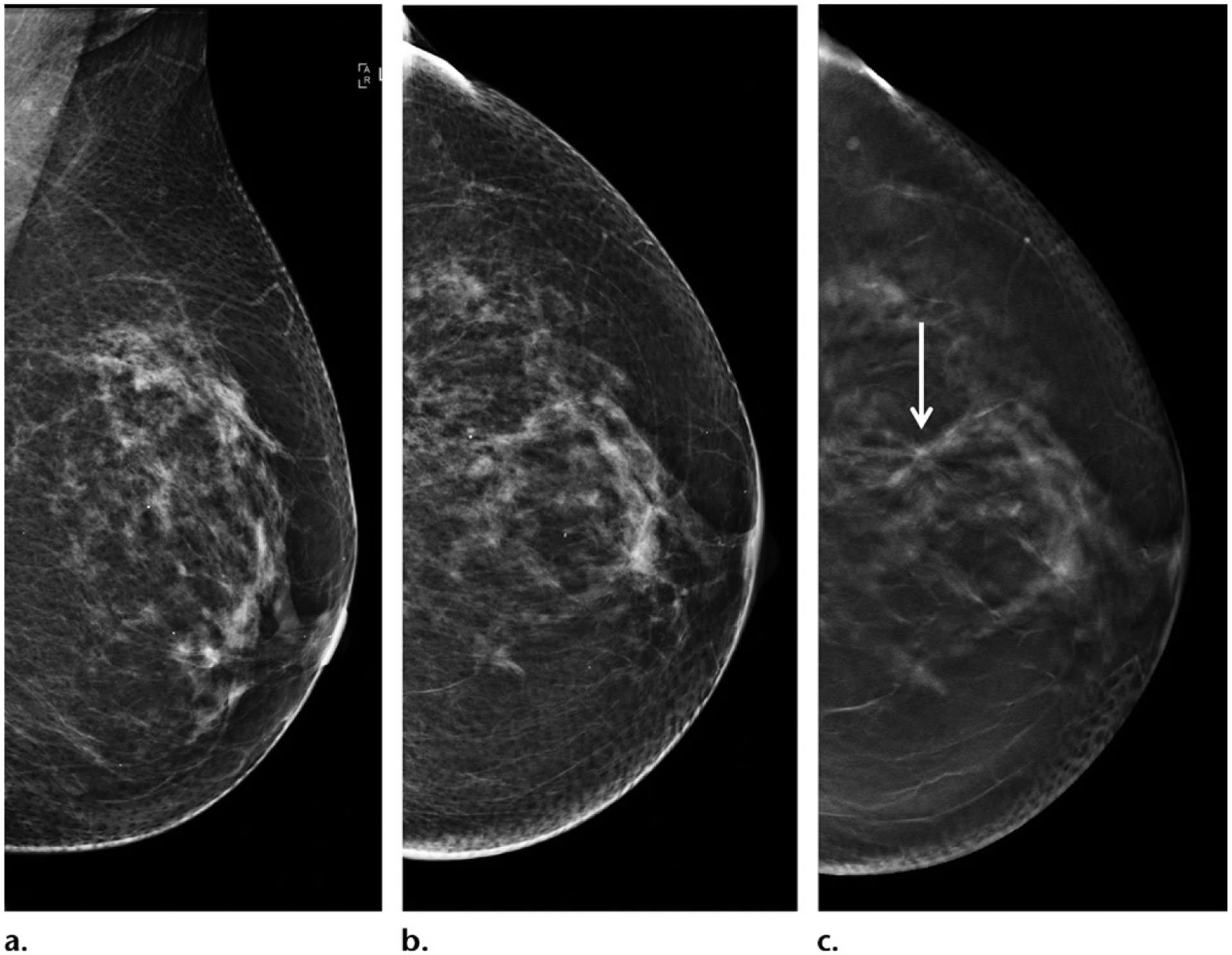
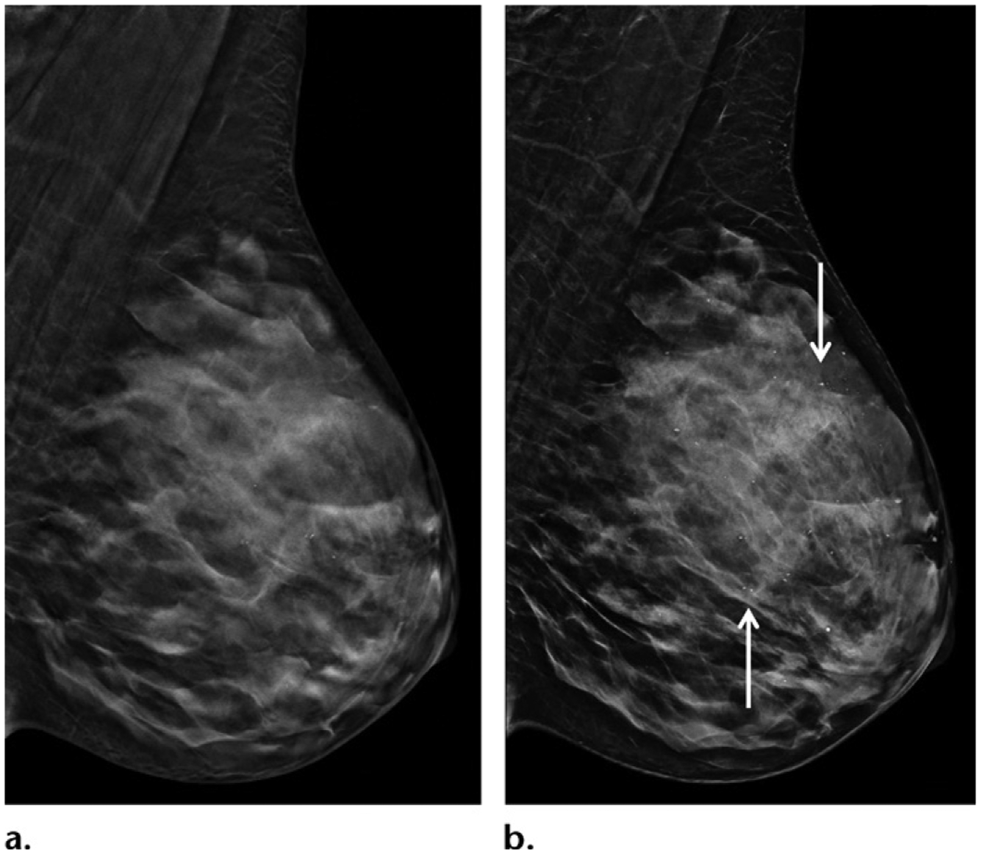
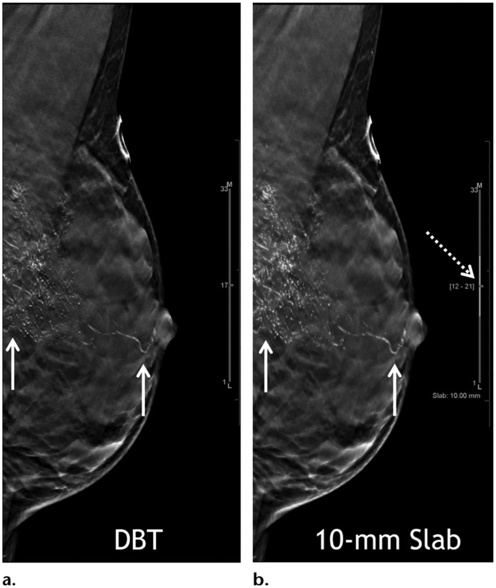
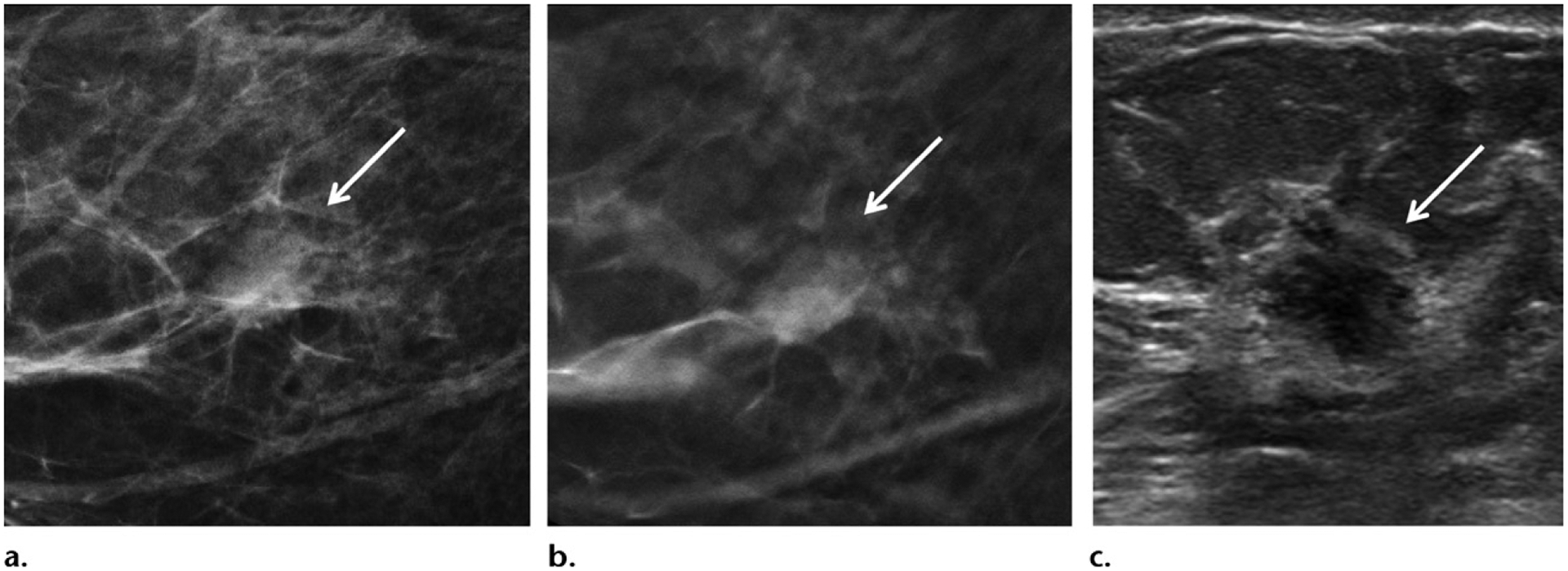
Digital breast tomosynthesis (DBT) has been widely adopted in breast imaging in both screening and diagnostic settings. The benefits of DBT are well established. Compared with two-dimensional digital mammography (DM), DBT preferentially increases detection of invasive cancers without increased detection of in-situ cancers, maximizing identification of biologically significant disease, while mitigating overdiagnosis. The higher sensitivity of DBT for architectural distortion allows increased diagnosis of invasive cancers overall and particularly improves the visibility of invasive lobular cancers. Implementation of DBT has decreased the number of recalls for false-positive findings at screening, contributing to improved specificity at diagnostic evaluation. Integration of DBT in diagnostic examinations has also resulted in an increased percentage of biopsies with positive results, improving diagnostic confidence. Although individual DBT examinations have a longer interpretation time compared with that for DM, DBT has streamlined the diagnostic workflow and minimized the need for short-term follow-up examinations, redistributing much-needed time resources to screening. Yet DBT has limitations. Although improvements in cancer detection and recall rates are seen for patients in a large spectrum of age groups and breast density categories, these benefits are minimal in women with extremely dense breast tissue, and the extent of these benefits may vary by practice environment and by geographic location. Although DBT allows detection of more invasive cancers than does DM, its incremental yield is lower than that of US and MRI. Current understanding of the biologic profile of DBT-detected cancers is limited. Whether DBT improves breast cancer-specific mortality remains a key question that requires further investigation. ©RSNA, 2021.


Conflict of interest statement

Disclosures of Conflicts of Interest.—L.M. Activities related to the present article: disclosed no relevant relationships. Activities not related to the present article: board membership for iCAD and Lunit, grants/grants pending from Siemens. Other activities: disclosed no relevant relationships.

Figures

Figure 1.
Figure 1.
Screening-detected grade 1 node-negative estrogen receptor positive (ER+) , progesterone receptor positive (PR+) human epidermal growth factor receptor 2 negative (HER2−) breast cancer in a 44-year-old woman. (a) Two-dimensional mammogram shows that the cancer is obscured. (b) Mediolateral oblique projection in-plane DBT image clearly shows the cancer (arrow), with improved lesion conspicuity and depiction of tumor margins.
Figure 2.
Figure 2.
Images show distinct visual representations of the logarithm of two-dimensional noise power spectra calculated from the center of the reconstructed plane for each studied DBT system. (Reprinted, with permission, from reference .)
Figure 3.
Figure 3.
Images from the Hologic Dimensions DBT system, which was approved by the FDA in 2011. Mediolateral oblique two-dimensional mammogram (a), synthetic C-view image (b), and in-plane DBT image (c) of the left breast in a 40-year-old woman at baseline screening show diagnostic-quality images of a small spiculated mass (arrow) that was biopsy proven to be a grade 1 ER+/PR+/HER2− invasive ductal carcinoma.
Figure 4.
Figure 4.
Images from the GE SenoClaire DBT system, which was approved by the FDA in 2014. Mediolateral oblique diagnostic-quality two-dimensional (a), synthetic V-preview (b), and DBT (c) images of the right breast in a 78-year-old woman at diagnostic imaging show an irregular mass in the inferior breast (arrow), which is consistent with a biopsy proven ER+/PR+/HER2− invasive lobular carcinoma.
Figure 5.
Figure 5.
Images from the Siemens Mammomat Inspiration system, which was FDA approved in 2015. Mediolateral oblique DBT images at various depths through a normal left breast in a 50-year-old woman at a screening examination show diagnostic image quality.
Figure 6.
Figure 6.
Small spiculated grade 1 node-negative ER+/PR+/HER2− invasive ductal carcinoma in a 62-year-old woman. (a, b) Mediolateral oblique (a) and craniocaudal (b) two-dimensional mammograms show that the tumor is occult. (c) In-plane craniocaudal DBT image shows that the tumor is visible (arrow), but it is occult on other DBT views (not shown). Note that cancers may be better seen or only seen in one projection at DBT because of nonisotropic imaging; therefore, findings suspected to be cancer on only one DBT view require complete workup.
Figure 7.
Figure 7.
Juxtaposition of mediolateral oblique views of the left breast in two separate patients. High-resolution (70-μm pixel size) (a) and standard-resolution (140-μm pixel size) (b) SM images show that glandular details are seen to greater advantage on a than on b. High-resolution acquisition may compensate for the loss of resolution on SM images but is associated with a slightly higher dose of radiation.
Figure 8.
Figure 8.
High-resolution DBT (a) and SM (b) images in mediolateral oblique projection of the left breast in a 45-year-old woman show excellent image quality because of improved resolution (70-μm pixel size). There are scattered punctate microcalcifications in the left breast, shown with great clarity and without significant artifacts on b (arrows).
Figure 9.
Figure 9.
High-grade ductal carcinoma in situ at baseline screening in a 40-year-old woman. Mediolateral oblique in-plane DBT (a) and 10-mm slab (b) images show segmentally distributed fine linear branching calcifications in the left breast (solid arrows). Currently the standard section thickness for DBT reconstruction images is 1 mm. Slab viewing is possible (dashed arrow in b) with adjustable slab thickness to allow quicker review. In this patient, slab viewing also allows a maximum intensity projection–like capture of the overall extent of the abnormality. Stereotactic biopsy was performed and yielded a diagnosis of high-grade ductal carcinoma in situ.
Figure 10.
Figure 10.
Invasive ductal carcinoma in a 56-year-old woman at screening mammography. (a, b) Craniocaudal projection two-dimensional mammogram (a) and in-plane DBT image (b) show scattered fibroglandular tissue and a circumscribed solitary right breast mass (arrow). (c) Subsequent US image shows a corresponding mass with irregular borders (arrow). Biopsy was performed and yielded a grade 2 ER+/PR+/HER2+ node-negative invasive ductal carcinoma.

References

    1. Lowry KP, Coley RY, Miglioretti DL, et al. Screening performance of digital breast tomosynthesis vs digital mammography in community practice by patient age, screening round, and breast density. JAMA Netw Open 2020;3(7):e2011792. - PMC - PubMed
    1. Hovda T, Holen ÅS, Lång K, et al. Interval and consecutive round breast cancer after digital breast tomosynthesis and synthetic 2D mammography versus standard 2D digital mammography in BreastScreen Norway. Radiology 2020;294(2):256–264. - PubMed
    1. Conant EF, Toledano AY, Periaswamy S, et al. Improving accuracy and efficiency with concurrent use of artificial intelligence for digital breast tomosynthesis. Radiol Artif Intell 2019;1(4):e180096. - PMC - PubMed
    1. Zuckerman SP, Conant EF, Keller BM, et al. Implementation of synthesized two-dimensional mammography in a population-based digital breast tomosynthesis screening program. Radiology 2016;281(3):730–736. - PMC - PubMed
    1. Skaane P, Bandos AI, Niklason LT, et al. Digital mammography versus digital mammography plus tomosynthesis in breast cancer screening: The Oslo tomosynthesis screening trial. Radiology 2019;291(1):23–30. - PubMed

LinkOut - more resources