. 2021 Feb 5;16(2):e0244127.
doi: 10.1371/journal.pone.0244127. eCollection 2021.

Airborne aerosol olfactory deposition contributes to anosmia in COVID-19

Affiliations

Airborne aerosol olfactory deposition contributes to anosmia in COVID-19

Alan D Workman et al. PLoS One. .

Abstract

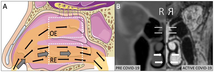
Introduction: Olfactory dysfunction (OD) affects a majority of COVID-19 patients, is atypical in duration and recovery, and is associated with focal opacification and inflammation of the olfactory epithelium. Given recent increased emphasis on airborne transmission of SARS-CoV-2, the purpose of the present study was to experimentally characterize aerosol dispersion within olfactory epithelium (OE) and respiratory epithelium (RE) in human subjects, to determine if small (sub 5μm) airborne aerosols selectively deposit in the OE.

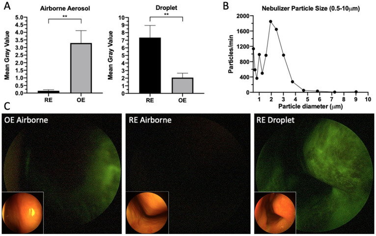
Methods: Healthy adult volunteers inhaled fluorescein-labeled nebulized 0.5-5μm airborne aerosol or atomized larger aerosolized droplets (30-100μm). Particulate deposition in the OE and RE was assessed by blue-light filter modified rigid endoscopic evaluation with subsequent image randomization, processing and quantification by a blinded reviewer.

Results: 0.5-5μm airborne aerosol deposition, as assessed by fluorescence gray value, was significantly higher in the OE than the RE bilaterally, with minimal to no deposition observed in the RE (maximum fluorescence: OE 19.5(IQR 22.5), RE 1(IQR 3.2), p<0.001; average fluorescence: OE 2.3(IQR 4.5), RE 0.1(IQR 0.2), p<0.01). Conversely, larger 30-100μm aerosolized droplet deposition was significantly greater in the RE than the OE (maximum fluorescence: OE 13(IQR 14.3), RE 38(IQR 45.5), p<0.01; average fluorescence: OE 1.9(IQR 2.1), RE 5.9(IQR 5.9), p<0.01).

Conclusions: Our data experimentally confirm that despite bypassing the majority of the upper airway, small-sized (0.5-5μm) airborne aerosols differentially deposit in significant concentrations within the olfactory epithelium. This provides a compelling aerodynamic mechanism to explain atypical OD in COVID-19.


Conflict of interest statement

The authors do not have competing interests that could bias this work.

Figures

Fig 1
Fig 1
A) Illustration of classic description of airborne aerosols (thin arrows) bypassing the nasal respiratory epithelium (RE) with proposed mechanism for a proportion of these aerosols penetrating into the olfactory epithelium (OE). Thick arrows depict larger-sized aerosolized droplets settling within RE (adapted from Servier Medical Art). B) Matched right sided CT and MRI (reflected) scan of same patient demonstrating new onset isolated inflammation of OE (thin arrows) during COVID-19 infection with sparing of RE (thick arrows).
Fig 2
Fig 2
A) Small airborne aerosol (0.5–5μm) deposition versus aerosolized droplet (30–100 μm) deposition of fluorescein solution in the respiratory epithelium (RE) and olfactory epithelium (OE) (small airborne aerosol average fluorescence: OE 2.3(IQR 4.5), RE 0.1(IQR 0.2), p<0.01, aerosolized droplet average fluorescence: OE 1.9(IQR 2.1), RE 5.9(IQR 5.9), p<0.01). B) Size distribution of airborne aerosols in the 0.5–10μm range produced during nebulizer use. C) Blue-light filtered endoscopic nasal images (with brightfield insets) of fluorescein airborne and droplet labeled distribution to the olfactory (OE) and respiratory epithelium (RE) in human subjects.

References

    1. Yan CH, Faraji F, Prajapati DP, Ostrander BT, DeConde AS. Self‐reported olfactory loss associates with outpatient clinical course in Covid‐19. Int Forum Allergy Rhinol. 2020; 10.1002/alr.22592 - DOI - PMC - PubMed
    1. Tong JY, Wong A, Zhu D, Fastenberg JH, Tham T. The Prevalence of Olfactory and Gustatory Dysfunction in COVID-19 Patients: A Systematic Review and Meta-analysis. Otolaryngol Neck Surg [Internet]. 2020. May 5;0194599820926473. 10.1177/0194599820926473 - DOI - PubMed
    1. Kaye R, Chang CWD, Kazahaya K, Brereton J, Denneny JC. COVID-19 Anosmia Reporting Tool: Initial Findings. Otolaryngol Neck Surg. 2020; 10.1177/0194599820922992 - DOI - PubMed
    1. Eliezer M, Hautefort C, Hamel A-L, Verillaud B, Herman P, Houdart E, et al. Sudden and Complete Olfactory Loss Function as a Possible Symptom of COVID-19. JAMA Otolaryngol Neck Surg. 2020. April; 10.1001/jamaoto.2020.0832 - DOI - PubMed
    1. Brann DH, Tsukahara T, Weinreb C, Logan DW, Datta SR. Non-neural expression of SARS-CoV-2 entry genes in the olfactory epithelium suggests mechanisms underlying anosmia in COVID-19 patients. bioRxiv. 2020; - PMC - PubMed